


Press release
Acute inactivation of the replicative helicase in human cells triggers MCM8–9-dependent DNA synthesis
Toyoaki Natsume, Kohei Nishimura, Sheroy Minocherhomji, Rahul Bhowmick, Ian D. Hickson, Masato T. Kanemaki
Genes & Development DOI:10.1101/gad.297663.117
Pressrelease (In Japanese only)
Dr. Toyoaki Natsume and Prof. Masato Kanemaki at National Institute of Genetics, ROIS, together with the group led by Prof. Ian D. Hickson at University of Copenhagen, reported a new system to deal with failure in DNA replication. The finding was published in Genes & Development in advance of the print journal.
In cell proliferation, genomic DNA has to be precisely copied into two (DNA replication) before equal distribution to two daughter cells. For doing this, double-stranded DNA is unwound, the process of which is similar to open a zipper of your clothes (Figure 1A). Similar to pulling the ‘slider’ for opening a zipper, the replicative helicase known as MCM2–7 moves on DNA for unwinding double-stranded DNA. However, because human genomic DNA is very long (approx. 2 m per cell), it is challenging to entirely unwind the genomic DNA. Occasionally, the MCM2–7 helicase falls off from DNA when it encounters a roadblock (such as DNA damage). Because reloading of MCM2–7 is strictly inhibited during S phase (when cells carry out DNA replication), this might lead to incompletion of DNA replication, which causes the loss of genetic information from daughter cells unless the cells have a mechanism to deal with the problem.
In this study, the research groups observed how human cells responded to artificial removal of the MCM2–7 helicase by using the auxin-inducible degron (AID) technology that they had developed previously (the information about this technology is described here). They revealed that the MCM8–9 helicase, which is evolutionally related to the MCM2–7 helicase, promotes a non-canonical DNA synthesis as a backup system after removal of MCM2–7 (Figure 1B).
Many anticancer drugs kills cancer cells by inducing DNA lesions, which enhance removal of MCM2–7 from DNA during DNA replication. An inhibitor of the MCM8–9 helicase might enhance the effect of existing anticancer drugs by shutting off this backup system (Figure 2).
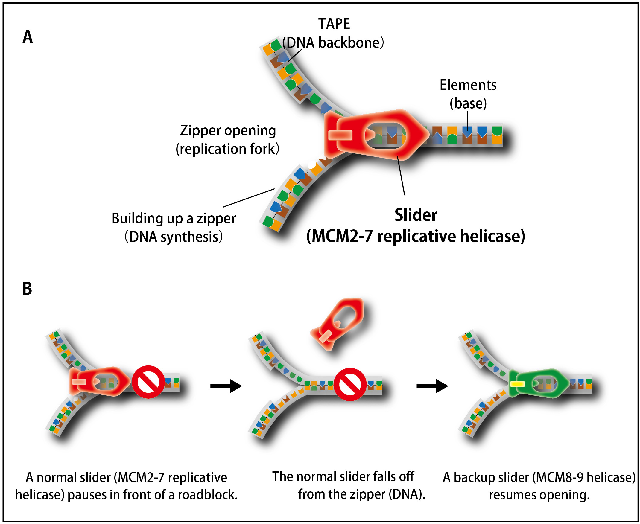
Figure 1. The process of unwinding double-stranded DNA during DNA replication is similar to that of opening a zipper of your clothes.
(A) Similar to the slider that opens a zipper, the MCM2–7 replicative helicase opens double-stranded DNA in cells.(B) When the replicative MCM2–7 helicase (a normal slider) encounters to a roadblock, it occasionally falls off from DNA. To continue DNA synthesis, cells recruit the MCM8–9 helicase as a backup slider.
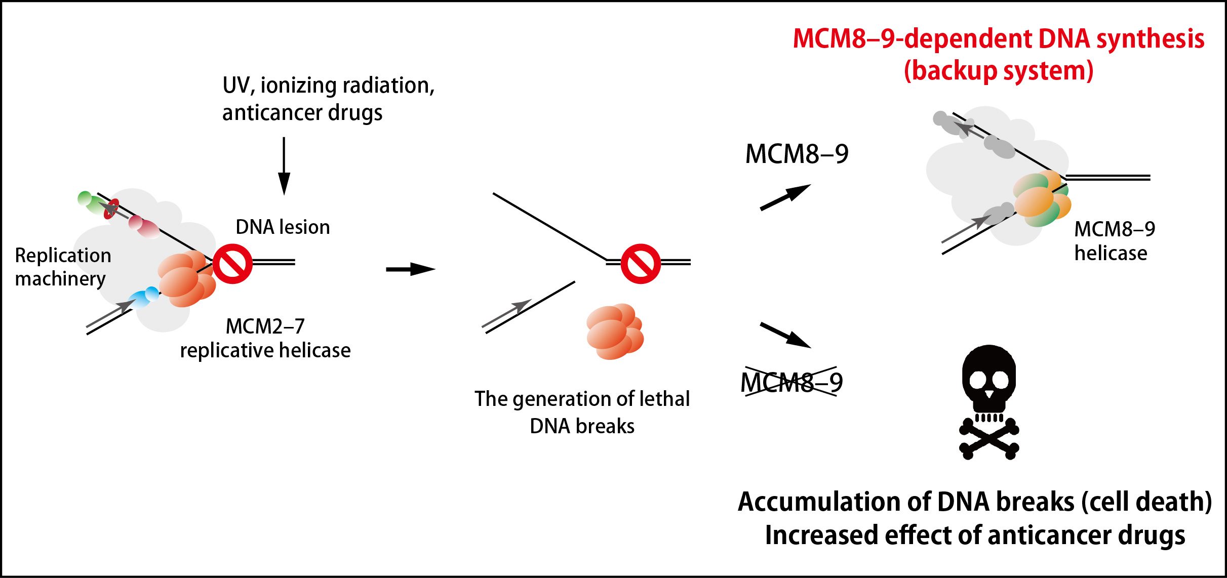
Figure 2. An MCM8–9-dependent backup system against failure in DNA replication.
The MCM2–7 replicative helicase falls off when encountered to a roadblock on DNA, leading to generation of DNA breaks. In this study, the research groups found that the MCM8–9 helicase continues DNA synthesis on behalf of the MCM2–7 (top right). If MCM8–9 does not work, the accumulation of DNA breaks results in cell death (bottom right).
Press release
A novel plasma membrane-anchored protein regulates xylem cell-wall deposition through microtubule-dependent lateral inhibition of Rho GTPase domains
Yuki Sugiyama, Mayumi Wakazaki, Kiminori Toyooka, Hiroo Fukuda, Yoshihisa Oda
Current Biology DOI:10.1016/j.cub.2017.06.059
Press release (In Japanese only)
Cellulose is an indispensable material in many applications for our life and industry. Cellulose is the main component of plant cell walls. However, the regulatory mechanism of cell wall synthesis is poorly understood. Our research group identified a novel gene named IQD13 that promotes cell wall synthesis in the model plant, Arabidopsis. We found that IQD13 products stabilizes microtubules that serve as a scaffold of cellulose synthesis. We also found that IQD13 increases area that are active for cell wall synthesis at the cell surface.
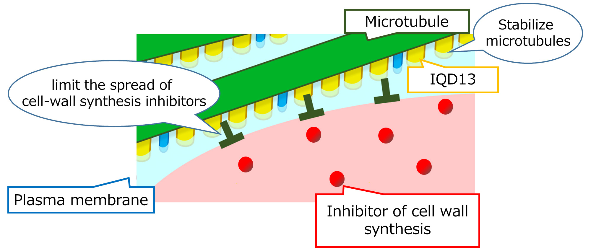
Figure: IQD13 prevents microtubule disruption and spread of the inhibitor of cell wall synthesis.
Press release
Acute inactivation of the replicative helicase in human cells triggers MCM8–9-dependent DNA synthesis
Toyoaki Natsume, Kohei Nishimura, Sheroy Minocherhomji, Rahul Bhowmick, Ian D. Hickson, Masato T. Kanemaki
Genes & Development DOI:10.1101/gad.297663.117
Pressrelease (In Japanese only)
Dr. Toyoaki Natsume and Prof. Masato Kanemaki at National Institute of Genetics, ROIS, together with the group led by Prof. Ian D. Hickson at University of Copenhagen, reported a new system to deal with failure in DNA replication. The finding was published in Genes & Development in advance of the print journal.
In cell proliferation, genomic DNA has to be precisely copied into two (DNA replication) before equal distribution to two daughter cells. For doing this, double-stranded DNA is unwound, the process of which is similar to open a zipper of your clothes (Figure 1A). Similar to pulling the ‘slider’ for opening a zipper, the replicative helicase known as MCM2–7 moves on DNA for unwinding double-stranded DNA. However, because human genomic DNA is very long (approx. 2 m per cell), it is challenging to entirely unwind the genomic DNA. Occasionally, the MCM2–7 helicase falls off from DNA when it encounters a roadblock (such as DNA damage). Because reloading of MCM2–7 is strictly inhibited during S phase (when cells carry out DNA replication), this might lead to incompletion of DNA replication, which causes the loss of genetic information from daughter cells unless the cells have a mechanism to deal with the problem.
In this study, the research groups observed how human cells responded to artificial removal of the MCM2–7 helicase by using the auxin-inducible degron (AID) technology that they had developed previously (the information about this technology is described here). They revealed that the MCM8–9 helicase, which is evolutionally related to the MCM2–7 helicase, promotes a non-canonical DNA synthesis as a backup system after removal of MCM2–7 (Figure 1B).
Many anticancer drugs kills cancer cells by inducing DNA lesions, which enhance removal of MCM2–7 from DNA during DNA replication. An inhibitor of the MCM8–9 helicase might enhance the effect of existing anticancer drugs by shutting off this backup system (Figure 2).
Figure 1. The process of unwinding double-stranded DNA during DNA replication is similar to that of opening a zipper of your clothes.
(A) Similar to the slider that opens a zipper, the MCM2–7 replicative helicase opens double-stranded DNA in cells.(B) When the replicative MCM2–7 helicase (a normal slider) encounters to a roadblock, it occasionally falls off from DNA. To continue DNA synthesis, cells recruit the MCM8–9 helicase as a backup slider.
Figure 2. An MCM8–9-dependent backup system against failure in DNA replication.
The MCM2–7 replicative helicase falls off when encountered to a roadblock on DNA, leading to generation of DNA breaks. In this study, the research groups found that the MCM8–9 helicase continues DNA synthesis on behalf of the MCM2–7 (top right). If MCM8–9 does not work, the accumulation of DNA breaks results in cell death (bottom right).
Biological Macromolecules Laboratory / Maeshima Group
Density imaging of heterochromatin in live cells using orientation-independent-DIC microscopy
Ryosuke Imai, Tadasu Nozaki, Tomomi Tani, Kazunari Kaizu, Kayo Hibino, Satoru Ide, Sachiko Tamura, Koichi Takahashi, Michael Shribak, and Kazuhiro Maeshima
Molecular Biology of the Cell, 2017 DOI:10.1091/mbc.E17-06-0359
In eukaryotic cells, highly condensed inactive/silenced chromatin has long been called “heterochromatin.” However, recent research suggests that such regions are in fact not fully transcriptionally silent, and that there exists only a moderate access barrier to heterochromatin. To further investigate this issue, it is critical to elucidate the physical properties of heterochromatin such as its total density in live cells. Here, using orientation-independent differential interference contrast (OI-DIC) microscopy, which is capable of mapping optical path differences, we investigated the density of the total materials in pericentric foci, a representative heterochromatin model, in live mouse NIH3T3 cells. We demonstrated that the total density of heterochromatin (208 mg/mL) was only 1.53-fold higher than that of the surrounding euchromatic regions (136 mg/mL) while the DNA density of heterochromatin was 5.5- to 7.5-fold higher. This surprisingly small difference may be due to that non-nucleosomal materials (proteins/RNAs)(∼120 mg/mL) are dominant in both chromatin regions. Monte Carlo simulation suggested that non-nucleosomal materials contribute to creating a moderate access barrier to heterochromatin, allowing minimal protein access to functional regions. Our OI-DIC imaging offers insight into the density of live cellular environments.
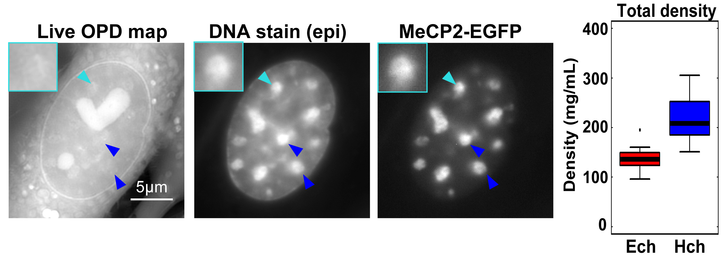
Typical images of the Optical Path Distance (OPD) map (reflecting density at each pixel) by OI-DIC, DNA staining, and MeCP2-EGFP (heterochromatin marker) signals in live NIH3T3 cells. Large foci seen in DNA staining and MeCP2-EGFP images (arrowheads) were assumed to be heterochromatin. Note that the OPD of the foci was similar or only slightly higher than that of the surrounding euchromatin. (Right) The analyzed total densities of pericentric heterochromatin foci (Hch, 208 mg/mL) and euchromatin (Ech, 136 mg/mL). The median density ratio between them was 1.53.
NIG will be closed from August 14 and August 15, 2017 for summer holiday.
Thank you for your understanding and cooperation.
Press release
Hierarchy in the home cage affects behaviour and gene expression in group-housed C57BL/6 male mice
Yasuyuki Horii, Tatsuhiro Nagasawa, Hiroyuki Sakakibara, Aki Takahashi, Akira Tanave, Yuki Matsumoto, Hiromichi Nagayama, Kazuto Yoshimi, Michiko T. Yasuda, Kayoko Shimoi, Tsuyoshi Koide
Scientific Reports Article number: 6991 (2017) DOI:10.1038/s41598-017-07233-5
Pressrelease (In Japanese only)
Social stress is one of the major causes of depression in humans. It is therefore important that the effects of social stress on behaviour and gene expression in the brain are studied. We performed experiments using male mice to analyse social hierarchy in groups of mice in cages and investigated emotional behaviour and hippocampal gene expression in the mice. We found significantly different emotional behaviour and expression of genes associated with the serotonergic system in dominant and subordinate mice. Treating the mice with a selective serotonin reuptake inhibitor antidepressant restored gene expression in subordinate mice and caused the emotional behaviour of the subordinate mice to be recovered, suggesting that alterations to the serotonergic system are key factors in phenotypic changes caused by the social ranks of subordinate mice. We believe that the results of this study are important because they improve our understanding of how social stress induces mental health problems in humans.
Press release
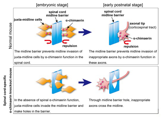
Spinal RacGAP α-chimaerin is required to establish the midline barrier for proper corticospinal axon guidance
Shota Katori, Yukiko Noguchi-Katori, Shigeyoshi Itohara, Takuji Iwasato
Journal of Neuroscience 26 July 2017, 3123-16; DOI:10.1523/JNEUROSCI.3123-16.2017
Pressrelease (In Japanese only)
The midline barrier plays a critical role in midline axon guidance, which is fundamental to the formation of neural circuits that are responsible for proper left-right coordination of our body. Studies have revealed some of the mechanisms underlying how the midline barrier navigates axons. In contrast, the establishment of the midline barrier during embryonic development remains unclear. In this study, Katori et al. determined that α-chimaerin is required for the formation of an intact midline barrier. Spinal cord-specific α-chimaerin knockout mice had spinal midline barriers with numerous breaks (holes), through which corticospinal axons aberrantly crossed the midline. Katori et al. propose that α-chimaerin protects the midline barrier by mediating cell repulsive signaling in juxta-midline cells, which prevents these cells from invading the midline.
Source: Journal of Neuroscience 26 July 2017, 3123-16;
DOI:10.1523/JNEUROSCI.3123-16.2017
Division of Human Genetics / Inoue Group
Systematic Identification and Characterization of Regulatory Elements Derived from Human Endogenous Retroviruses.
Jumpei Ito, Ryota Sugimoto, Hirofumi Nakaoka, Shiro Yamada, Tetsuaki Kimura, Takahide Hayano, and Ituro Inoue.
PLoS Genetics. Jul 12;13(7):e1006883. 2017. DOI:10.1371/journal.pgen.1006883
Human endogenous retroviruses (HERVs) have regulatory elements that possibly influence the transcription of host genes. We systematically identified these HERV regulatory elements (HERV-REs) based on publicly available datasets of ChIP-Seq. Overall, 794,972 HERV-REs were identified. Clustering analysis showed that HERVs can be grouped according to the TF binding patterns; HERV groups bounded by pluripotent TFs (e.g., SOX2, POU5F1, and NANOG), endoderm TFs (e.g., GATA4/6, SOX17, and FOXA1/2), hematopoietic TFs (e.g., SPI1, GATA1/2, and TAL1), and CTCF were identified. Regulatory elements of HERVs tended to locate nearby genes involved in immune responses, indicating that the regulatory elements play an important role in controlling the immune regulatory network. Finally, we constructed dbHERV-REs, an interactive database of HERV regulatory elements (http://herv-tfbs.com/). This study provides fundamental information in understanding the impact of HERVs on host transcription, and offers insights into the transcriptional modulation systems of HERVs.
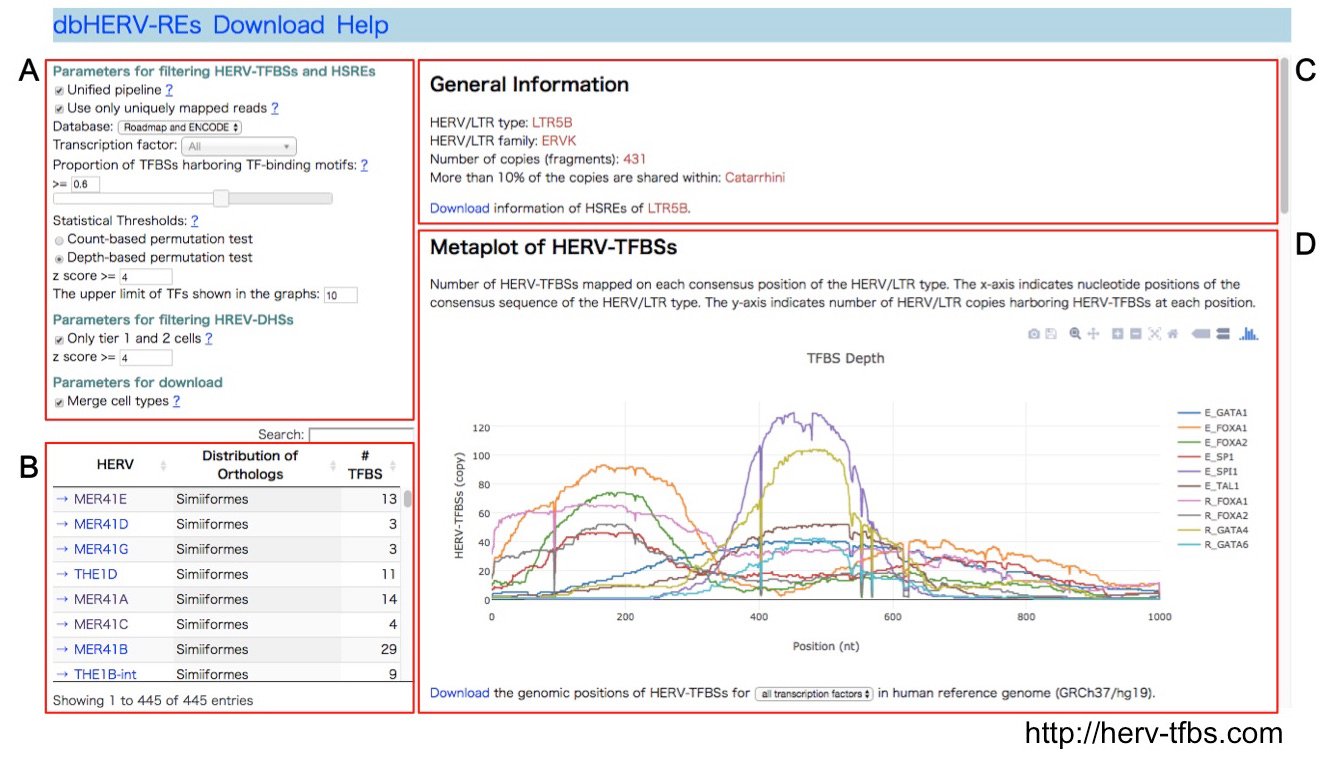
dbHERV-RE (http://herv-tfbs.com/). First, users choose a transcription factor and other parameters (A). Second, users select a type of HERVs (B). dbHERV-REs displays general information of the HERVs (phylogenetic classification, copy number, and insertion date) (C) and visualizes genetic positions of HERV-REs on the HERV sequence and the human reference genome (D).
Press release
Dynamic organization of chromatin domains revealed by super-resolution live-cell imaging
Tadasu Nozaki, Ryosuke Imai, Mai Tanbo, Ryosuke Nagashima, Sachiko Tamura, Tomomi Tani, Yasumasa Joti, Masaru Tomita, Kayo Hibino, Masato T. Kanemaki, Kerstin S. Wendt, Yasushi Okada, Takeharu Nagai, and Kazuhiro Maeshima
Molecular Cell Published: July 13, 2017 DOI:10.1016/j.molcel.2017.06.018
Press release (In Japanese only)
The eukaryotic genome is organized within cells as chromatin. For proper information output, higher-order chromatin structures can be regulated dynamically. How such structures form and behave in various cellular processes remains unclear. Here, by combining super-resolution imaging (photoactivated localization microscopy [PALM]) and single-nucleosome tracking, we developed a nuclear imaging system to visualize the higher-order structures along with their dynamics in live mammalian cells. We demonstrated that nucleosomes form compact domains with a peak diameter of ∼160 nm and move coherently in live cells. The heterochromatin-rich regions showed more domains and less movement. With cell differentiation, the domains became more apparent, with reduced dynamics. Furthermore, various perturbation experiments indicated that they are organized by a combination of factors, including cohesin and nucleosome-nucleosome interactions. Notably, we observed the domains during mitosis, suggesting that they act as building blocks of chromosomes and may serve as information units throughout the cell cycle.
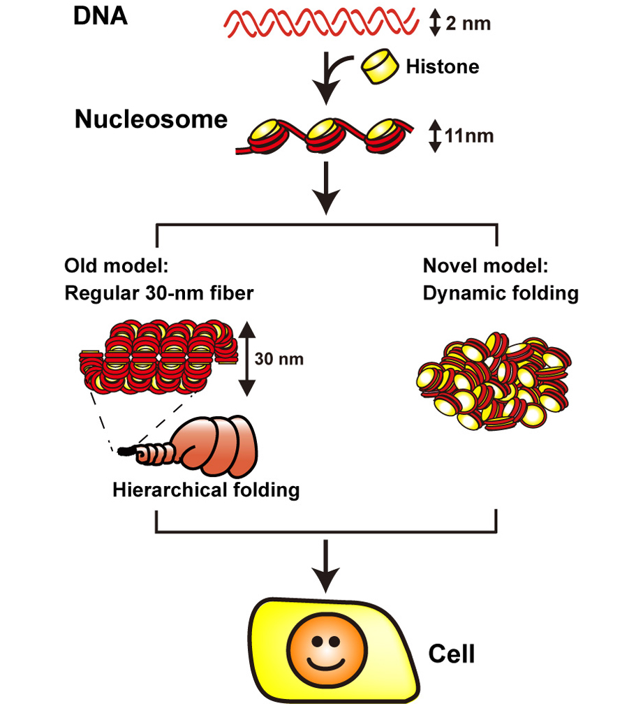
1. DNA (in red) is wrapped around core histone (in yellow) and forms a nucleosome structure (or 10-nm fiber). In the old model (left), the nucleosome fiber had long been assumed to fold into a 30-nm chromatin fiber, and subsequently into helically folded larger fibers (Hierarchical folding). In new current model (right), chromatin is composed of irregularly folded 10-nm fibers, without 30-nm chromatin fibers (dynamic folding) and stored in the cell.
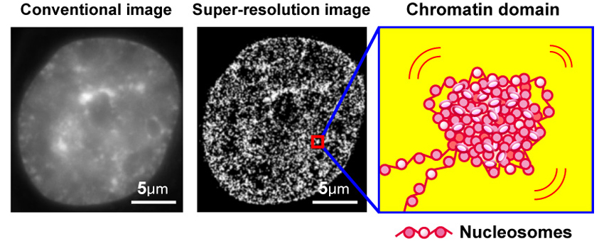
2. Left, conventional DNA staining image; center, super-resolution image of chromatin; a model of chromatin domain.
Press release
Selective breeding and selection mapping using a novel wild-derived heterogeneous stock of mice revealed two closely-linked loci for tameness
Yuki Matsumoto, Tatsuhiko Goto, Jo Nishino, Hirofumi Nakaoka, Akira Tanave, Toshiyuki Takano-Shimizu, Richard F Mott, Tsuyoshi Koide
Scientific Reports 7, Article number: 4607 (2017) DOI:10.1038/s41598-017-04869-1
Pressrelease (In Japanese only)
Tameness play important role during the process of domestication, and can be divided into two potential components: motivation to approach humans (active tameness) and reluctance to avoid them (passive tameness). To understand the genetic basis associated with active tameness in mice we applied selective breeding of a genetically heterogeneous population that we founded by crossing eight wild mouse strains. As a result of selective breeding, the level of active tameness increased over the generations, compared to an unselected control experiment. We performed two selection and two control experiments. Genetic differences between the selected and control groups, measured using a high-dense array of single-nucleotide polymorphisms, were assessed using a computer simulation experiment. In one selection experiment we found a significant increase in the occurrence of a particular genomic segment present in just one of the founder strains, compared to the control groups. This selected region contains two loci related to active tameness and is syntenic to genomic regions which are known to be a region selected during dog domestication, suggesting that responsible genes in these loci are associated with active tameness in both mouse and dog.
Yasuto MURAYAMA joined the Center for Frontier Science as of July 1, 2017.
MURAYAMA, Yasuto:Center for Frontier Research,Chromosome Biochemistry Laboratory

Center for Frontier Research is an incubation center to simultaneously develop two elements: human resources and new research fields. Promising young scientists conduct research as principal investigator (tenure-track associate professor) to explore new frontiers in genetics and related areas, taking advantage of NIG’s research infrastructure and various support systems.
Mammalian Development Laboratory / Saga Group
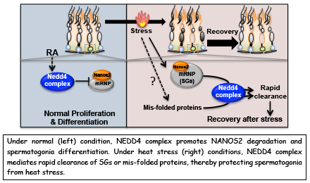
NEDD4 controls spermatogonial stem cell homeostasis and stress response by regulating messenger ribonucleoprotein complexes
Zhi Zhou, Hiroshi Kawabe, Atsushi Suzuki, Kaori Shinmyozu & Yumiko Saga
NATURE COMMUNICATIONS. 8, Article number: 15662 (2017) DOI:10.1038/ncomms15662
P bodies (PBs) and stress granules (SGs) are conserved cytoplasmic aggregates of cellular messenger ribonucleoprotein complexes (mRNPs) that are implicated in mRNA metabolism and play crucial roles in adult stem cell homeostasis and stress responses. However, the mechanisms underlying the dynamics of mRNP granules are poorly understood. Here, we report NEDD4, an E3 ubiquitin ligase, as a key regulator of mRNP dynamics that controls the size of the spermatogonial progenitor cell (SPC) pool. We find that NEDD4 targets an RNA-binding protein, NANOS2, in spermatogonia to destabilize it, leading to cell differentiation. In addition, NEDD4 is required for SG clearance. NEDD4 targets SGs and facilitates their rapid clearance through the endosomal–lysosomal pathway during the recovery period. Therefore, NEDD4 controls the turnover of mRNP components and inhibits pathological SG accumulation. Accordingly, we propose that a NEDD4-mediated mechanism regulates mRNP dynamics, and facilitates SPC homeostasis and viability under normal and stress conditions.This research was mainly conducted by Zhi Zhou who was a NIG postdoctoral fellow 2012-2014.
Press release
Acute inactivation of the replicative helicase in human cells triggers MCM8–9-dependent DNA synthesis
Toyoaki Natsume, Kohei Nishimura, Sheroy Minocherhomji, Rahul Bhowmick, Ian D. Hickson, Masato T. Kanemaki
Genes & Development DOI:10.1101/gad.297663.117
Pressrelease (In Japanese only)
Dr. Toyoaki Natsume and Prof. Masato Kanemaki at National Institute of Genetics, ROIS, together with the group led by Prof. Ian D. Hickson at University of Copenhagen, reported a new system to deal with failure in DNA replication. The finding was published in Genes & Development in advance of the print journal.
In cell proliferation, genomic DNA has to be precisely copied into two (DNA replication) before equal distribution to two daughter cells. For doing this, double-stranded DNA is unwound, the process of which is similar to open a zipper of your clothes (Figure 1A). Similar to pulling the ‘slider’ for opening a zipper, the replicative helicase known as MCM2–7 moves on DNA for unwinding double-stranded DNA. However, because human genomic DNA is very long (approx. 2 m per cell), it is challenging to entirely unwind the genomic DNA. Occasionally, the MCM2–7 helicase falls off from DNA when it encounters a roadblock (such as DNA damage). Because reloading of MCM2–7 is strictly inhibited during S phase (when cells carry out DNA replication), this might lead to incompletion of DNA replication, which causes the loss of genetic information from daughter cells unless the cells have a mechanism to deal with the problem.
In this study, the research groups observed how human cells responded to artificial removal of the MCM2–7 helicase by using the auxin-inducible degron (AID) technology that they had developed previously (the information about this technology is described here). They revealed that the MCM8–9 helicase, which is evolutionally related to the MCM2–7 helicase, promotes a non-canonical DNA synthesis as a backup system after removal of MCM2–7 (Figure 1B).
Many anticancer drugs kills cancer cells by inducing DNA lesions, which enhance removal of MCM2–7 from DNA during DNA replication. An inhibitor of the MCM8–9 helicase might enhance the effect of existing anticancer drugs by shutting off this backup system (Figure 2).
Figure 1. The process of unwinding double-stranded DNA during DNA replication is similar to that of opening a zipper of your clothes.
(A) Similar to the slider that opens a zipper, the MCM2–7 replicative helicase opens double-stranded DNA in cells.(B) When the replicative MCM2–7 helicase (a normal slider) encounters to a roadblock, it occasionally falls off from DNA. To continue DNA synthesis, cells recruit the MCM8–9 helicase as a backup slider.
Figure 2. An MCM8–9-dependent backup system against failure in DNA replication.
The MCM2–7 replicative helicase falls off when encountered to a roadblock on DNA, leading to generation of DNA breaks. In this study, the research groups found that the MCM8–9 helicase continues DNA synthesis on behalf of the MCM2–7 (top right). If MCM8–9 does not work, the accumulation of DNA breaks results in cell death (bottom right).
Division of Microbial Genetics / Araki Group
Flexible DNA Path in the MCM Double Hexamer Loaded on DNA
Kohji Hizume, Hiroaki Kominami, Kei Kobayashi, Hirofumi Yamada, and Hiroyuki Araki
Biochemistry. Publication Date (Web): May 1, 2017 DOI:10.1021/acs.biochem.6b00922
The formation of the pre-replicative complex (pre-RC) during the G1 phase, which is also called the licensing of DNA replication, is the initial and essential step of faithful DNA replication during the subsequent S phase. It is widely accepted that in the pre-RC, double-stranded DNA passes through the holes of two ring-shaped minichromosome maintenance (Mcm) 2–7 hexamers; however, the spatial organization of the DNA and proteins involved in the pre-RC formation is unclear.
A research group lead by Drs. Hiroyuki Araki and Kohji Hizume at NIG reconstituted pre-RC from purified DNA and proteins and visualized the complex using atomic force microscopy (AFM). Higher-resolution imaging of the pre-RC was performed by using FM-AFM in collaboration with the group of Professor Hirofumi Yamada at Kyoto University, and successfully detected two globules (two MCM hexamers) of the elliptical particle formed on DNA.
Analyses through AFM observation revealed that the DNA does not completely pass through both holes of the MCM hexamers, possibly because the DNA exited from the gap between Mcm2 and Mcm5. A DNA loop fastened by the MCM double hexamer was detected in pre-RC samples reconstituted from purified proteins as well as those purified from yeast cells. These results suggest that a higher-order architecture of the loaded MCM hexamers and DNA strands during licensing of DNA replication, which have not been proposed before.
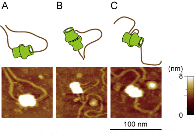
Loaded MCM–DNA complex visualized at higher resolution using FM-AFM. It was detected that the DNA passes through two MCM hexamers (A) as it is widely accepted. However, these were also detected that the DNA does not completely pass through both the MCM hexamers (B) and that DNA was fastened by the MCM double hexamer and form loop (C).
Press release
Activation of the hypothalamic feeding centre upon visual prey detection
Akira Muto, Pradeep Lal, Deepak Ailani, Gembu Abe, Mari Itoh, Koichi Kawakami
Nature Communications 8, Article number: 15029 (2017) DOI:10.1038/ncomms15029
Pressrelease (In Japanese only)
Have you ever wondered why just seeing food can make your mouth start to water? By visualizing neuronal activity in specific areas of the zebrafish brain, scientists at the National Institute of Genetics (NIG) in Japan have revealed a direct link between visual perception of food and feeding motivation. The study, published in the April 20, 2017 issue of Nature Communications, suggests that “eating with the eyes” is deeply rooted in evolution.
This study was supported by JSPS KAKENHI Grant Numbers JP25290009 and JP25650120, and also partly supported by JSPS KAKENHI Grant Numbers JP15H02370 and JP16H01651, and NBRP from Japan Agency for Medical Research and Development (AMED). This work was also supported in part by the Center for the Promotion of Integrated Sciences (CPIS) of SOKENDAI.
Click here for article
https://www.eurekalert.org/pub_releases/2017-04/rooi-wt041717.php
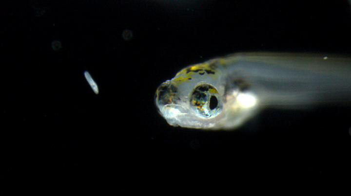
THIS IS A ZEBRAFISH LARVA TRYING TO CATCH PREY.
▶This study is based on the previous study.
Experimental Farm / Nonomura Group
Mammalian Genetics Laboratory / Shiroishi Group
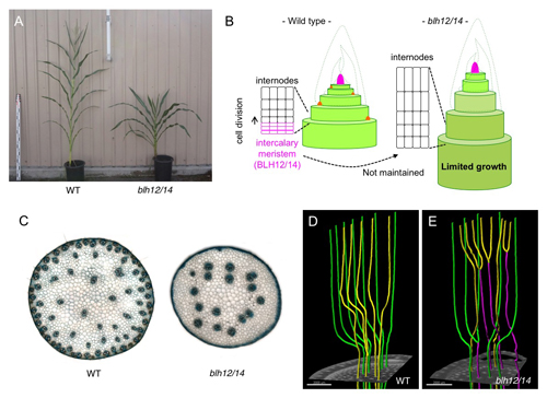
KNOTTED1 Cofactors, BLH12 and BLH14, Regulate Internode Patterning and Vein Anastomosis in Maize
Katsutoshi Tsuda, Maria-Jazmin Abraham-Juarez, Akiteru Maeno, Zhaobin Dong, Dale Aromdee, Robert Meeley, Toshihiko Shiroishi, Ken-ichi Nonomura and Sarah Hake.
The Plant Cell. published online April 5, 2017 DOI:10.1105/tpc.16.00967
Monocot stems lack the vascular cambium and instead have characteristic structures in which intercalary meristems generate internodes and veins remain separate and scattered. Developmental processes of these unique structures, however, have been poorly described. We found that maize BELL1-like homeodomain transcription factors, BLH12 and BLH14, have redundant but important roles in stem development. BLH12/14 interact with the shoot meristem regulator, KNOTTED1 (KN1) in vivo, and accumulate in overlapping domains in shoot meristems, young stems and provascular bundles. In addition to defects in the maintenance and development of various shoot meristems, blh12/14 double mutant showed unique abnormalities in the stem including shortened internodes and the reduced vein number. Detailed observation of BLH12/14 accumulation patterns and of stem inner structures using micro-computed tomography (CT) suggested that BLH12/14 (1) maintain intercalary meristems at the bottom of internodes to provide internodal cells as differentiated progenies, and (2) prevent precocious anastomosis of provascular bundles in young stems to ensure the production of sufficient independent veins. This work is the collaborative work between Katsutoshi Tsuda in Experimental Farm and Prof. Sarah Hake in University of California, Berkeley, and was supported by JSPS KAKENHI JP16K18637. Micro-CT scanning performed by Akiteru Maeno in Mammalian Genetics Lab enabled to capture the detailed view of inner stem structures and vein networks.
Three new faculty have joined NIG as of April 1, 2017.
professor
SAITO, Kuniaki : Invertebrate Genetics Laboratory
Associate Professor
KAWAMOTO, Shoko : Genetic Informatics Laboratory


Assistant Professor


