


Press release
Real-Time Visualization of Neuronal Activity during Perception
Akira Muto, Masamichi Ohkura, Gembu Abe, Junichi Nakai and Koichi Kawakami Current Biology, 23(4), Feb 18, 2013. DOI: 10.1016/j.cub.2012.12.040The visual world is first projected onto the retina, and the visual information is further transmitted to the brain. In this initial stage of visual processing, the visual world is mapped on the brain, which is called visuotopy. This is a common feature found in the brains of all animals with eyes. While visuotopy is a well-established notion, no one has ever demonstrated this in real time in a natural condition. In this study we developed an improved version of GCaMP, a calcium sensor, in collaboration with Prof. Nakai at Saitama University. Using this new highly sensitive calcium probe, we could visualize neuronal activity in a zebrafish larva during visual perception in prey capture behavior.
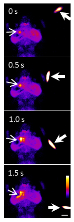
A swimming paramecium (arrowheads) evoked Ca2+ transients (arrows) in the neuropil and cell bodies of the left tectum of a one-week old larva embedded in agarose. Ratio images were created and pseudo-colored. Scale bar represents 100 μm.
▶This method is one of the basis of these researches. A virtual reality system to analyze neural activity and behavior in adult zebrafish Appetite control via hunger and satiety Neural signatures of sleep in zebrafish Glia-neuron interactions underlie state transitions to generalized seizures ‘Eating with the eyes’ is hard-wired in the brain


