Koide Group / Mouse Genomics Resource Laboratory
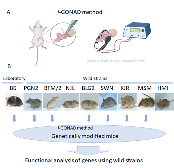
Efficient genome editing in wild strains of mice using the i-GONAD method
Yuji Imai, Akira Tanave, Makoto Matsuyama, and Tsuyoshi Koide
Scientific Reports (2022) 12, 13821 DOI:10.1038/s41598-022-17776-x
At the National Institute of Genetics, nine wild mouse strains have been established. These strains have characteristics not found in laboratory strains, such as marked genetic differences between different strains and behavior characteristic of wild mice.
These series of wild strains, named the Mishima Battery, are provided to researchers in Japan and overseas as highly unique resources, and are used for various research in the fields of cancer, immunology, development, and behavior. However, in spite of these excellent characteristics, wild strains have the problem of being difficult to apply genetic engineering technology.
In collaboration with Shigei Medical Research Institute and RIKEN, technical staff Yuji Imai and Associate Professor Tsuyoshi Koide of the Mouse Genomics Resource Laboratory have applied genome-editing using i-GONAD method, which does not involve any ex vivo manipulation of unfertilized or fertilized egg. The group showed that it is possible to efficiently modify genes in most wild strains by using this method.
First, in vitro fertilization was performed on the experimental strain C57BL/6 (B6 strain) and nine wild strains to investigate the efficiency of in vitro fertilization necessary for general genome editing experiments. As a result, it was found that only two wild strains were able to perform in vitro fertilization with the similar efficiencies as the B6 strain, and the other strains were extremely inefficient. Therefore, we applied a method called i-GONAD, developed by Professor Masato Otsuka of Tokai University, to wild strains, which performs genome editing without ex vivo manipulation of fertilized eggs. As a result, we succeeded in performing genome editing in 7 out of the 9 strains examined. This result indicates that it has become possible to efficiently perform genetic modification using wild strains in the future, and the use of wild strains in many research fields can be expected.
Kimura Group / Cell Architecture Laboratory
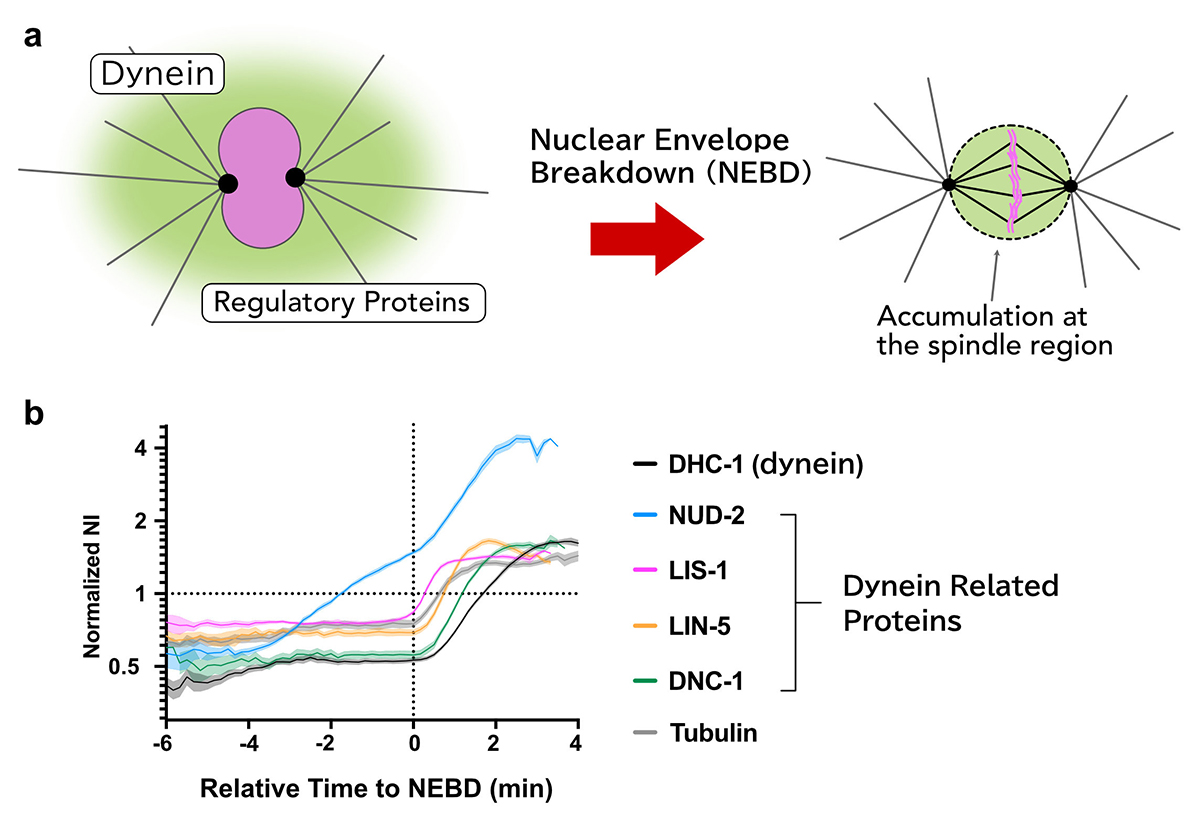
Sequential accumulation of dynein and its regulatory proteins at the spindle region in the Caenorhabditis elegans embryo
Takayuki Torisawa, & Akatsuki Kimura
Scientific Reports (2022) 12, 11740 DOI:10.1038/s41598-022-15042-8
Cytoplasmic dynein is a molecular motor responsible for various cellular activities, including intracellular transport and cell division. To achieve various functions, dynein needs to be regulated by other proteins, and it has still been elusive how these regulations are achieved in many cellular contexts.
Using the early embryos of Caenorhabditis elegans, we focused on the spatiotemporal regulation of dynein during mitosis, where dynein and its regulatory proteins translocated from the cytoplasm to the spindle region, and observed the dynamics of dynein and the regulatory proteins.
We revealed that there are i) selectivity, ii) varieties in the accumulation sites, and iii) the order of accumulation in the accumulation dynamics in the spindle region.
Furthermore, we found that the accumulation of NUD-2 was unique among the dynein regulators we analyzed. NUD-2 started to accumulate before NEBD (pre-NEBD accumulation). Using a protein injection approach, we revealed that the C-terminal helix of NUD-2 was responsible for pre-NEBD accumulation. These findings suggest a fine temporal control of the subcellular localization of regulatory proteins.
This work was supported by JSPS Grants-in-Aid for Scientific Research JP19K16094, JP18H02414, JP18H05529, and JP18KK0202.
Maeshima Group / Genome Dynamics Laboratory
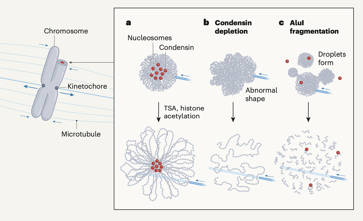
A phase transition for chromosome transmission when cells divide
Kazuhiro Maeshima
Nature 2022 August 03 DOI:10.1038/d41586-022-01925-3
Mitotic chromosomes are the structure where DNA is tightly compacted, and they carry genetic information to be passed on to the next generation. These chromosomes are transmitted from the mother cell onto the daughter cell with physical force from microtubes and other factors. In other words, chromosomes should have mechanical resistance to endure such forces.
Recently, Daniel W. Gerlich and his colleagues have shown that chromosomes condense and gain such mechanical resistance by phase transition(Schneider et al. “A chromatin phase transition protects mitotic chromosomes against microtubule perforation” Nature 2022 doi: 10.1038/ s41586-022-05027-y). In this paper, Schneider et al. have shown that global histone deacetylation cause phase transition in mitotic chromosomes, which makes them condensed and resistant to mechanical forces. The authors have also shown that a protein complex called condensin, which was previously thought to be essential for chromosome condensation, is not involved in the condensation process itself.
Professor Kazuhiro Maeshima at Genome Dynamics Laboratory wrote a commentary on this paper in the News & Views section of Nature. Maeshima discussed how global histone deacetylation causes chromosome condensation and the role of condensin in shaping rod-like chromosomes (chromatin loop formation mechanism).
July 25, 2022
Research Organization of Information and Systems
National Institute of Genetics
The announcement of new coronavirus infections after July 25 will be made on the following website instead of the Information section.
NIG Measures for the Novel Coronavirus Infectious Disease
NIG will be closed from August 15 to August 16, 2022 for summer holiday.
Thank you for your understanding and cooperation.
MURAYAMA, Yasuto tenure track associate professor at the Center for Frontier Research, has been awarded tenure as of July 1, 2022.
MURAYAMA, Yasuto: Chromosome Biochemistry Laboratory

New assistant professor joins NIG as of July 1, 2022.
KAWAGUCHI,Akane : Kuraku Group • Molecular Life History Laboratory
A stepwise route to domesticate rice by controlling seed shattering and panicle shape
Ryo Ishikawa, Cristina Cobo Castillo, Than Myint Htun, Koji Numaguchi, Kazuya Inoue, Yumi Oka, Miki Ogasawara, Shohei Sugiyama, Natsumi Takama, Chhourn Orn, Chizuru Inoue, Ken-Ichi Nonomura, Robin Allaby, Dorian Q Fuller and Takashige Ishii
PNAS (2022) 119, e2121692119 DOI:10.1073/pnas.2121692119
![]() Press release (In Japanese only)
Press release (In Japanese only)
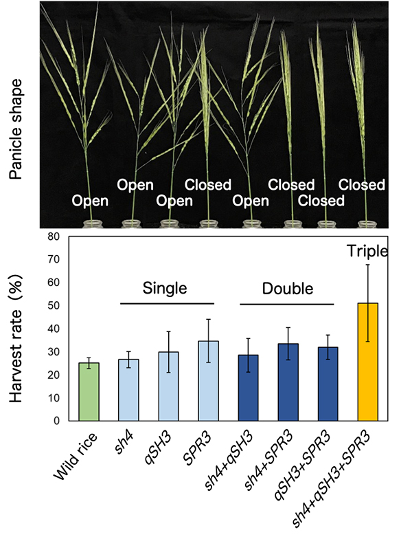
The international collaborative group of Kobe University, National Institute of Genetics (NIG), University of Warwick, University College London, Yezin Agricultural University, Cambodian Agricultural Research and Development Institute, successfully unveiled that the Asian wild rice (O. rufipogon Griff.) dramatically had increased grain yields with mutations at three genes in an initial step of domestication.
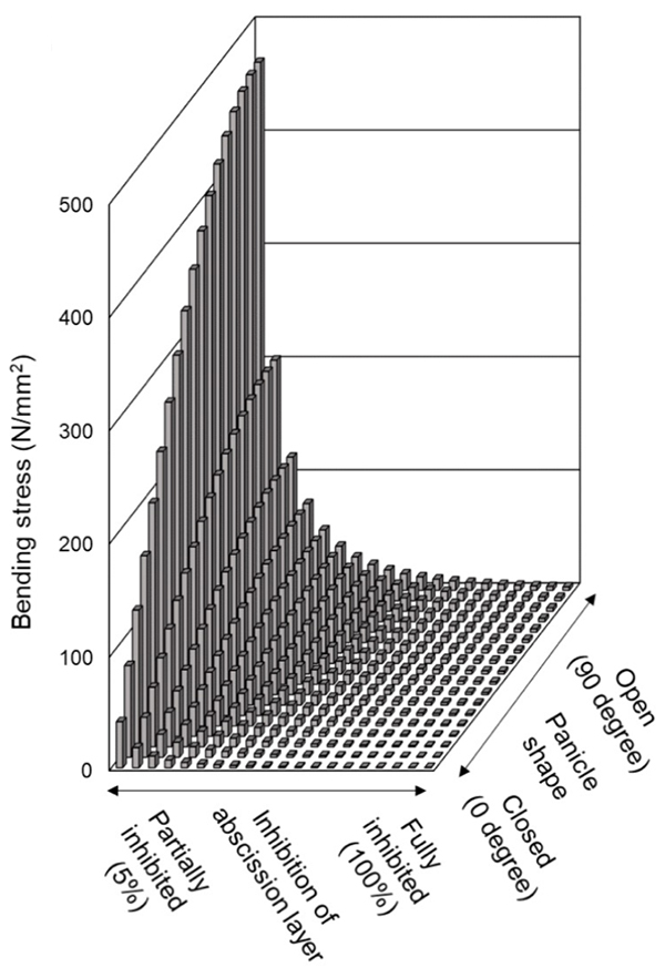
The loss of seed-shattering behaviour is an important step of domestication enabling humans to harvest more grains. At first, to experimentally reproduce an earlier process of rice domestication, several domestication-related genes were replaced with those of cultivated rice in the genetic background of wild rice, O. rufipogon. The replacement alone with a cultivated rice-type mutation of sh4, a rice gene with a major impact on seed shattering, was insufficient, but the replacement with cultivated rice-type mutations of both sh4 and qSH3, an allele within the seed-shattering gene OsSh1, caused the partial loss of abscission layer formation in O. rufipogon. However, the combination of two mutations of sh4 and qSH3 was not sufficient to increase yield under natural conditions. To further increase yield, our group found that the replacement together with cultivated rice-type mutations of three genes, sh4, qSH3 and SPR3, dramatically increased yields (Fig. 1). SPR3 is a rice gene controlling a panicle shape, and the closed panicle property enhances long awn-retaining seeds entangled, and in addition, reduces a bending moment which is a predominant factor affecting seed dispersal (Fig. 2), resulting in increased harvests.
We propose a stepwise route in the earliest phase of rice domestication, in which SPR3-controlled closed panicle morphology was instrumental in addition to sequential recruitments of sh4- and qSH3-dependent loss of shattering.
This study was supported partly by NIG-JOINT program 83A2016-2018. The wild rice accession, maintained by NIG in the support of National Bioresource Project (NBRP) Rice, was used in this study.
Inoue Group / Human Genetics Laboratory
Detection of Ancient Viruses and Long-Term Viral Evolution
Luca Nishimura, Naoko Fujito, Ryota Sugimoto, and Ituro Inoue*
Viruses (2022) 14, 1336 DOI:10.3390/v14061336
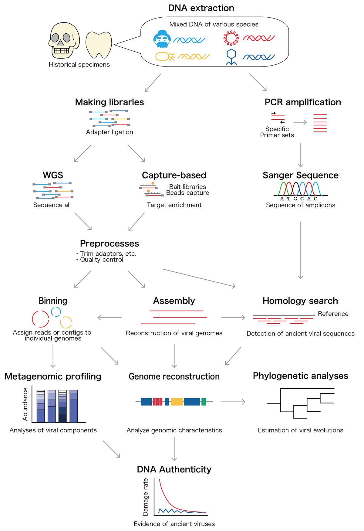
Archaeological remains contain Ancient DNA and RNA, and those nucleic acids provide genomic information about ancient people together with ancient microbiomes and viruses that infected ancient individuals. Since 1997, more than 20 ancient viral species including influenza virus and hepatitis B virus have been discovered from historical samples such as bones and mummified tissues. Those ancient viral genomes have been utilized to estimate the past pandemics of pathogenic viruses within the ancient human population and long-term evolutionary events. In our review article, we overview the ancient viral studies and experimental and analytical techniques and discuss the long-term viral evolutionary studies using ancient viral genomes.
Maeshima Group / Genome Dynamics Laboratory
Chromatin behavior in living cells: lessons from single-nucleosome imaging and tracking
*Satoru Ide, Sachiko Tamura, *Kazuhiro Maeshima *corresponding author
BioEssays 2022 June 03 DOI:10.1002/bies.202200043
Eukaryotic genome DNA is wrapped around core histones and forms a nucleosome structure. Together with associated proteins and RNAs, these nucleosomes are organized three-dimensionally in the cell as chromatin. Emerging evidence demonstrates that chromatin consists of rather irregular and variable nucleosome arrangements without the regular fiber structure and that its dynamic behavior plays a critical role in regulating various genome functions. Single-nucleosome imaging is a promising method to investigate chromatin behavior in living cells. It reveals local chromatin motion, which reflects chromatin organization not observed in chemically fixed cells. The motion data is like a gold mine. Data analyses from many aspects bring us more and more information that contributes to better understanding of genome functions. In this review article, we describe imaging of single-nucleosomes and their tracked behavior through oblique illumination microscopy. We also discuss applications of this technique, especially in elucidating nucleolar organization in living cells.
This work was supported by JSPS Grants and MEXT KAKENHI grants (21H02535, 20H05936 and 21H02453) and the Uehara Memorial Foundation.
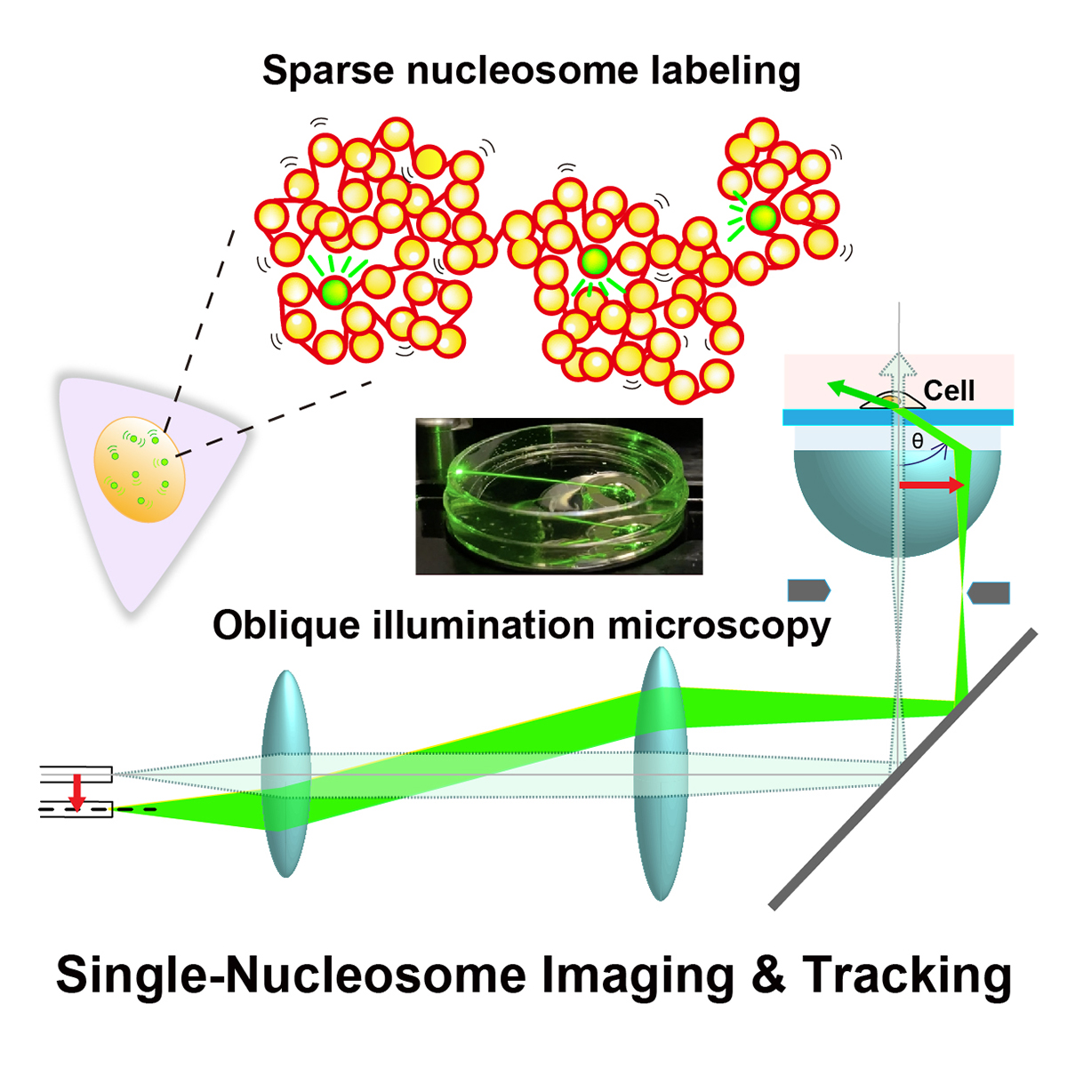
Figure: Single-nucleosome imaging and tracking is a promising technique to investigate dynamic chromatin behavior in living cells. We describe how this imaging works using sparse nucleosome labeling and oblique illumination microscopy, and how information on chromatin dynamics can be extracted from the obtained motion data.
Video1: How laser beams traveled through a glass dish filled with medium using an oblique illumination microscopy. As the incident laser beam shifts from the center axis, higher refraction angles of the beam from the glass are obtained. At the last 2 frames, total internal reflection (TIR) occurs and only the glass surface is illuminated.
Video2: Movie data (50 ms/frame) of single nucleosomes (basic units of chromatin) fluorescently labeled in a living human cell. Note that clear, well-separated dots and their movements were visualized.
Press release
Single-nucleosome imaging reveals steady-state motion of interphase chromatin in living human cells
Shiori Iida, Soya Shinkai, Yuji Itoh, Sachiko Tamura, Masato T. Kanemaki, Shuichi Onami, Kazuhiro Maeshima
Science Advances (2022) 8, eabn5626 DOI:10.1126/sciadv.abn5626
![]() Press release (In Japanese only)
Press release (In Japanese only)
The human body is composed of over forty trillion cells. Each of these cells has totally two meters of tightly packaged genomic DNA, the blueprint of life. Recently, there have been many advances in understanding how DNA is packaged and organized as chromatin in the cell. In contrast, how chromatin behaves in living cells remains unclear.
SOKENDAI graduate student Shiori Iida, JSPS Fellow Yuji Itoh, Technical Stuff Sachiko Tamura, and Professor Kazuhiro Maeshima of Genome Dynamics Laboratory (NIG), together with Research Scientist Soya Shinkai and Team Leader Shuichi Onami of RIKEN BDR, and Professor Masato T. Kanemaki of Molecular Cell Engineering Laboratory (NIG), have investigated the local movements of chromatin in living human cells using super-resolution fluorescence microscopy (Movie 1).
Both DNA amount and nuclear size become double during the preparation period for the cell division (interphase). Previously, it has been suggested that these drastic changes in the nuclear environment would affect chromatin movements. However, Iida et al. have revealed that chromatin motion keeps a steady state throughout the interphase. Chromatin motion is directly related to the accessibility of the DNA (readability of genomic information). Steady-state chromatin motion allows cells to conduct housekeeping tasks under similar environments during interphase.
This work was supported by JSPS and MEXT KAKENHI grants (20H05550、21H05763、19K23735、 20J00572、18H05412、19H05273、20H05936), a Japan Science and Technology Agency CREST grant (JPMJCR15G2), JST SPRING(JPMJSP2104), the Takeda Science Foundation, and the Uehara Memorial Foundation.
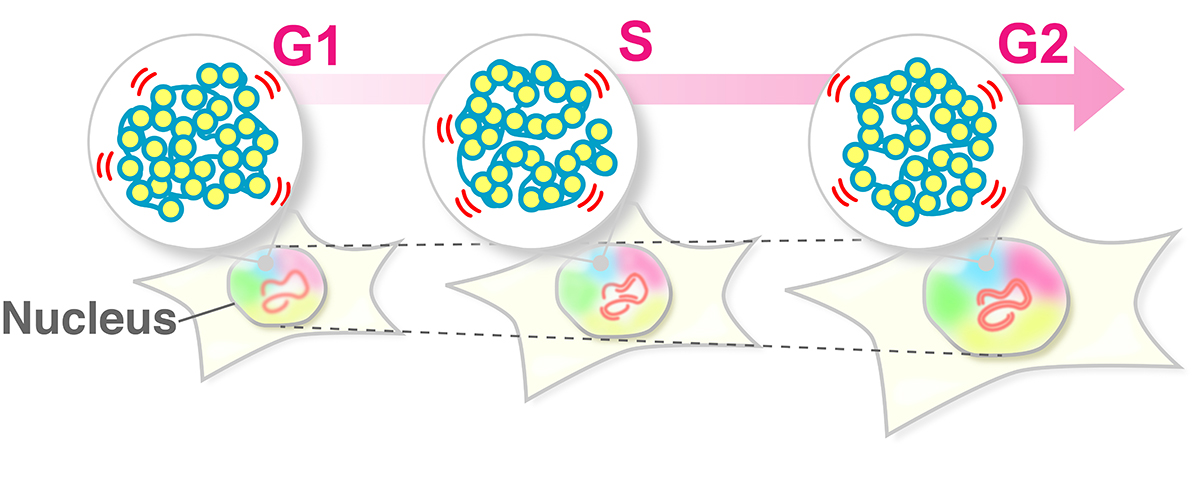
Figure: The swaying motion of chromatin keeps a steady state throughout the interphase (G1, S, G2 phases) despite increases in DNA amount and nuclear volume. Steady-state chromatin motion allows cells to conduct housekeeping tasks under similar environments during interphase.
Video1: Movie data (50 ms/frame) of single nucleosomes (basic units of chromatin) fluorescently labeled in a living human cell. Note that clear, well-separated dots and their movements were visualized.
Video2: Movie data (50 ms/frame) of single nucleosomes fluorescently labeled in living human cells; (left) G1-phase, (right) G2-phase. Note that there is not much difference in nucleosome motion between G1 and G2 phase, even as nuclear size increases.
▶ The article of EurekAlert! is here.
Press release
The extra-embryonic space and the local contour are critical geometric constraints regulating cell arrangement
*Sungrim Seirin-Lee, Kazunori Yamamoto, *Akatsuki Kimura
*Corresponding authors
Development (2022) 149, dev200401 DOI:10.1242/dev.200401
![]() Press release (In Japanese only)
Press release (In Japanese only)
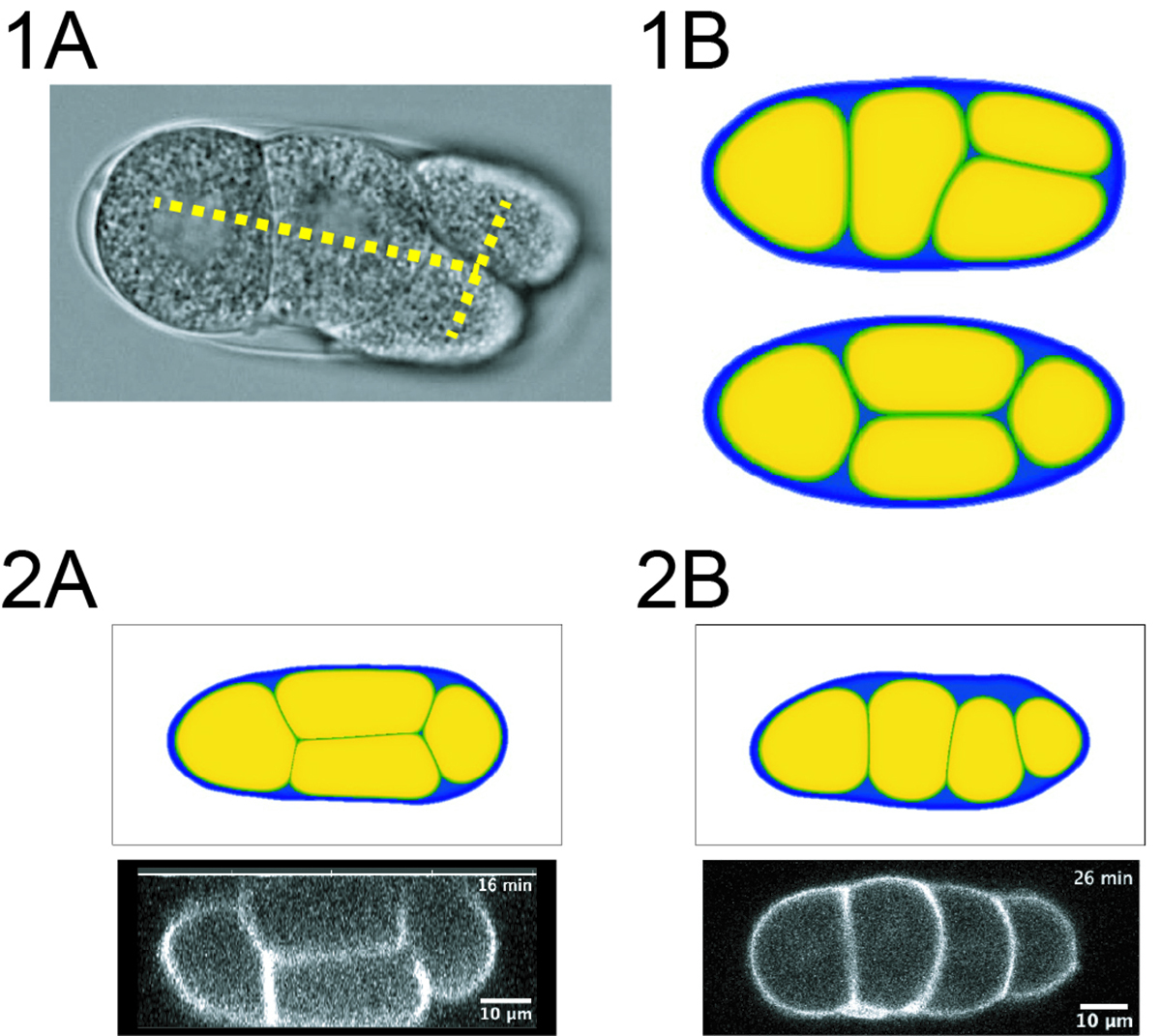
Arrangement of cells which defines how cells contact each other is important in developmental processes. The mechanisms determining cell arrangement can be classified into three factors: orientation of cell division, interaction between cells, and geometrical constraints provided by surrounding structures where cells are confined such as the eggshell. Among these factors, the contribution of geometrical constrains have been less explored. Many of theoretical and experimental approaches exploring cell arrangements inside the eggshell have assumed the eggshell as a pure ellipsoidal shape. In this study, the authors developed a computational model based on a phase-field method that can incorporate the real shape of the eggshells. Using this model and the experimental data obtained with the nematode Caenorhabditis elegans embryo, the authors showed that an arrangement observed in vivo can be explained by the real shape but not by an ellipsoid. Extending this finding, the authors found the amount of the extra-embryonic space (ES), the empty space within the eggshell not occupied by embryonic cells, is critical to define cell arrangement. The study proposed that the local features of geometric constraints such as the ES, play important roles in cell arrangement, which should be important for any multicellular systems.
Prof. Kanemaki’s News and Views on the recent finding on DNA replication was published in Nature.
A rethink about enzymes that drive DNA replication
Comment from Prof. Kanemaki:
I wrote a short review article about a new paper published in Nature. It has been believed that two kinases, CDC7 and CDK2, drive DNA replication. However, this paper showed that either CDC7 or CDK1 suffices for driving DNA replication in mouse and human cells. In addition, the auxin-inducible degron (AID) that my group developed was used in this research.
Kanemaki Group • Molecular Cell Engineering Laboratory
A special issue on Cell Nucleus of Current Opinion in Cell Biology edited by Prof. Kazuhiro Maeshima at Genome Dynamics Laboratory and Prof. Eran Meshorer, Hebrew University of Israel, has been published.
There are 11 review articles on cell nuclei in Vol. 74, published in February 2022, and 4 articles in Vol. 75, published in April 2022.
https://www.sciencedirect.com/journal/current-opinion-in-cell-biology/vol/74/suppl/C
https://www.sciencedirect.com/journal/current-opinion-in-cell-biology/vol/75/suppl/C
Vol. 74 includes “Ligand-induced degrons for studying nuclear functions” by NIG Prof. Masato Kanemaki and also a review paper on plant chromatin organization by NIG Visiting Professor Frederic Berger.
https://www.sciencedirect.com/science/article/pii/S0955067421001228
https://www.sciencedirect.com/science/article/pii/S0955067421001186
Press release
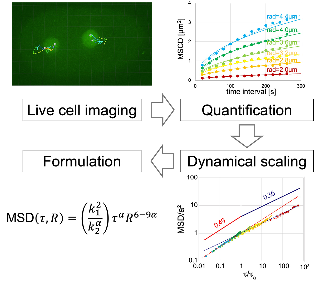
Formulation of chromatin mobility as a function of nuclear size during C. elegans embryogenesis using polymer physics theories.
Aiya K. Yesbolatova, Ritsuko Arai, *Takahiro Sakaue, and *Akatsuki Kimura. *Corresponding authors
Physical Review Letters (2022) 128, 178101 DOI:10.1103/PhysRevLett.128.178101
![]() Press release (In Japanese only)
Press release (In Japanese only)
By combining theory and experiment, Aiya K. Yesbolatova and her collaborators provided unambiguous evidence that the chromatin in early embryos obeys the universal dynamics predicted by the polymer physics. Although the chromatin dynamics is believed to play a key role in controlling the gene expression, its quantitative characterization has been elusive mainly due to the complexity in living cells. The findings and formulation help researchers quantify the chromatin motion in living cells, thus laying the foundation for future research in this biologically important problem.