Press release
Dazl is a target RNA suppressed by mammalian NANOS2 in sexually differentiating male germ cells
Yuzuru Kato, Takeo Katsuki, Hiroki Kokubo, Aki Masuda, Yumiko Saga
Nature Communications 7, Article number: 11272 DOI:10.1038/NCOMMS11272
Press Release (Only in Japanese)
Evolutionally conserved Nanos RNA-binding proteins play crucial roles in germ cell development. While a mammalian Nanos family protein, NANOS2, is required for sexual differentiation of male (XY) germ cells in mice, the underlying mechanisms and the identities of its target RNAs in vivo remain elusive. Using comprehensive microarray analysis and a bacterial artificial chromosome transgenic system, we identify Dazl, a germ cell-specific gene encoding an RNA-binding protein implicated in translation, as a crucial target of NANOS2. Importantly, removal of the Dazl 3′ UTR in XY germ cells stabilises the Dazl mRNA, resulting in elevated meiotic gene expression, abnormal resumption of the cell cycle, and impaired processing-body formation, reminiscent of Nanos2-knockout phenotypes. Furthermore, our data suggest that NANOS2 acts as an antagonist of the DAZL protein. We propose a dual system of NANOS2-mediated suppression of Dazl expression as a pivotal molecular mechanism promoting sexual differentiation of XY germ cells. This work was supported by a Grant-in-Aid for Young Scientists (B) from the Japan Society for the Promotion of Science (JSPS) to Y.K. (Grant number 25840091) and a Grant-in-Aid for Scientific Research (S) from JSPS to Y.S. (Grant number 21227008).
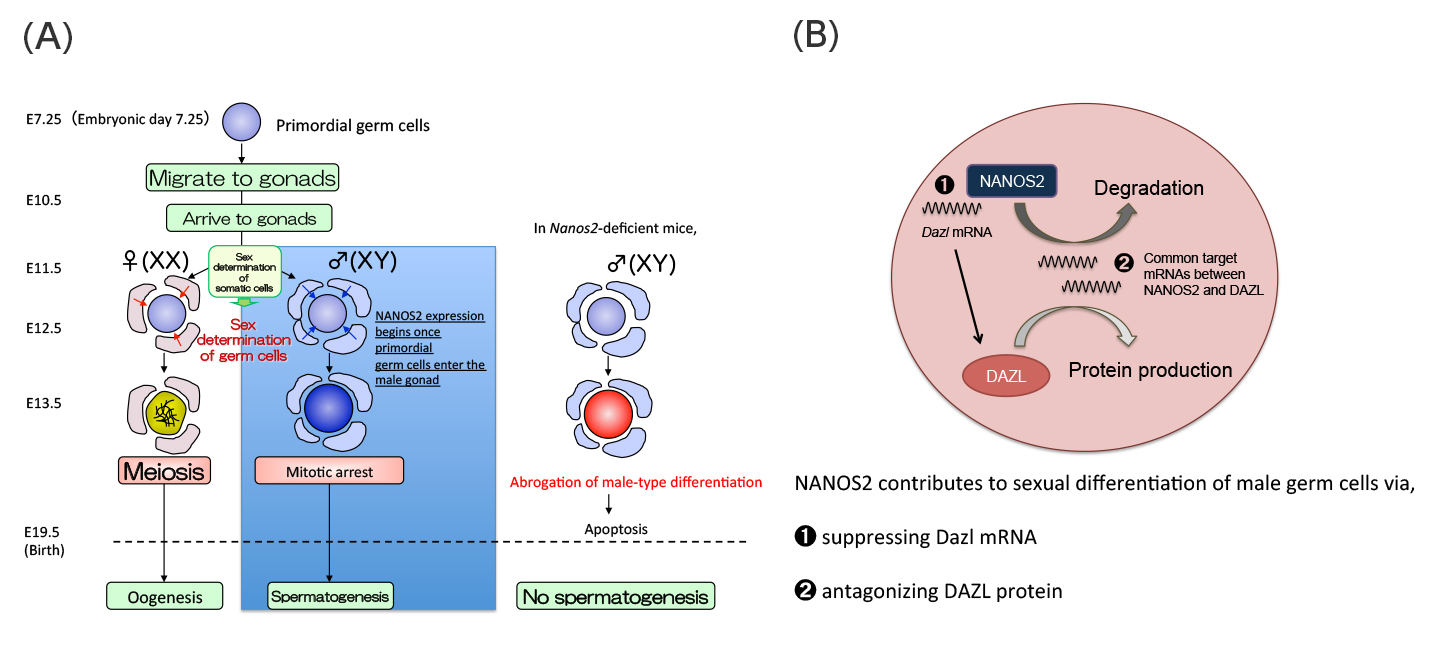
(A)Sex of mouse germ cells is determined after they are enclosed by gonadal somatic cells at E11.5. NANOS2 expression begins once primordial germ cells enter the sex-specific process of sexual differentiation in male germ cells. Germ cells lacking the Nanos2 gene die by apoptosis due to the abrogated male-type differentiation.
(B)Working model of NANOS2-dependent Dazl suppression in sexually differentiating male germ cells.
Press release
Nucleosomal arrays self-assemble into supramolecular globular structures lacking 30-nm fibers
Kazuhiro Maeshima, Ryan Rogge, Sachiko Tamura, Yasumasa Joti, Takaaki Hikima, Heather Szerlong, Christine Krause, Jake Herman, Erik Seidel, Jennifer DeLuca, Tetsuya Ishikawa, and Jeffrey C. Hansen
The EMBO Journal Published online 12.04.2016 DOI:10.15252/embj.201592660
The existence of a 30-nm fiber as a basic folding unit for DNA packaging has remained a topic of active discussion. Here we characterize the supramolecular structures formed by reversible Mg2+-dependent self-association of linear 12-mer nucleosomal arrays using microscopy and physicochemical approaches. These structures, which we call ’oligomers’, were globular throughout all stages of the cooperative assembly process, and ranged in size from ~50 nm to a maximum diameter of ~1000 nm. The nucleosomal arrays were packaged within the oligomers as interdigitated 10-nm fibers, rather than folded 30-nm structures. Linker DNA was freely accessible to micrococcal nuclease, although the oligomers remained partially intact after linker DNA digestion. The organization of chromosomal fibers in human nuclei in situ was stabilized by 1 mM MgCl2 but became disrupted in the absence of MgCl2, conditions that also dissociated the oligomers in vitro. These results indicate that a 10-nm array of nucleosomes has the intrinsic ability to self-assemble into large chromatin globules stabilized by nucleosome-nucleosome interactions, and suggest that the oligomers are good in vitro model for investigating the structure and organization of interphase chromosomes.
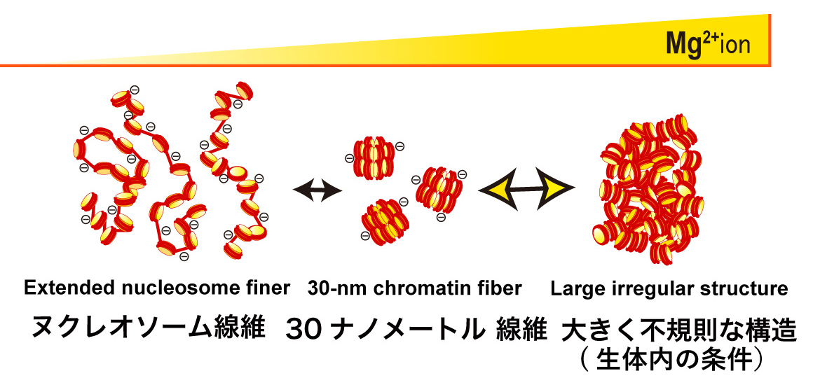
The 12-mer nucleosomal array is a well-defined model chromatin system. Without Mg2+, the nucleosomal array is extended by repulsion (Left). In 1-2 mM Mg2+, the nucleosomal array folds into a regular 30-nm chromatin fiber structure (Center). With further increases in Mg2+, the nucleosome arrays assemble into supramolecular structures (Right). The large structures are not assemblies of the 30-nm chromatin fibers, but interdigitated and melted structures of 10-nm nucleosomal arrays, reflecting on the native chromatin structure in the cell.
Press release
Allelic Imbalance in Regulation of ANRIL through Chromatin Interaction at 9p21 Endometriosis Risk Locus
Hirofumi Nakaoka, Aishwarya Gurumurthy, Takahide Hayano, Somayeh Ahmadloo, Waleed H Omer, Kosuke Yoshihara, Akihito Yamamoto, Keisuke Kurose, Takayuki Enomoto, Shigeo Akira, Kazuyoshi Hosomichi, Ituro Inoue
PLOS Genetics Published: April 7, 2016 DOI:10.1371/journal.pgen.1005893
Press Release (Only in Japanese)
A large number of variants associated with human complex diseases have been discovered by genome-wide association studies (GWASs). These discoveries have been anticipated to be translated into the definitive understanding of disease pathogeneses; however, functional characterization of the disease-associated SNPs remains a formidable challenge. Here we explored regulatory mechanism of a variant on chromosome 9p21 associated with endometriosis, a common gynecological disorder. By scrutinizing linkage disequilibrium structure and DNase I hypersensitive sites across the risk locus, we prioritized rs17761446 as a candidate causal variant. The results of our “allele-specific” functional genomic approaches sheds light on regulatory mechanisms underlying 9p21 endometriosis risk locus, in which preferential bindings of TCF7L2 and its coactivator EP300 to the protective G allele of rs17761446 lead to stronger chromatin interaction with the promoter of ANRIL, which in turn activate transcription of the non-coding RNA. Motivated by the fact that TCF7L2 was a key transcription factor of Wnt signaling pathway, we postulated that the induction of Wnt signaling activated expression levels of ANRIL and cell cycle inhibitors, CDKN2A/2B. Functional genomics on common disease will unlock functional aspect of genotype-phenotype correlations in the post-GWAS stage.
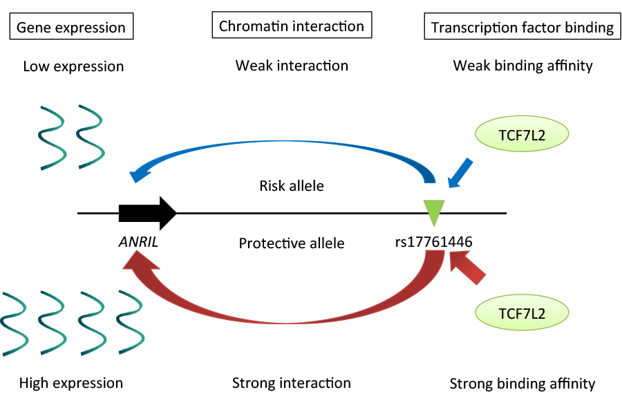
Regulatory mechanisms underlying 9p21 endometriosis risk locus. Preferential bindings of TCF7L2 and its coactivator EP300 to the protective G allele of rs17761446 lead to stronger chromatin interaction with the promoter of ANRIL, which in turn activate transcription of the non-coding RNA.
Cell Architecture Laboratory / Kimura Group
Shape–motion relationships of centering microtubule asters
Hirokazu Tanimoto, Akatsuki Kimura*, and Nicolas Minc*
J. Cell Biol. 212: 777-787, 2016. DOI:10.1083/jcb.201510064
(* corresponding authors)
A joint group of Drs. Nicolas Minc at Institut Jacques Monod (France) and Akatsuki Kimura at National Institute of Genetics/SOKENDAI analyzed the centering of microtubule asters in the sea urchin egg, and constructed a quantitative model that dictates the speed and trajectory of the centering in normal cell as well as shape-manipulated cells. The study was initiated when Prof. Kimura was a visiting researcher at the Minc lab through the SOKENDAI Young Faculty Overseas Visit Program.
In this study, Dr. Hirokazu Tanimoto et al. utilized multiple approaches such as laser ablation and pharmacological assays to demonstrate that the centering is accomplished by the forces pulling on asters from the cytoplasm. An interesting feature of the centering was to find that asters moved at a constant speed. Considering the speed was comparable to that of the growth speed of the aster, Tanimoto et al. proposed a theoretical model for the centering that explains the constant speed independent on the actual size of the aster. The model predicts well the change of the speed and trajectory of asters when the shape of the cells was manipulated in micro-chambers. Overall, this study advanced our understanding on the long-standing debate on the mechanism of the centering of microtubule asters and their associating nucleus.
See also: Ben Short “How asters find their center” J Cell Biol 212, 743 (2016) DOI: 10.1083/jcb.2127if
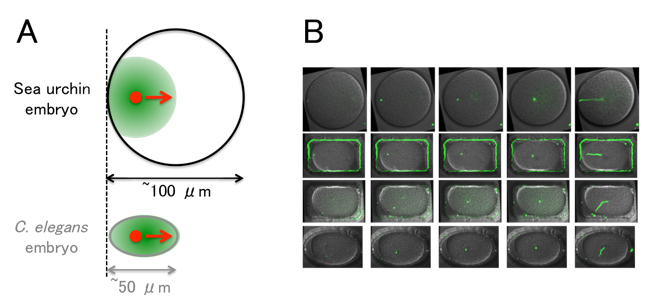
(A) The centering of microtubule asters (green) in the sea urchin embryo. The size of the embryo is large compared to C. elegans. The aster does not cover the entire cell during the centering, and the center of the aster (red) approaches the cell center with a speed comparable to the growth speed of the aster.
(B) Examples of shape manipulation using micro-chambers. The sperm pronucleus (i.e. the center of the aster, green) moves toward the center. (The entire trajectories are shown on the rightmost panels.) The theoretical model constructed in the study reproduces the speed and trajectory of aster movements even in the shape-manipulated embryos.
Time & Date
9:00 ~ 16:00, Saturday, April 2th, 2016 (No Reservations Required, Free Admission)
Access
Free Shuttle Buses Available to the National Institute of Genetics from the North Exit of Mishima Station. ( Service Time: 8:50 ~ 15:00)
Map
Map Free Shuttle Buses Available Parking around Mishima Station Available for Car Visitors.
No Pets Allowed except Service Dogs, No General Parking*
(*Disabled Parking Available on the Institute Premises)
Information
1111 Yata, Mishima, Shizuoka 411-8540, JAPAN TEL:+81-55-981-5873
Two professors and three assistant professors have joined NIG as of April 1, 2016.
professor
Yutaka SATO:Genetic Resource Center,Plant Genetics Laboratory
Ken KUROKAWA:Center for Information Biology, Genome Evolution Laboratory

assistant professor
Takayuki FUJIWARA: Division of Symbiosis and Cell Evolution, Miyagishima Group
Tomotaka MATSUMOTO: Division of Evolutionary Genetics, Akashi Group
Toshihiro KAWASAKI: Model Fish Genomics Resource Laboratory, Sakai Group
In addition, Masato KANEMAKI, tenure track associate professor at the Center for Frontier Research, has been awarded tenure and promoted to professor.
Masato KANEMAKI: Division of Molecular Cell Engineering, Department of Molecular Genetics
On March 8, 2016 Mr. Isaac Adeyemi Babarinde (Division of Population Genetics) received FY2015 Morishima Award from the Department of Genetics, SOKENDAI. The Morishima Award is given to students in the Department of Genetics to honor their outstanding performances during PhD studies and to encourage further achievements.

Pioneers in Genetics
Genetic Informatics Laboratory / Yamazaki Group
Transcription profile of Escherichia coli: genomic SELEX search for regulatory targets of transcription factors
Akira Ishihama, Tomohiro Shimada, and Yukiko Yamazaki
Nucleic Acids Research (2016) DOI:10.1093/nar/gkw051
Bacterial genome is transcribed by a single species of DNA-dependent RNA polymerase (RNAP). The genome transcription pattern is determined by controlling the utilization of a limited number of RNAP, of which the gene selectivity is modulated through two-steps of protein-protein interaction with two groups of regulatory protein, sigma factors with promoter recognition activity and transcription factors (TFs). The identification of regulatory targets for all these regulatory proteins is absolutely needed towards understanding the genome regulation at molecular level. Using the newly developed Genomic SELEX screening system, Ishihama, A. (NIG Emeritus Professor) and colleagues in Hosei University performed the systematic search of regulatory targets for all 7 sigma factors and all 300 transcriptions of the model prokaryote Escherichia coli. This is the first report on this line, describing the assembly and analysis of regulatory targets for the first group of TFs. The experiments have been carried out using a single and the same E. coli strain and using the same experimental systems in a single laboratory. To facilitate sharing these accumulated data sets with the research community, we compiled a database, “Transcription Profile of Escherichia coli” (TEC)(www.shigen.nig.ac.jp/ecoli/tec/). This pioneering research of identification of the whole set of regulatory targets for all regulatory proteins from a single organism will contribute for creation of a new paradigm in modern molecular genetics.
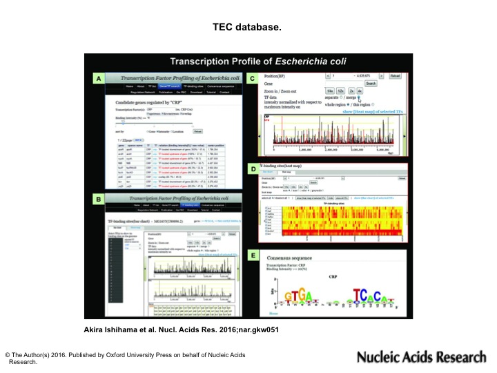
Using the TEC database, various types of analysis are possible:(A) search for the regulatory targets of TF. (B) genomic view of TF binding sites/intensity (C) comparison of binding sites between different TFs (D) heat map view of TF binding sites/intensity (E) Search for the consensus sequence of TF binding region.
Model Fish Genomics Resource Laboratory / Sakai Group
Differentiation of zebrafish spermatogonial stem cells to functional sperm in culture.
Toshihiro Kawasaki, Kellee R Siegfried and Noriyoshi Sakai.
Development, 2016 143: 566-574; DOI:10.1242/dev.129643
Molecular dissection of spermatogenesis could be facilitated by cell culture approaches that allow easy access for experimental manipulation and live imaging of specific molecules; however, technical limitations have thus far prevented the complete reconstruction of spermatogenic events in cell culture. Here, we established cell culture systems for production of functional sperm from self-renewing spermatogonial stem cells (SSCs), using zebrafish testicular hyperplasias that accumulate SSCs. By serially transplanting into immuno-deficient mutant zebrafish, we succeeded in long-term and efficient production of hyperplasias. Through improvements of culture conditions, we achieved efficient propagation of the SSCs and differentiation to sperm that gave rise to offspring. Oxygen at the concentration of air proved to be detrimental for sperm differentiation from SSCs. These results indicate that the whole spermatogenic process can be represented in cell culture, facilitating analyses of molecular mechanisms of spermatogenesis in vertebrates. This work was supported by JSPS KAKENHI (23013023, 25251034, 25114003).
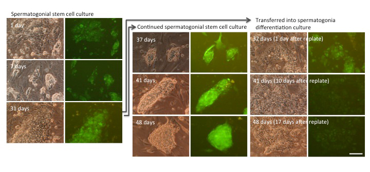
Propagation and differentiation of SSCs in culture. SSCs that express green fluorescent protein grow in propagation culture (left and middle panels), while they differentiated into sperm in differentiation culture (the right panel).
Biological Macromolecules Laboratory / Maeshima Group
Liquid-like behavior of chromatin
Kazuhiro Maeshima, Satoru Ide, Kayo Hibino, and Masaki Sasai.
Current Opinion in Genetics & Development, 2016, 37: 37-45. DOI:10.1016/j.gde.2015.11.006
Eukaryotic chromatin is a negatively charged long polymer composed of genomic DNA, histones, and various proteins. The charged property causes the chromatin structure to be dynamically changed. These dynamic changes are critical for genome functions such as gene expression because they directly govern the degree of DNA accessibility. Although the chromatin structure is not yet fully understood, currently increasing evidence suggests that chromatin has a dynamic liquid-like structure based on the 10-nm fiber but not the 30-nm fiber. This liquid-like property can drive the process of ‘scanning and targeting genomic DNA,’ which contributes to various genome functions including gene expression and DNA replication, repair, and recombination. Here, we discuss the liquid-like behavior of chromatin and its physical and biological relevance.
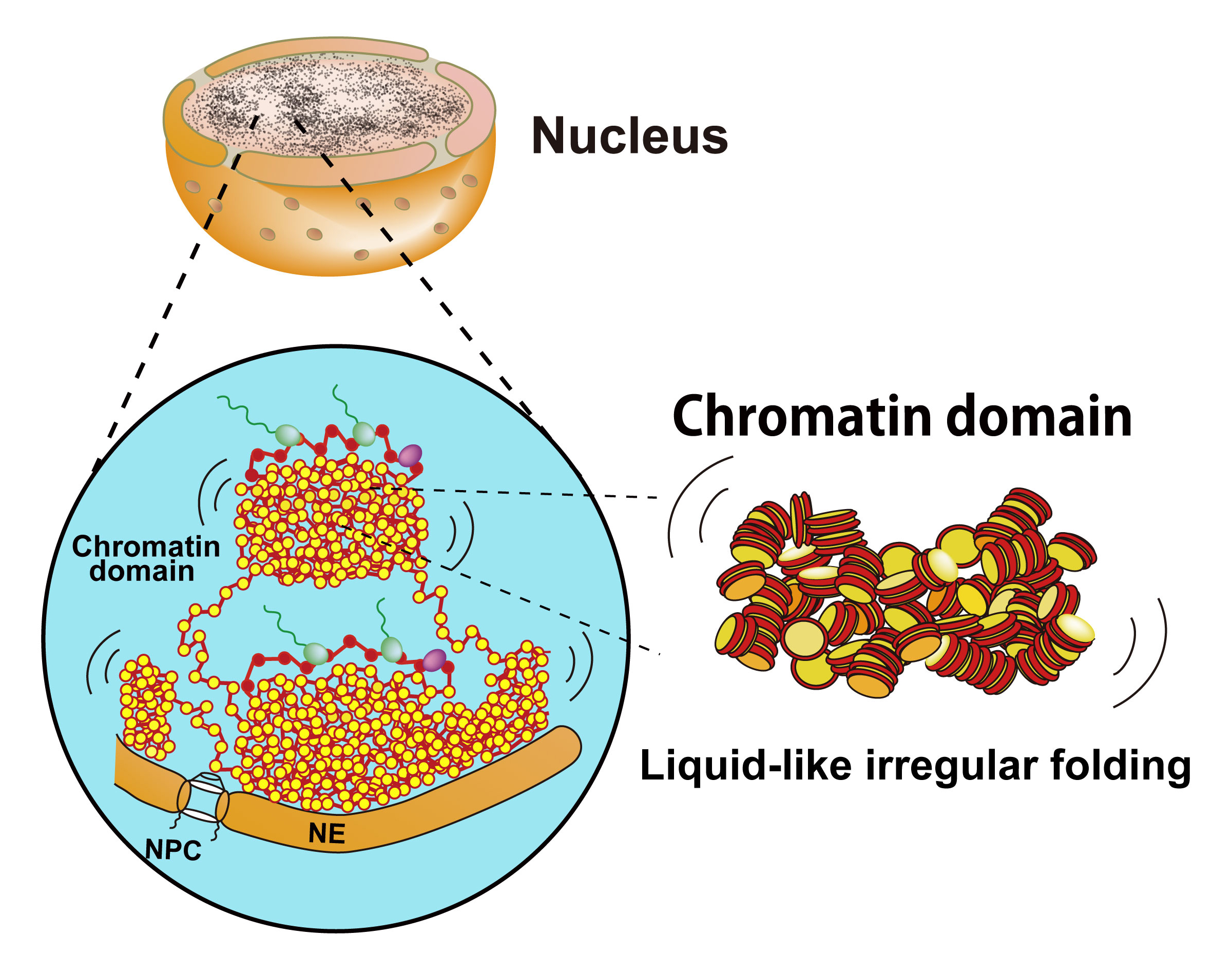
Chromatin consists of irregularly folded 10-nm fibers and forms numerous chromatin domains (e.g., topologically associating domains). The liquid-like movement of chromatin should bring about fluctuation of the chromatin domain. NPC, nuclear pore complex; NE, nuclear envelope.
Department of Molecular Genetics Division of Centrosome Biology•Kitagawa Laboratory
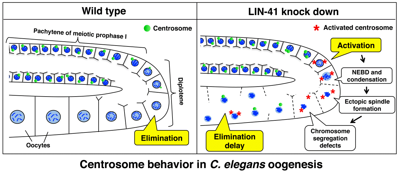
LIN-41 inactivation leads to delayed centrosome elimination and abnormal chromosome behavior during female meiosis in Caenorhabditis elegans
Rieko Matsuura, Tomoko Ashikawa, Yuka Nozaki and Daiju Kitagawa
Molecular Biology of the Cell, 2016 DOI:10.1091/mbc.E15-10-0713
The centrosome serves as the major microtubule-organizing center (MTOC) in most animal cells. MTOC activity of centrosomes is crucial for proper chromosome segregation in mitosis and therefore for genome integrity. Centrosome dysfunction is known to be associated with a variety of diseases including cancer and infertility.
On the other hand, during oogenesis in most metazoans, maternal centrosomes are inactivated and thereafter eliminated during meiotic prophase I; hence, two successive meiotic cell divisions occur without centrosomes. The elimination of maternal centrosomes and the inheritance of paternal centrosomes to the progeny are crucial for maintaining the precise number of centrosomes in the fertilized zygote and thus for proper sexual reproduction. Despite being a conserved phenomenon in most metazoans, the means by which this centrosome behavior is controlled during female meiosis remain elusive.
The gonad of the Caenorhabditis elegans hermaphrodite is a well-suited model for analyzing the mechanisms governing centrosome behavior during oogenesis because all stages of oogenesis can be seen in a continuous fashion within a single gonad. In this study, we conducted a targeted RNAi screening in the Caenorhabditis elegans gonad to identify novel regulators of centrosome behavior during oogenesis. We screened ~500 genes known to be essential for embryo production and directly visualized GFP-γ-tubulin to monitor centrosome behavior at all stages of oogenesis. In the screening, we found that RNAi-mediated inactivation of 33 genes delayed the elimination of GFP-γ-tubulin at centrosomes during oogenesis, whereas inactivation of nine genes accelerated the process. Depletion of the TRIM-NHL protein LIN-41 led to a significant delay in centrosome elimination and to the separation and reactivation of centrosomes during oogenesis. Upon LIN-41 depletion, meiotic chromosomes were abnormally condensed and pulled toward one of the two spindle poles around late pachytene even though the spindle microtubules emanated from both centrosomes. Overall, our work provides new insights into the regulation of centrosome behavior to ensure critical meiotic events and the generation of intact oocytes.
This work was supported by JSPS KAKENHI (24687026 24113003), Takeda Science Foundation and the NAITO foundation.