


Press release
Orientation-Independent-DIC imaging reveals that a rise in depletion attraction contributes to mitotic chromosome condensation
Shiori Iida , Satoru Ide , Sachiko Tamura, Masaki Sasai, Tomomi Tani , Tatsuhiko Goto , *Michael Shribak , *Kazuhiro Maeshima
* Corresponding Author
Proceedings of the National Academy of Sciences (2024) 121(36), e2403153121 DOI:10.1073/pnas.240315312
![]() Press release (In Japanese only)
Press release (In Japanese only)
Mitotic chromosome condensation is an essential process to transmit replicated chromosomes into two daughter cells during cell division. To study the underlying physical principles of this process, we focused on depletion attraction/macromolecular crowding, which is a force that attracts large structures in crowded cell environments. Using special light microscopy, which can image the molecular density of cellular environments, we found that crowding around chromosomes increases during cell division. In vitro, higher concentrations of macromolecules condense chromatin and make it stiffer and more solid-like. Our results suggest that the rise in depletion attraction renders chromosomes more rigid, ensuring accurate chromosome transmission during cell division.
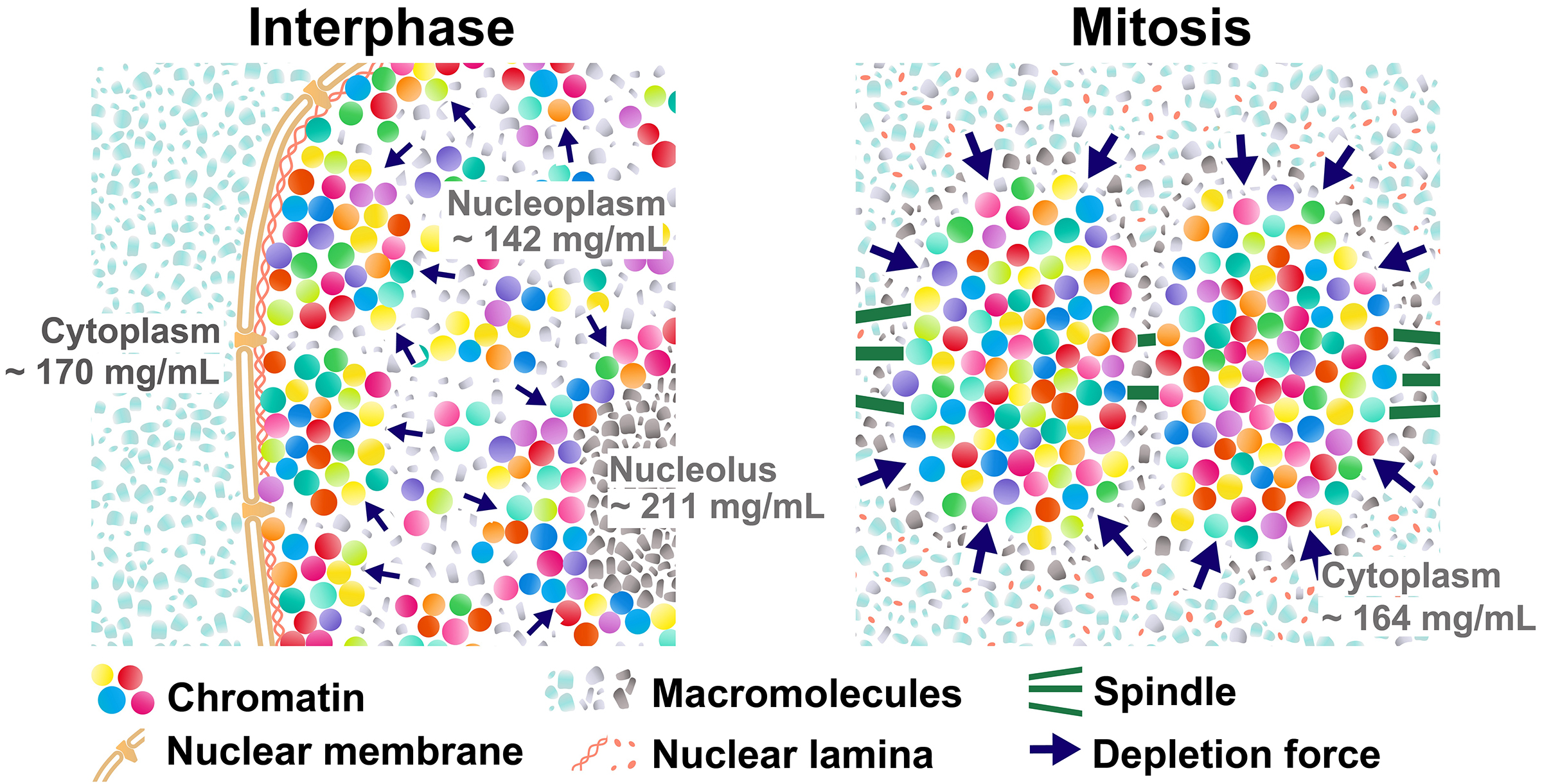
Figure: (Left) Schematic of depletion attraction in interphase live cells with soluble macromolecules in the cytoplasm (light blue), nucleoplasm (gray), and nucleolus (dark gray). The cytoplasm, nucleus, and nucleolus are compartmentalized and do not mix during interphase. Molecular densities of the nucleoplasm are lower than the cytoplasm and nucleolus. (Right) After NEBD, soluble macromolecules that were localized to the cytoplasm, nucleus, and nucleolus at interphase are now mixed. Molecular density of chromosome environment increases, making the depletion attraction stronger, and contributes to local condensation of chromosomes. These schematics are highly simplified models, and depletion attraction also works in interphase chromatin (smaller navy arrows).
Press release
Combination of AID2 and BromoTag expands the utility of degron-based protein knockdowns
Yuki Hatoyama*, Moutushi Islam*, Adam G. Bond, Ken-ichiro Hayashi, Alessio Ciulli and Masato T. Kanemaki.
EMBO Reports (2024) Aug 23. DOI:10.1038/s44319-024-00224-4
![]() Press release (In Japanese only)
Press release (In Japanese only)
Prof. Kanemaki at the NIG and his colleagues have developed the Auxin-Inducible Degron (AID) technology, which enables rapid protein degradation for functional analysis of the proteins of interest. The group further refined it with the AID2 (Nishimura et al., Nat. Meth., 2009; Yesbolatova et al., Nat. Commun., 2020). However, they noted that some proteins remain even after degradation by AID2. In response, the group developed a double-degron technology by combining AID2 with another degron, BromoTag, achieving super-efficient and rapid degradation of target proteins. This advancement allows for the functional evaluation of proteins that were previously difficult to analyze. Additionally, they demonstrated the independent control of degradation for two different target proteins using AID2 and BromoTag.
In an application of this double-degron technology, the group successfully achieved complete inhibition of DNA replication by depleting DNA replication factors. They found that cells can progress through the cell cycle to mitosis without DNA replication, suggesting that cells may lack the system to recognize whether DNA has doubled. This finding not only demonstrates the versatility of degron technology but also offers deeper insights into the relationship between DNA replication and the cell cycle.
This study was carried out by Prof. Masato Kanemaki’s group at NIG, in collaboration with Prof. Alessio Ciulli at the University of Dundee and Prof. Kenichiro Hayashi at Okayama University of Science.
This study was supported by JSPS (21H04719, 23H04925), and JST CREST (MJCR21E6). Yuki Hatoyama and Moutushi Islam at SOKENDAI were supported by JSPS Research Fellowship (DC2) and MEXT Scholarship, respectively.
This study was published in EMBO Reports on August 23, 2024.
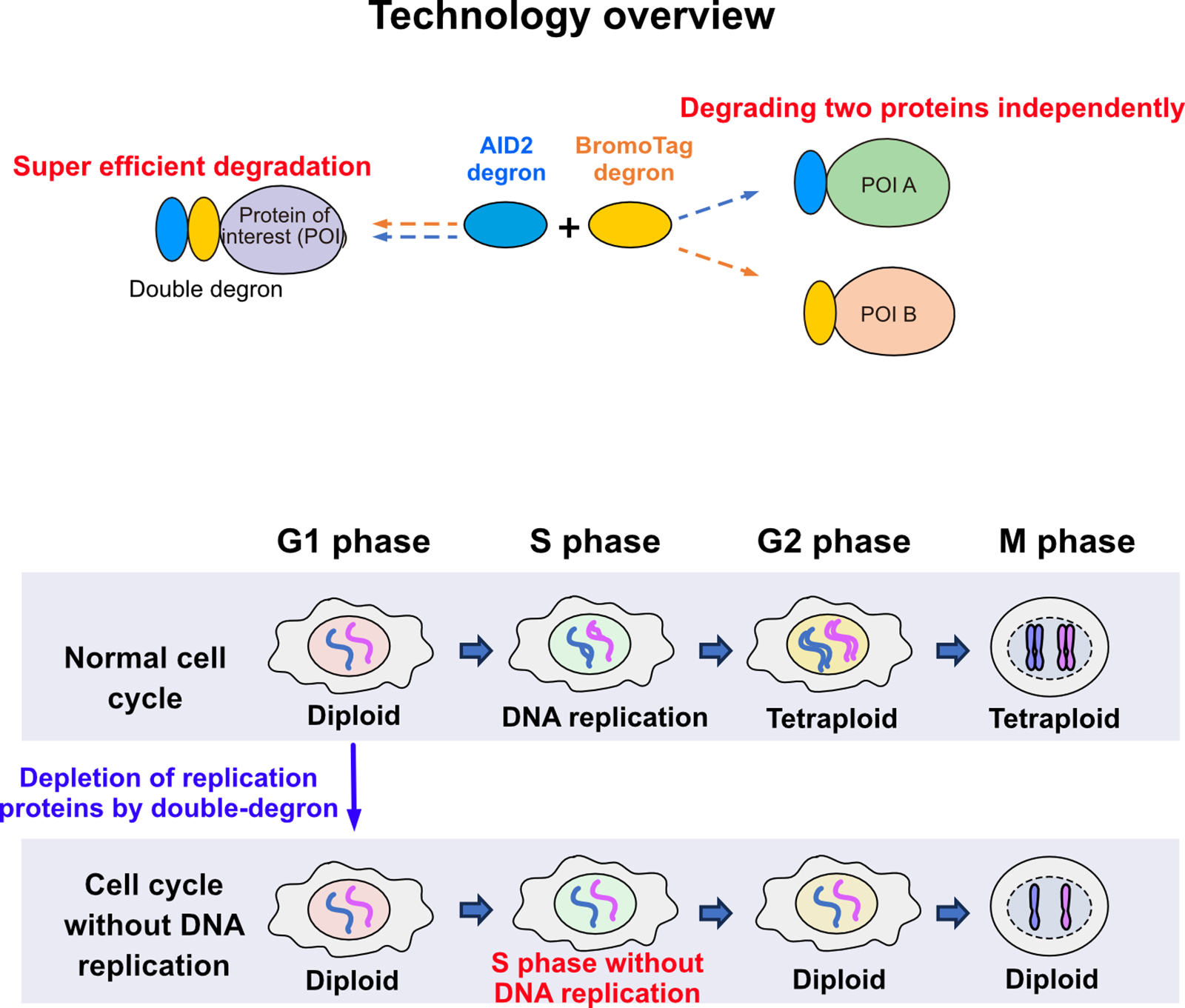
Figure: An overview of the expanded versatility achieved by combining degron technologies. The double-degron approach induced cell cycle progression to mitosis without DNA replication.
Ayjan Urazbayeva, D4 SOKENDAI student and MEXT Scholar in Multiscale Sensory Structure Laboratory, received the Best Presentation Award at “Shaping the Future of Early-Career Zebrafish Researchers” Workshop in the 18th International Zebrafish Conference Conference held in Kyoto, August 17th – 21st.

Ayjan-san
Press release
Single-nucleosome imaging unveils that condensins and nucleosome-nucleosome interactions differentially constrain chromatin to organize mitotic chromosomes.
Kayo Hibino, Yuji Sakai, Sachiko Tamura, Masatoshi Takagi, Katsuhiko Minami, Masa A. Shimazoe, Toyoaki Natsume, Masato T. Kanemaki, Naoko Imamoto, Kazuhiro Maeshima*
* Corresponding Author
Nature Communications (2024) 15, 7152 DOI:10.1038/s41467-024-51454-y
![]() Press release (In Japanese only)
Press release (In Japanese only)
For accurate mitotic cell division, replicated chromatin must be assembled into chromosomes and faithfully segregated into daughter cells. While protein factors like condensin play key roles in this process, it is unclear how chromosome assembly proceeds as molecular events of nucleosomes in living cells and how condensins act on nucleosomes to organize chromosomes. To approach these questions, we investigate nucleosome behavior during mitosis of living human cells using single-nucleosome tracking, combined with rapid-protein depletion technology and computational modeling. Our results show that local nucleosome motion becomes increasingly constrained during mitotic chromosome assembly, which is functionally distinct from condensed apoptotic chromatin. Condensins act as molecular crosslinkers, locally constraining nucleosomes to organize chromosomes. Additionally, nucleosome-nucleosome interactions via histone tails constrain and compact whole chromosomes. Our findings elucidate the physical nature of the chromosome assembly process during mitosis.
This study was published online as open access in Nature Communications on August 21, 2024.
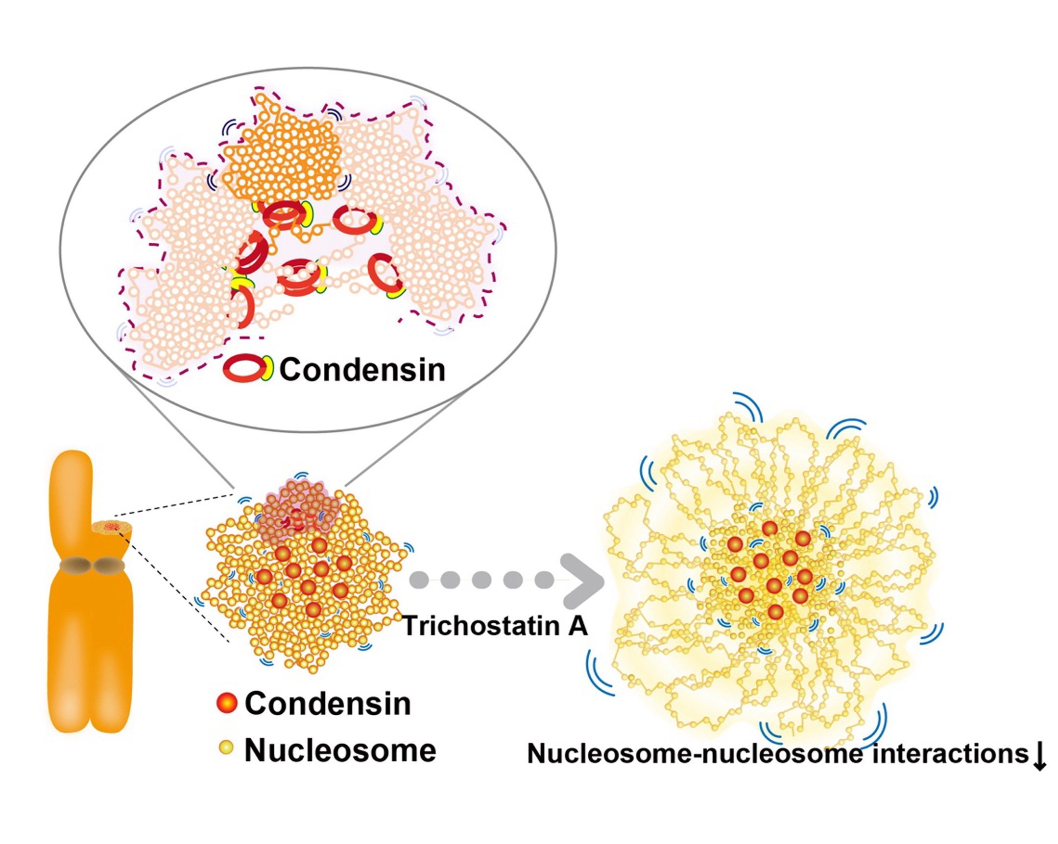
Figure: Condensins seem to act as a molecular crosslinkersto make loops. (Bottom, left) Condensins (red) locate around chromosome center. (Bottom, right) Nucleosomes around the periphery (those mostly free from condensins) in the Trichostatin A-treated chromosomes, whose nucleosome–nucleosomes interactions are weakened, are less constrained and have higher mobility than those around the axis.
Video: Single nucleosomes fluorescently labeled in a living HeLa interphase cell (left) and mitotic cell (center)(50 ms/frame). Each dot represents a single nucleosome. (Right) Calculated chromatin motion of the simulated chromosome. Chromatin beads (blue) and condensins (green and red) are shown.
Nonomura Group / Plant Cytogenetics Laboratory
Koide Group / Mouse Genomics Resource Laboratory
Callose Deficiency Modulates Plasmodesmata Frequency and Extracellular Distance in Rice Pollen Mother and Tapetal cells.
Harsha Somashekar, Keiko Takanami, Yoselin Benitez-Alfonso, Akane Oishi, Rie Hiratsuka, Ken-Ichi Nonomura
Annals of Botany (2024) mcae137. DOI:10.1093/aob/mcae137
Fertilization relies on pollen mother cells able to transit from mitosis to meiosis to supply gametes. This process involves remarkable changes at the molecular, cellular and physiological levels including (but not limited to) remodelling of the cell wall. During the meiosis onset, cellulose content at the pollen mother cell walls gradually declines with the concurrent deposition of the polysaccharide callose in anther locules. We aim to understand the biological significance of cellulose-to-callose turnover in pollen mother cells walls using electron microscopic analyses of rice flowers. Our observations indicate that in wild type rice anthers, the mitosis-to-meiosis transition coincides with a gradual reduction in the number of cytoplasmic connections called plasmodesmata. A mutant in the Oryza sativa callose synthase GSL5 (Osgsl5-3), impaired in callose accumulation in premeiotic and meiotic anthers, displayed a greater reduction in plasmodesmata frequency among pollen mother cells and tapetal cells suggesting a role for callose in plasmodesmata maintenance. In addition, a significant increase in extracellular distance between pollen mother cells and impaired premeiotic cell shaping was observed in the Osgsl5-3 mutant. The results suggest that callose-to-cellulose turnover during mitosis-meiosis transition is necessary to maintain cell-to-cell connections and optimal extracellular distance among the central anther locular cells. Findings of this study contribute to our understanding of the regulatory influence of callose metabolism during meiosis initiation in flowering plants.

Figure: PD frequency is differentially regulated upon entry to meiosis and in Osgsl5-3 mutant anthers
(A) A diagram of the cross section of rice anther (left), and transmission electron microscopic images of PDs (yellow arrows) between neighbouring pollen mother cells (PMCs) in wild type (WT) (middle) and Osgsl5-3 mutant (right).
(B) Quantification of PD frequency in premeiotic interphase and early meiosis I for WT and Osgsl5-3 anthers. PD frequency was gradually decreased from premeiosis to early meiosis in WT, but significantly reduced in Osgsl5-3 mutant compared to WT.
Kitano Group / Ecological Genetics Laboratory
Records of Neocaridina denticulata from Uku Island, N. davidi from Fukue Island, Goto Islands, and N. ikiensis from mainland Kyushu, Japan
Yusuke Fuke, Shota Kunimatsu, Jun Nakajima
Cancer (2024) 33: 47–55. DOI:10.18988/cancer.33.0_47
A freshwater shrimp, the genus Neocaridina has a land-locked life history and lives its entire life in freshwater. Two Neocaridina species are distributed in Honshu and the surrounding islands: Neocaridina denticulata and Neocaridina ikiensis, which is endemic to Iki Island. Recently, the invasive alien species Neocaridina davidi has become established in various sites in Japan, causing ecological problems.
We reported the first records of N. denticulata from Uku Island, Goto Islands, Nagasaki Prefecture, and N. ikiensis from mainland Kyushu, based on specimens. The results of mitochondrial DNA analysis suggest that these newly discovered populations are native. On the other hand, an invasive species, N. davidi was confirmed at eight sites in Fukuoka, Nagasaki, Oita, and Kumamoto prefectures. Our findings are useful for the conservation of native species and the management of invasive species.
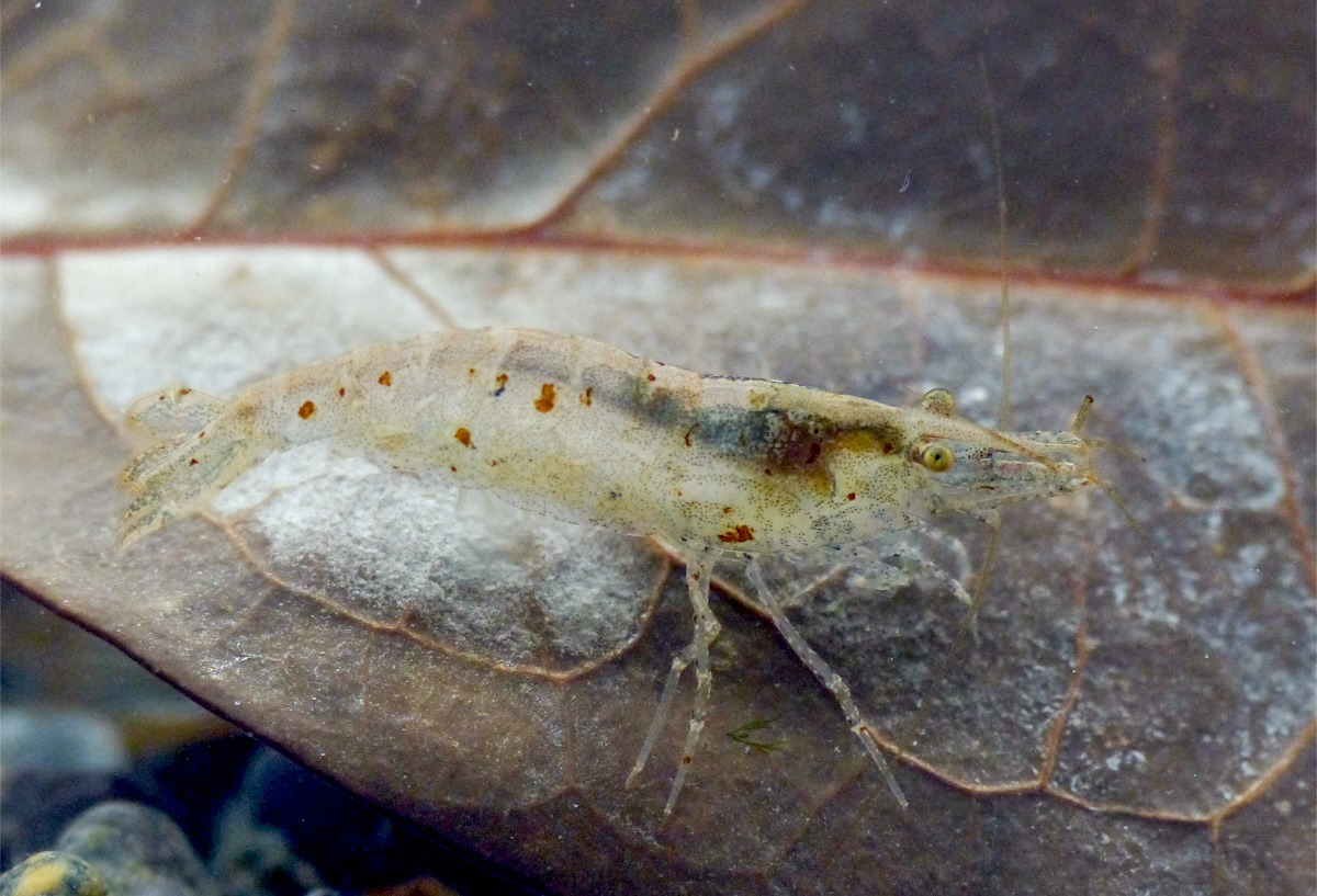
Figure: Neocaridina denticulata collected from Uku Island. They would be a native population because they have a unique mitochondrial DNA haplotype. Photo by Jun Nakajima.
Kitano Group / Ecological Genetics Laboratory
Additional records of the freshwater prawn Macrobrachium ustulatum (Crustacea: Decapoda: Palaemonidae) from the Ryukyu Islands, Japan
Yusuke Fuke, Taigi Sato, Naoto Shimizu, Naoto Inui
Cancer (2024) 33: 41–46. DOI:10.18988/cancer.33.0_41
Macrobrachium ustulatum is the sister species of Macrobrachium australe, and they have been historically confused because of their morphological similarity. The northern edge of the distribution range of both species is Japan: M. australe has been recorded from the Ryukyu Islands to a wide area of Honshu, while M. ustulatum has only two records in Japan. Therefore, the distributional records and habitat information of M. ustulatum are lacking. We report a total of 10 specimens of M. ustulatum collected from three localities in the Ryukyu Islands. The environments in which these specimens were collected suggest that the habitat of this species is a weakly flowing environment in the middle reach of a river. This differs slightly from M. australe, which prefers stagnant environments. Furthermore, we have succeeded in proposing new distinguishing characteristics for both species.
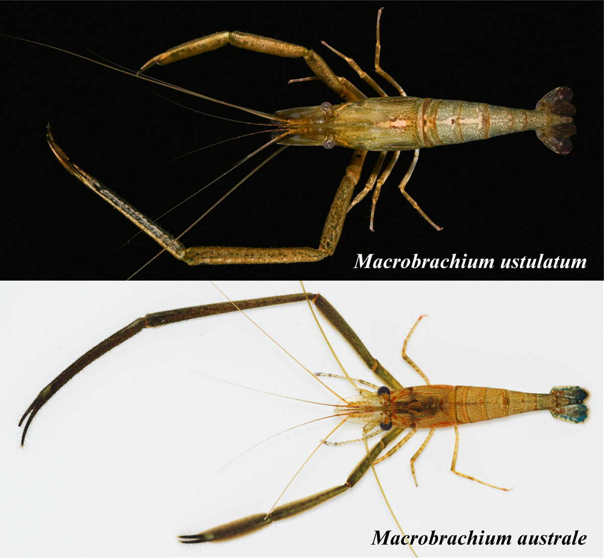
Figure: Macrobrachium ustulatum (top) and M. australe (bottom). Photo by the authors.
Kitano Group / Ecological Genetics Laboratory
Confusion between Caridina laoagensis and Caridina tupaia in Japan
Yusuke Fuke & Tomoaki Maruyama
Cancer (2024) 33: 15–23. DOI:10.18988/cancer.33.0_15
The morphological characters of the freshwater shrimp called “Ryugu-hime-ebi” in Japan are not always consistent with those of Caridina laoagensis, which has been given its Japanese name. Recently, a cryptic species of C. weberi species group that includes C. laoagensis was newly described as Caridina tupaia. We aimed to resolve the historical confusion between C. laoagensis and C. tupaia in Japan. For DNA barcoding and morphological examination, we recognized the two taxa in “Ryugu-hime-ebi,” i.e. C. laoagensis and C. tupaia. Based on some diagnostic characters, we reviewed previous records and found that the taxon that was given the Japanese name “Ryugu-hime-ebi” was most likely C. tupaia. We proposed to give the standard Japanese name “Ryugu-hime-ebi” to C. tupaia and the new standard Japanese name “Un-mon-hime-ebi” to C. laoagensis.
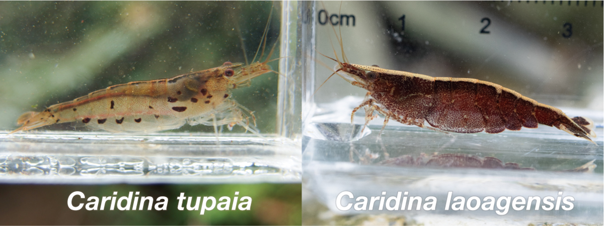
Figure: Caridina tupaia and C. laoagensis. Photo by the authors.
Adilgazy Semeigazin, D4 student and MEXT Scholar in Genome Dynamics Laboratory, received the Second-place Poster Award at the FASEB Research Conference ”Biology of Acetylation in Health and Disease“ held in Rome, Italy, on August 4th – 8th. He also received the Travel Award.
Semeigazin was also supported by SOKENDAI Student Dispatch Program to attend the conference.

Semeigazin-san


