


SCHENGEN receptor module drives localized ROS production and lignification in plant roots
Satoshi Fujita, Damien De Bellis, Kai H Edel, Philipp Köster, Tonni Grube Andersen, Emanuel Schmid-Siegert, Valérie Denervaud Tendon, Alexander Pfister, Peter Marhavý, Robertas Ursache, Verónica G. Doblas, Marie Barberon, Jean Daraspe, Audrey Creff, Gwyneth Ingram, Jörg Kudla, Niko Geldner
EMBO J (2020)e103894 DOI:10.15252/embj.2019103894
Reactive oxygen species (ROS) impact many physiological processes in animals or plants, but its production by NADPH oxidases is strictly regulated because of extremely high reactivity. Many plant receptor pathways are known as critical regulators of ROS production, but how they spatially control ROS production is yet to be clarified.
Fujita et al. (2020) established a phospho-signaling pathway that integrates direct, rapid activation of ROS production with positional information and transcriptional changes to form proper diffusion barriers. Firstly, the authors presented a direct connection from a peptide-receptor complex to NADPH oxidases via a membrane-anchored kinase by biochemically. Next, the authors focused on the membrane-anchored kinase that localizes on the plasma membrane in a polar fashion. Manipulation of the kinase localization successfully showed that this biased distribution of the kinase protein gave the positional cue, which enables the SCHENGEN pathway to activate only one side of the Casparian strip at micrometer-scale precision. This work would contribute to highlight how other receptor pathways could control local ROS production.
This work was mainly done by Dr. Satoshi Fujita (former postdoctoral fellow in University of Lausanne, currently NIG research fellow) in Prof. Niko Geldner’s group with a collaboration with the Central imaging, Electron microscope, and Genomics facilities of University of Lausanne, Swiss Institute of bioinformatics, Prof. Jörg Kudla’s group at University of Munster and Dr. Gwyneth Ingram’s group at University of Lyon.
This work was aided by grants no. 31003A_156261 and 310030E_176090 (N.G.), from the Swiss National Science Foundation, an ERC Consolidator Grant (616228-ENDOFUN) (N.G.), DFG grant (Ku931/14-1 (J.K.)) FEBS long-term fellowship (P.M.), EMBO long-term fellowship (R.U., M.B.), Marie Curie postdoctoral fellowship (T.G.A,), Fundacion Alfonso Martin Escudero fellowship (V.G.D.), and a Japan Society for the Promotion of Science (JSPS) fellowship (S.F.).
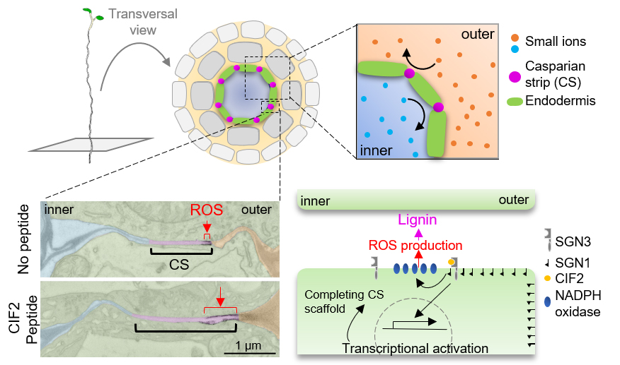
Figure: Plant roots have a lignin-based diffusion barrier, namely Casparian strips. Localized ROS production (EM pictures) by asymmetrical signal activation (schematic model) sustains functional barrier formation.
Gastrointestinal Neurons Expressing HCN4 Regulate Retrograde Peristalsis
Kensuke Fujii, Koichi Nakajo, Yoshihiro Egashira, Yasuhiro Yamamoto, Kazuya Kitada, Kohei Taniguchi, Masaru Kawai, Hideki Tomiyama, Koichi Kawakami, Kazuhisa Uchiyama, and Fumihito Ono
Cell Reports 30(9), 2879-2888 (2020). DOI:10.1016/j.celrep.2020.02.024
Peristalsis is indispensable for physiological function of the gut. The enteric nervous system (ENS) plays an important role in regulating peristalsis. While the neu- ral network regulating anterograde peristalsis, which migrates from the oral end to the anal end, is charac- terized to some extent, retrograde peristalsis re- mains unresolved with regards to its neural regula- tion. Using forward genetics in zebrafish, we reveal that a population of neurons expressing a hyperpo- larization-activated nucleotide-gated channel HCN4 specifically regulates retrograde peristalsis. When HCN4 channels are blocked by an HCN channel inhibitor or morpholinos blocking the protein ex- pression, retrograde peristalsis is specifically attenu- ated. Conversely, when HCN4(+) neurons expressing channelrhodopsin are activated by illumination, retrograde peristalsis is enhanced while anterograde peristalsis remains unchanged. We propose that HCN4(+) neurons in the ENS forward activating sig- nals toward the oral end and simultaneously stimu- late local circuits regulating the circular muscle.
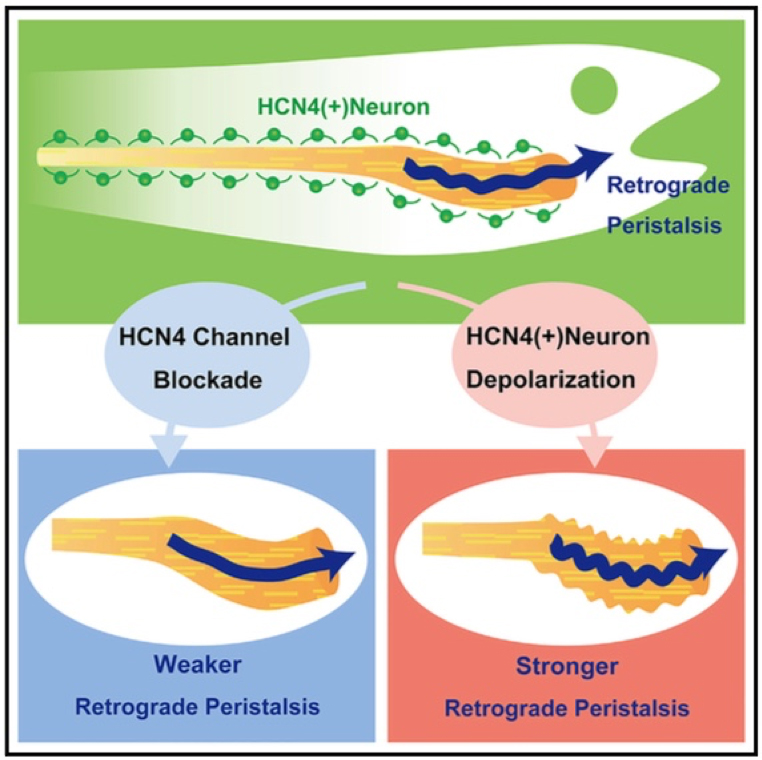
Figure1: Gastrointestinal neurons expressing a hyperpolarization-activated nucleotide- gated channel HCN4 regulate retrograde peristalsis, which migrates from the anal end to the oral end of the gut.
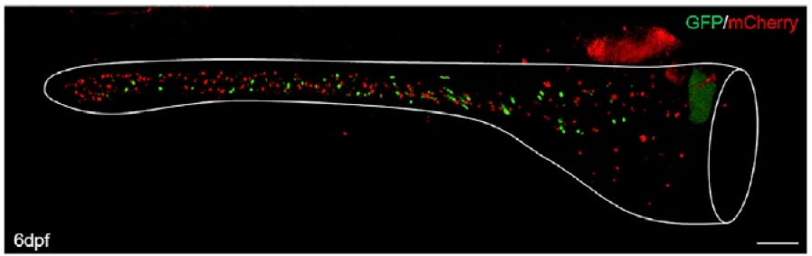
Figure2: Visualization of neurons (red) and HCN4-expressing neurons (green) in the zebrafish intestine.
Press release
A virtual reality system to analyze neural activity and behavior in adult zebrafish
Kuo-Hua Huang, Peter Rupprecht, Thomas Frank, Koichi Kawakami, Tewis Bouwmeester and Rainer W. Friedrich
Nature methods 02 March 2020 DOI:10.1038/s41592-020-0759-2
Press release (In Japanese only)
Virtual realities are powerful tools to analyze and manipulate interactions between animals and their environment and to enable measurements of neuronal activity during behavior. In many species, however, optical access to the brain and/or the behavioral repertoire are limited. We developed a high-resolution virtual reality for head-restrained adult zebrafish, which exhibit cognitive behaviors not shown by larvae. We noninvasively measured activity throughout the dorsal telencephalon by multiphoton calcium imaging. Fish in the virtual reality showed regular swimming patterns and were attracted to animations of conspecifics. Manipulations of visuo-motor feedback revealed neurons that responded selectively to the mismatch between the expected and the actual visual consequences of motor output. Such error signals were prominent in multiple telencephalic areas, consistent with models of predictive processing. A virtual reality system for adult zebrafish therefore provides opportunities to analyze neuronal processing mechanisms underlying higher brain functions including decision making, associative learning, and social interactions.
Source: Kuo-Hua Huang , et al., Published: 02 March 2020
DOI: 10.1038/s41592-020-0759-2
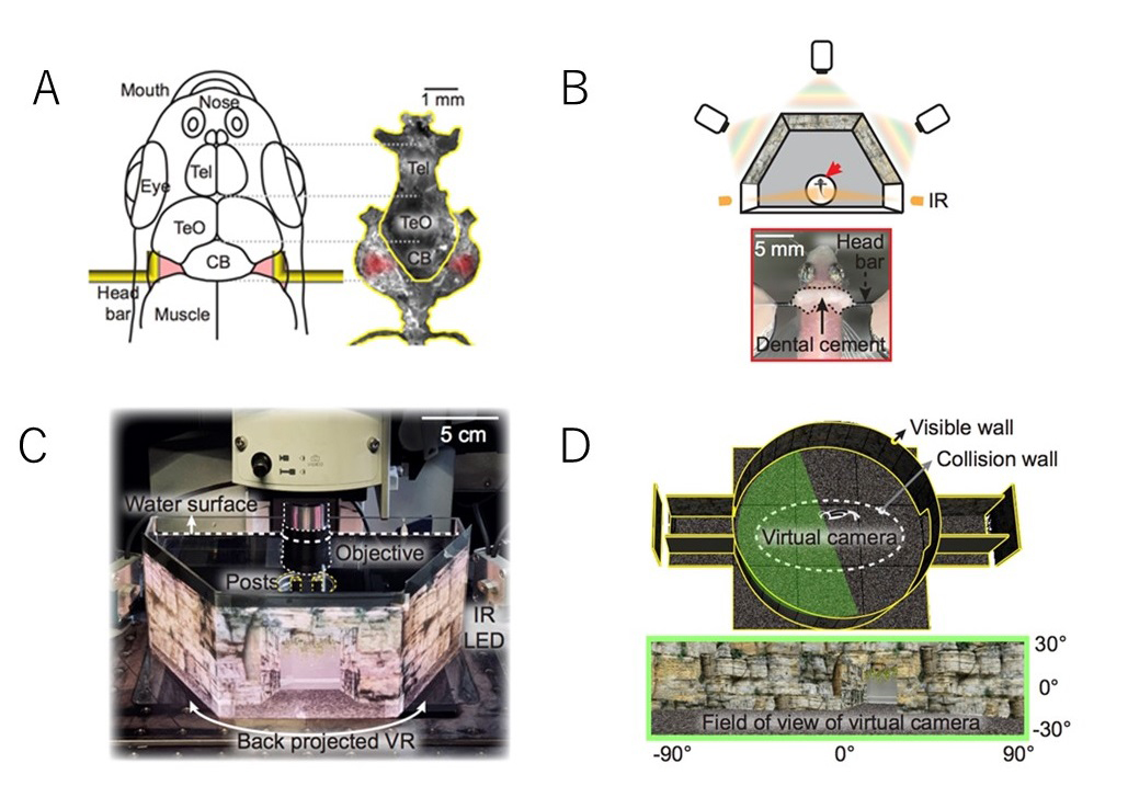
Fig: Virtual reality system to analyze neural activity and behavior in adult zebrafish
A: Head fixation with L-shaped bars
B: Virtual reality projected by three projectors onto a panoramic screen
C: Setup for virtual reality and two-photon imaging
D: Virtual reality seen by the head-fixed zebrafish
Press release
Optogenetic modulation of TDP-43 oligomerization accelerates ALS-related pathologies in the spinal motor neurons
Kazuhide Asakawa, Hiroshi Handa, Koichi Kawakami
Nature Communications 11, 1004 (2020) DOI:10.1038/s41467-020-14815-x
Press release (In Japanese only)
A joint research group in Japan has succeeded in reproducing key ALS symptoms in a small tropical fish by remote controlling a disease-associated protein molecule using light illumination.
In amyotrophic lateral sclerosis (ALS), also known as Lou Gehrig’s disease or motor neuron disease, nerve cells called motor neurons progressively degenerate. These motor neurons accumulate inclusions containing an aggregated form of TDP-43 protein.
In human body, motor neurons align along the spinal cord length and extend along the cables called axons to connect with muscles covering the body surface. This anatomical feature makes motor neurons one of the most difficult cells to observe. Consequently, we do not fully understand when and how healthy motor neurons begin to become abnormal and pathological in ALS. Read More>
Video: An optogenetic ALS zebrafish showing motor decline after blue light illumination (right).
EurekAlert!, the online, global news service operated by AAAS, the science society, PUBLIC RELEASE: 26-FEB-2020
A bidirectional network for appetite control in zebrafish. Caroline Lei Wee
Erin Yue Song, Robert Evan Johnson, Deepak Ailani, Owen Randlett, Ji-Yoon Kim, Maxim Nikitchenko, Armin Bahl, Chao-Tsung Yang, Misha B Ahrens, Koichi Kawakami, Florian Engert, and Sam Kunes.
eLife 8:e43775 (2019). DOI:10.7554/eLife.43775
Medial and lateral hypothalamic loci are known to suppress and enhance appetite, respectively, but the dynamics and functional significance of their interaction have yet to be explored. Here we report that, in larval zebrafish, primarily serotonergic neurons of the ventromedial caudal hypothalamus (cH) become increasingly active during food deprivation, whereas activity in the lateral hypothalamus (LH) is reduced. Exposure to food sensory and consummatory cues reverses the activity patterns of these two nuclei, consistent with their representation of opposing internal hunger states. Baseline activity is restored as food-deprived animals return to satiety via voracious feeding. The antagonistic relationship and functional importance of cH and LH activity patterns were confirmed by targeted stimulation and ablation of cH neurons. Collectively, the data allow us to propose a model in which these hypothalamic nuclei regulate different phases of hunger and satiety and coordinate energy balance via antagonistic control of distinct behavioral outputs.
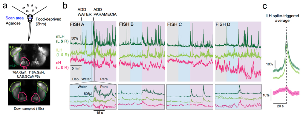
Figure: Calcium imaging of cH and LH by using transgenic zebrafish. By addition of paramecia (food), cH activity was reduced and LH activity was increased.
▶This study is based on the previous study.
Essential roles of autophagy in metabolic regulation in endosperm development during rice seed maturation
Yuri Sera, Shigeru Hanamata, Shingo Sakamoto, Seijiro Ono, Kentaro Kaneko, Yuudai Mitsui, Tomoko Koyano, Naoko Fujita, Ai Sasou, Takehiro Masumura, Hikaru Saji, Ken-Ichi Nonomura, Nobutaka Mitsuda, Toshiaki Mitsui, Takamitsu Kurusu, Kazuyuki Kuchitsu
Scientific Reports 9, 18544 (2019) DOI:10.1038/s41598-019-54361-1
Autophagy, the recycling system of metabolites in eukaryotic cells, plays crucial roles in developmental processes, reproduction and biotic/abiotic-stress responses. However, the role in plants has been largely elusive.
We found that rice autophagy-deficient mutants set smaller seeds with chalky endosperms, in which starch granules were smaller and sparser than in the wild type (Figure). The activity of α-amylases, starch degradation enzymes, was abnormally activated in the mutant endosperm, and in contrast, the level of starch synthesis-promoting enzymes was reduced. The levels of heat shock-, oxidation- or high temperature-responsible proteins were elevated in the mutant endosperm. These results suggest the importance of autophagy in starch degradation and/or stress response pathways in rice endosperm development, and will be useful for breeding of high yielding varieties tolerant to environmental changes.
This work was achieved by collaboration of Tokyo University of Science, Suwa University of Science, Niigata University, National Institute of Advanced Industrial Science and Technology, National Institute of Genetics, Akita Prefectural University, Kyoto Prefectural University and National Institute for Environmental Studies, and was supported by NIG-JOINT (84A2018).
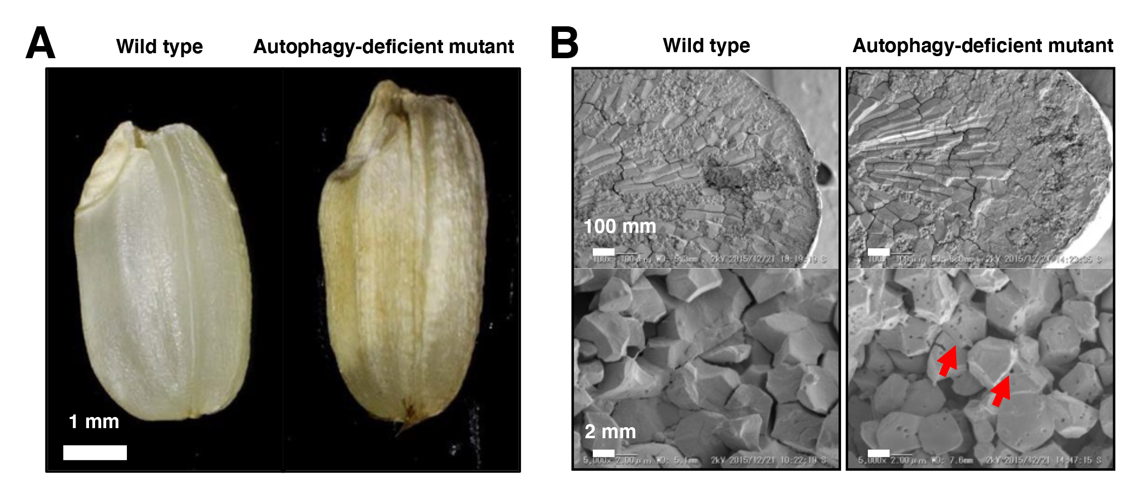
Figure: Mutant phenotypes in the endosperm of autophagy-deficient rice seeds
(A) The autophagy-deficient grain displays chalky appearance. (B) Scanning electron microscopic (top) and electron probe microscopic (bottom) images of endosperm sections. The starch granules had a lot of small pits in autophagy-deficient seeds (red arrows).
Molecular mechanism for the recognition of sequence-divergent CIF peptides by the plant receptor kinases GSO1/SGN3 and GSO2
Satohiro Okuda*, Satoshi Fujita*, Andrea Moretti, Ulrich Hohmann, Verónica G. Doblas, Yan Ma, Alexandre Pfister, Benjamin Brandt, Niko Geldner, Michael Hothorn
*These authors are equally contributed to this work
Proceedings of the National Academy of Sciences PNAS first published January 21, 2020 DOI:10.1073/pnas.1911553117
Higher plants develop Casparian strips in their roots to restrict simple diffusion of small molecules in their extracellular spaces to maintain homeostasis of whole plant bodies. Previous studies showed that a pair of receptor-kinase, GSO1(SGN3), and 21a.a.-sulfated peptides, CIF1/2, were required for Casparian strip maturation, but detail molecular mechanisms remained unclear.
Okuda and Fujita et al. presented a crystal structure of CIF2- GSO1(SGN3) peptide-receptor complex at 3 Å resolution. Quantitative analysis revealed that the GSO1(SGN3) and CIF2 pair had one of the highest affinities among known peptide-receptor pairs. Structure-based prediction uncovered Ile81 of CIF2 as a critical residue to form a tri-complex with newly identified co-receptors, SERK family proteins, to activate the downstream of the signaling pathway. Moreover, the structure-guided analysis identified homologs of CIF1/2, namely CIF3/4. CIF3/4 directly binds to GSO1(SGN3) and its homolog GSO2, but their binding properties are different from CIF1/2. Additionally, neither CIF3 nor 4 are expressing in root endodermal cells, where Casparian strips are made, implying these newly identified peptides play roles in different contexts. Our biochemical and developmental approaches provide a molecular framework to understand the recognition of diverse peptide hormones in plants.
This work was aided by grants no. 31003A_156261 and 310030E_176090 (N.G.), 31003A_176237 and 31CP30_180213 (M.H.) from the Swiss National Science Foundation, an ERC Consolidator Grant (616228-ENDOFUN) (N.G.), and an International Research Scholar Award from the Howard Hughes Medical Institute (M.H.), a Human Frontier Science Program Organization (HFSPO) postdoctoral fellowship no. LT000567/2016-L (S.O.) and a Japan Society for the Promotion of Science (JSPS) fellowship (S.F.).
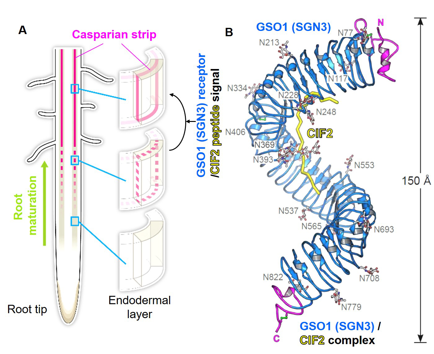
Figure: (A) Schematic model of Casparian strips (magenta). After establishing the connectivity (third row), this ring-like structure functions as an extracellular barrier. (Illustrated by Hiroko Uchida http://uchidahiroko.com/)
(B) Crystal structure of the GSO1(SGN3)/CIF2 receptor-peptide complex.
New assistant professor joins NIG as of January 1, 2020.
SASAKI, Takema : Oda Group • Cell Dynamics and Signaling Laboratory


