


Illuminating ALS Motor Neurons With Optogenetics in Zebrafish
Kazuhide Asakawa, Hiroshi Handa, Koichi Kawakami
Frontiers in Cell Developmental Biology 9, 640414 (2021) DOI:10.3389/fcell.2021.640414
Amyotrophic lateral sclerosis (ALS) is a fatal neurological disorder characterized by progressive degeneration of motor neurons in the brain and spinal cord. Spinal motor neurons align along the spinal cord length within the vertebral column, and extend long axons to connect with skeletal muscles covering the body surface. Due to this anatomy, spinal motor neurons are among the most difficult cells to observe in vivo. Larval zebrafish have transparent bodies that allow non-invasive visualization of whole cells of single spinal motor neurons, from somas to the neuromuscular synapses. This unique feature, combined with its amenability to genome editing, pharmacology, and optogenetics, enables functional analyses of ALS-associated proteins in the spinal motor neurons in vivo with subcellular resolution. Here, we review the zebrafish skeletal neuromuscular system and the optical methods used to study it. We then introduce a recently developed optogenetic zebrafish ALS model that uses light illumination to control oligomerization, phase transition and aggregation of the ALS-associated DNA/RNA-binding protein called TDP-43. Finally, we will discuss how this disease-in-a-fish ALS model can help solve key questions about ALS pathogenesis and lead to new ALS therapeutics.
This review article is a collaborative work conducted by Tokyo Medical University and NIG, and supported by the SERIKA FUND and KAKENHI (JP19K06933 and JP20H05345).
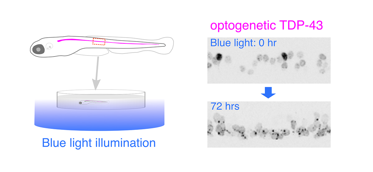
Figure: Optogenetic stimulation of TDP-43 phase transition in the zebrafish spinal motor neurons. Optogenetic TDP-43 forms abnormal aggregates by blue light illumination.


