


Division of Microbial Genetics • Araki Group
Dpb11/Cut5/TopBP1 is evolutionarily conserved and is essential for the initiation of DNA replication in eukaryotes. The Dpb11 of the budding yeast Saccharomyces cerevisiae has four BRCT domains (BRCT1 to -4). The N-terminal pair (BRCT1 and -2) and the C-terminal pair (BRCT3 and -4) bind to cyclin-dependent kinase (CDK)-phosphorylated Sld3 and Sld2, respectively. These phosphorylation-dependent interactions trigger the initiation of DNA replication. BRCT1 and -2 and BRCT3 and -4 of Dpb11 are separated by a short stretch of ~100 amino acids. It is unknown whether this inter-BRCT region functions in DNA replication. Here, we showed that the inter-BRCT region is a GINS interaction domain that is essential for cell growth and that mutations in this domain cause replication defects in budding yeast. We found the corresponding region in the vertebrate ortholog, TopBP1, and showed that the corresponding region also interacts with GINS and is required for efficient DNA replication. We propose that the inter-BRCT region of Dpb11 is a functionally conserved GINS interaction domain that is important for the initiation of DNA replication in eukaryotes.
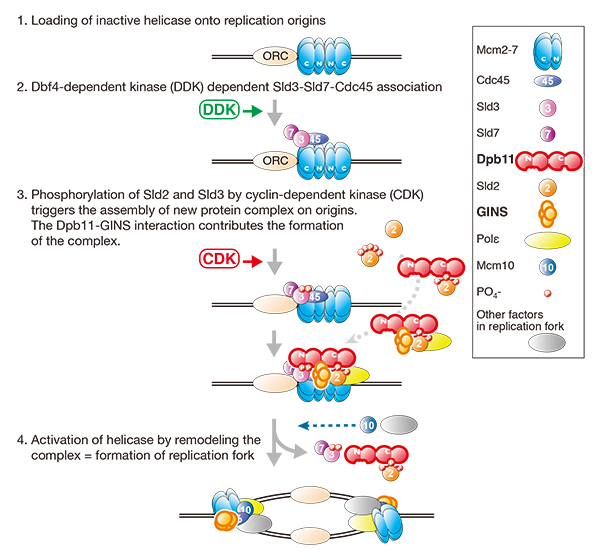
Schematic drawings of the initiation of DNA replication in eukaryotes. Dpb11-GINS interaction is important for the assembly of protein complex, which is required for the activation of replicative helicase.
Division of Molecular and Developmental Biology・Kawakami Group
The spatio-temporal regulation of muscle contractions that generate body movements critically depends on the exquisitely precise innervation of each muscle type by the appropriate motoneuron subtype. Therefore, delineating the identities of motoneurons and their connectivity to target muscles is fundamental to an understanding of the motor control by the central nervous system. In this study, by taking advantage of the optical and genetic accessibility, we dissected the spinal cord motor column of zebrafish larvae at the cellular level. By using the BAC for the Mnx homeodomain gene mnr2b, we established the mnGFF7 transgenic line expressing the Gal4FF transcription factor in a large part of the motor column. Single cell labelling of Gal4FF-expressing cells in the mnGFF7 larvae enabled a detailed investigation of the morphological characteristics of individual spinal motoneurons, as well as the overall organisation of the motor column in a spinal segment. The transgenic fish established here should facilitate an understanding of the cellular and molecular architecture of the spinal cord motor column and its connection to muscles in vertebrates.
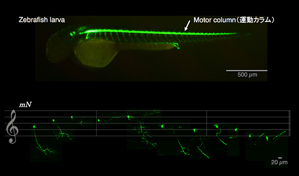
Lateral view of a zebrafish larva (top). Eleven different types of spinal motoneurons identified in the single cell labeling experiment.
Press release
The ancestor of extant Japanese fancy mice contributed to the mosaic genomes of classical inbred strains
Takada, T., Ebata, T., Noguchi, H., Keane, K., Adams, D., Narita, T., Shin-I, T., Fujisawa, H., Toyoda, A., Abe, K., Obata, Y., Sakaki, Y., Moriwaki, K., Fujiyama, A., Kohara, Y. and Shiroishi, T.We resequenced the genomes of Mus musculus molossinus-derived two inbred strains, MSM/Ms and JF1/Ms. MSM/Ms originated from Japanese wild mice, and ancestry of JF1/Ms was originally found in Europe and then transferred to Japan. We compared the characteristics of these sequences to those of the C57BL/6J reference sequence and the recent datasets from the resequencing of 17 inbred strains in the Mouse Genome Project.
The major outcomes of this study are summarized below.
1. Over 10 million SNPs and 1 million short indels are identified between the MSM/Ms or JF1/Ms sequence and the B6 reference sequence.
2. In comparison with the B6 reference sequence, the MSM and JF1 sequences contain 38,182 and 38,124 non-synonymous SNPs in 11,489 and 11,313 genes, respectively.
3. Genome introgression from M. m. molossinus into M. m. domesticus is primary framework for the mosaic genomes of classical inbred strains.
4. The genomes of B6 and other classical inbred strains have long consecutive segments with extremely high similarity (>99.998%) to the JF1/Ms strain. This indicates that the ancestor of JF1/Ms was direct origin of M. m. molossinus genome in classical inbred strains.
5. Roughly 30 to 40% of the SNPs detected in pairwise comparisons of classical inbred strains are attributable to the M. m. molossinus genome introgression.
The sequence data of MSM/Ms and JF1/Ms are available through the NIG mouse genome database, with side-by-side comparison to the B6 reference sequence.
This work was carried out in collaboration with National Institute of Genetics (Mammalian Genetics Laboratory, Comparative Genomics Laboratory and Genome Biology Laboratory), RIKEN (BioResource Center and Genome Science Center) and the Wellcome Trust Sanger Institute, and was supported by a Grant-in-Aid for Scientific Research on Priority Areas “Comparative Genomics” and the National BioResource Projects “the Genome Information Upgrading Program” from the Ministry of Education, Culture, Sports, Science and Technology of Japan. This work was also supported in part by the Biodiversity Research Project of the Transdisciplinary Research Integration Center, Research Organization of Information and Systems.
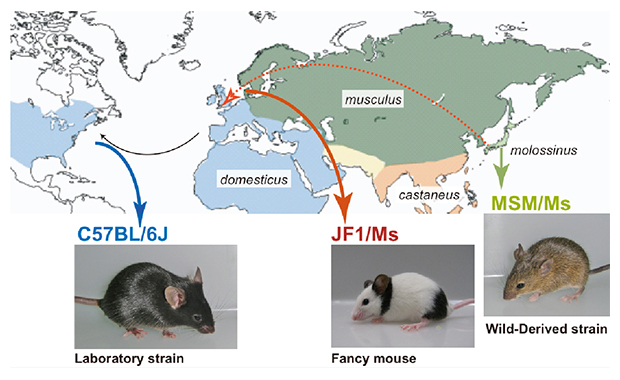
Three mouse strains mainly used in this study. C57BL/6J (B6) is a representative classical inbred strain, which was derived mostly from M. m. domesticus (west European subspecies). MSM/Ms and JF1/Ms originated from M. m. molossinus (Japanese subspecies).
Mammalian Genetics Laboratory・Shiroishi Group
Human chromosomal disorders, such as trisomy or monosomy, occur in over 1% of newborns, and are often associated with developmental failures including congenital heart disease and mental retardation. Though these disorders are likely caused by imbalance in genes involving the trisomic or monosomic region, the molecular bases are unknown in most cases.
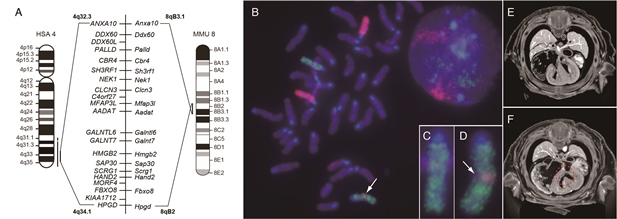
In this study, we demonstrated that Recombination-induced mutation 4 (Rim4) is a model animal of human chromosomal disorder, Partial trisomy distal 4q (4q+). Rim4 genome has extra fragment of 6.5Mb from mouse chromosome 8, which is syntenic to the distal end of human Chr4, 4q32.3 to 4q34.1, and contains 17 genes including basic helix-loop-helix transcription (bHLH) factor, Hand2. Rebalancing the gene dosage by genetic cross with Hand2 knockout mouse rescued symptoms of the heart and limb deformities of Rim4. These results suggest that over-dosage of Hand2 causes heart and limb deformities in Rim4 and 4q+, and Rim4 provides a unique animal model to understand the molecular bases underlying the complex phenotypes of 4q+.
This work was carried out in collaboration with National Institute of Genetics (Dr. Shiroishi group) and National Hospital Organization, Niigata Hospital (Dr. Tomizawa group).
Chromosome aberration and ventricular septal defect observed in Rim4 mutant mouse.
(A) Mouse Chr8 (MMU) 8qB2–B3.1 is syntenic to human Chr4 (HSA4) 4q32.3–34.1, and genes and gene order are well conserved in the syntenic regions of the two species. (B) Dual-color whole chromosome FISH image of Chr6 (green color) and Chr8 (magenta color). Magnified images of Wild type and Rim4/+ Chr6 are shown in insets, (C) and (D), respectively. Arrow in (B) and (D) indicates Chr8-derived insertion fragment. μ-CT images of E14.5 embryos of wild type (E) and Rim4/Rim4 mouse (F) are shown. Dotted circle in (F) indicates ventricular septal defect.
Experimental Farm・Nonomura Group
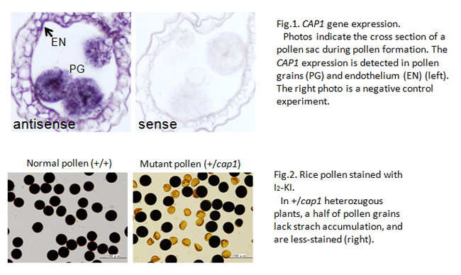
The genome of cultivated rice contains 32,000 genes, and more than 20,000 are expressed in developing anther and pollen. However, there are few genes whose function is evidenced in pollen development. In this study, we succeeded to identify the rice gene with its function specifically in pollen formation, and named COLLAPSED ABNORMAL POLLEN1 (CAP1).
In angiosperms, the pollen has a special, tricellular structure of a pair of sperm cells involved within the vegetative cell. The CAP1 gene is strongly expressed in anthers at the bicellular pollen stage (Fig.1). The pollen grains lacking CAP1 function lose most cellular components except for outer pollen wall, exine (Fig.2), and are unable to elongate the pollen tube at all. Homozygous cap1 mutant plants exhibit no remarkable aberration in other developmental stages, indicating the specific function of CAP1 in pollen development.
The amino acid alignment of rice CAP1 protein is similar to that of the plant L-arabinokinase. The function of this enzyme is supposed in phosphorylation of free L-arabinoses, generated during the cell-wall metabolism, for reuse in de novo wall formation. It is likely in rice cap1 mutant that the arabinoses unable to be reused accumulate aberrantly or the cell-wall metabolism is disrupted in pollen grains. One of Arabidopsis L-arabinokinase-like genes shows a similar expression pattern to rice CAP1-gene expression. This result suggests that the CAP1 function is conserved broadly among angiosperm species, and plays an important role in pollen development.
This is a collaborative work with the Akita Prefectural University and the National Institute of Agribiological Sciences, Japan, and is supported by the NIG Collaborative Research Funding.
Division of Molecular and Developmental Biology・Kawakami Group
Transcription factors (TFs) are able to regulate differentiation related processes, including dedifferentiation and direct conversion, through the regulation of cell type-specific transcriptional profiles. However, the functional interactions between the TFs regulating different transcriptional profiles are not well understood. Here, we show that the TFs capable of inducing cell type-specific transcriptional profiles prevent the dedifferentiation induced by TFs for pluripotency. Of the large number of TFs expressed in a neural lineage cell line, we identified a subset of TFs that, when overexpressed, strongly interfered with the dedifferentiation triggered by the procedure to generate induced pluripotent stem cells. This interference occurred through a maintenance mechanism of the cell type-specific transcriptional profile. Strikingly, the maintenance activity of the interfering TF set was strong enough to induce the cell line-specific transcriptional profile when overexpressed in a heterologous cell type. Our results suggest that dedifferentiation suppresses a cell type-specific transcriptional profile, which is primarily maintained by a small subset of TFs capable of inducing direct conversion. We anticipate that this functional correlation might be applicable in various cell types and might facilitate the identification of TFs with induction activity in efforts to understand differentiation.
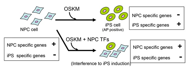
NPC (neural progenitor) cells are dedifferentiated by introduction of OSKM. This dedifferentiation was inhibited by expression n of NPC-specific transcription factors.
Division of Cytogenetics・Kobayashi Group
Gene amplification is one of the major strategies used by cells to increase the abundance of gene products. We have been studying amplification of the rDNA cluster in yeast with a focus on distinguishing the contributions of unequal sister-chromatid recombination vs rolling circle-type amplification as observed in the early developmental stage in amphibian oogenesis. It is not known how these two modes of amplification switch. We screened for yeast mutants in which rDNA copy number was unstable and found and characterized an rtt109 mutant that has an abnormally high rDNA copy number. Evidence is presented that the rolling circle-type amplification occurs in this mutant. Therefore, RTT109 plays a key role in regulating mode of rDNA amplification.
Variation in gene copy number (amplification) has been widely detected in various organisms, contributing to both beneficial adaptation and pathology (e.g., cancer). Our results shed new light on molecular mechanisms of gene amplification.
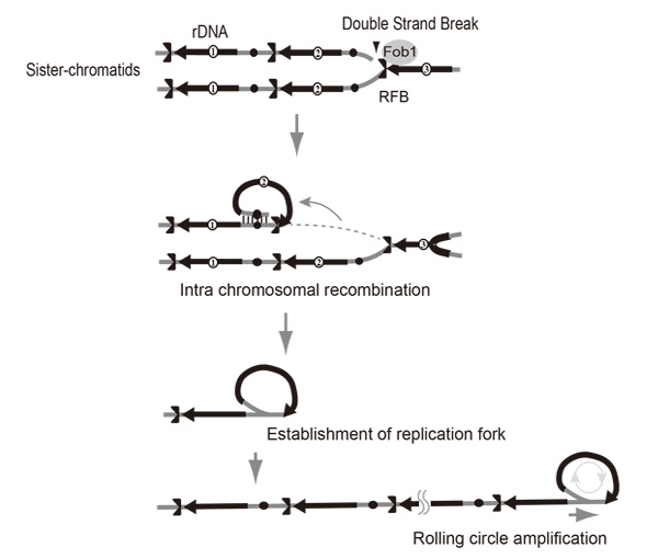
Rolling circle replication by intra-sister chromatid recombination.The broken end at the RFB (Replication Fork Barrier site) recombines via intra-sister chromatid exchange followed by rolling circle replication.
Press release
Chromosome engineering allows the efficient isolation of vertebrate neocentromeres
Wei-Hao Shang, Tetsuya Hori, Nuno M.C. Martins, Atsushi Toyoda, Sadahiko Misu, Norikazu Monma, Ichiro Hiratani, Kazuhiro Maeshima, Kazuho Ikeo, Asao Fujiyama, Hiroshi Kimura, William C. Earnshaw, and Tatsuo FukagawaCentromeres are specified by sequence-independent epigenetic mechanisms in most organisms. Rarely, centromere repositioning results in neocentromere formation at ectopic sites. However, the mechanisms governing how and where neocentromeres form are unknown.
Here, we established a chromosome-engineering system in chicken DT40 cells that allowed us to efficiently isolate neocentromere-containing chromosomes.
Neocentromeres appear to be structurally and functionally equivalent to native centromeres. Chromatin immunoprecipitation sequencing (ChIP-seq) analysis with 18 neocentromeres revealed that the centromere-specific histone H3 variant CENP-A occupies an ∼40 kb region at each neocentromere, which has no preference for specific DNA sequence motifs. Furthermore, we found that neocentromeres were not associated with histone modifications H3K9me3, H3K4me2, and H3K36me3 or with early replication timing. Importantly, low but significant levels of CENP-A are detected around endogenous centromeres, which are capable of seeding neocentromere assembly if the centromere core is removed.
Our experimental system provides valuable insights for understanding how neocentromeres form. This was performed by Wei-Hao Shang and Tetsuya Hori (Fukagawa Lab) in collaboration with TRIC of ROIS, Fujimaya Lab. (NIG, NII), Maeshima Lab (NIG), Ikeo Lab. (NIG), Kimura Lab (Osaka U.), and Eranshaw Lab (U. Edinburgh).
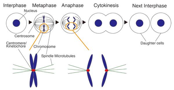
Cell division and centromere
During mitosis spindle microtubules capture a special structure of chromosome for faithful chromosome segregation. This structure is called “Kinetochore”. Centromere is a genome region in which kinetochore is formed. Dysfunction of kinetochore results in some diseases including cancer.
Division of Molecular and Developmental Biology・Kawakami Group
Various types of sense organs are distributed over the body surface. These organs are innervated by sensory axons, thereby transmit information to the central nervous system. Many sense organs emit molecules required for proper growth or guidance of the axons. However, roles of the axons for development of sense organs remain poorly understood. In this study, we reveal that proliferation of the mechanosensory organs (neuromasts) of fish is promoted by axonal innervation.
In adult zebrafish, the neuromasts give rise to new neuromasts by budding and generate a cluster of organs. The budding cells, that show high Wnt signaling activity, are associated by side branches of axons extended from the founder neuromast (Fig. 1A). To analyze the role of innervation, we ablated sensory neurons in larvae. The neuromast sent off budding cells normally, but subsequent cell proliferation to generate new sense organs did not take place (Fig. 1B,C). We propose that the axon promotes Wnt signaling activity, which is required for the proliferate phase that leads to sense organ formation.
This study was carried out in collaboration with Dr. Alain Ghysen (Montpellier University) and funded by the PRESTO of the Japan Science and Technology Agency.
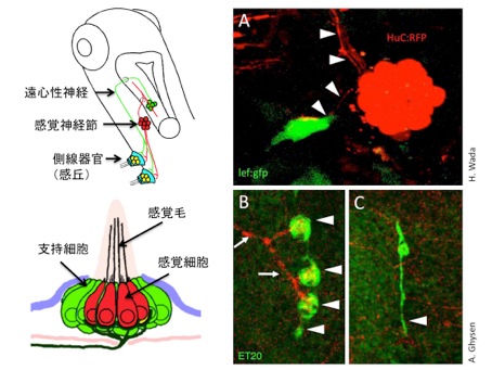
(A) The lateral line sense organ, neuromast, sends off proliferative bud cells, that show high Wnt signaling activity. Axons are indicated by arrowheads.
(B, C) The cell proliferation does not take place in the absence of the sensory axons.
Press release
Real-Time Visualization of Neuronal Activity during Perception
Akira Muto, Masamichi Ohkura, Gembu Abe, Junichi Nakai and Koichi Kawakami Current Biology, 23(4), Feb 18, 2013. DOI: 10.1016/j.cub.2012.12.040The visual world is first projected onto the retina, and the visual information is further transmitted to the brain. In this initial stage of visual processing, the visual world is mapped on the brain, which is called visuotopy. This is a common feature found in the brains of all animals with eyes. While visuotopy is a well-established notion, no one has ever demonstrated this in real time in a natural condition. In this study we developed an improved version of GCaMP, a calcium sensor, in collaboration with Prof. Nakai at Saitama University. Using this new highly sensitive calcium probe, we could visualize neuronal activity in a zebrafish larva during visual perception in prey capture behavior.
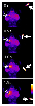
A swimming paramecium (arrowheads) evoked Ca2+ transients (arrows) in the neuropil and cell bodies of the left tectum of a one-week old larva embedded in agarose. Ratio images were created and pseudo-colored. Scale bar represents 100 μm.
▶This method is one of the basis of these researches. A virtual reality system to analyze neural activity and behavior in adult zebrafish Appetite control via hunger and satiety Neural signatures of sleep in zebrafish Glia-neuron interactions underlie state transitions to generalized seizures ‘Eating with the eyes’ is hard-wired in the brain
Microbial Genetics Laboratory・Niki Group
Comparative Genomics Laboratory・Fujiyama Group
Genome Biology Laboratory・Kohara Group
Wild-type E. coli are rod shape (Figure A, left). To make rod shaped cells, it is necessary that many proteins form complexes and function properly. E. coli cells are surrounded by rigid peptidoglycan (PG) layer and have to synthesize PG correctly. So far, we have identified RodZ required for determination of cell shape.
RodZ-deficient mutant is round shape (Figure A, middle) and grows slower than wild-type E. coli cell. To reveal function of RodZ protein, we isolated suppressor mutants that restore cell growth and shape of the rodZ mutant. To map the mutation sites in the suppressor mutants, we sequenced whole genome of twenty-nine mutants by a next-generation sequencer Solexa. This is the first report that mutation sites in ~30 mutants are determined by whole genome sequencing.
Most of the mutations were found in mreB, mrdA, or mrdB genes. It has been hypothesized that MreB, PBP2 encoded by mrdA gene, and RodA encoded by mrdB gene function with RodZ. Especially, twenty of twenty-nine mutants had a mutation in mreB gene. In addition, these mutations were clustered in domain 1A, one of the subdomain of MreB protein. These mutations change properties of MreB protein so that E. coli can form rod shape without RodZ protein. We also found that mutants of PBP2 and RodA change properties of MreB. Thus, we concluded that RodZ regulates function of MreB to form rod shape of E. coli.
This work was done by a collaboration of Niki lab, Fujiyama lab, and Kohara lab.
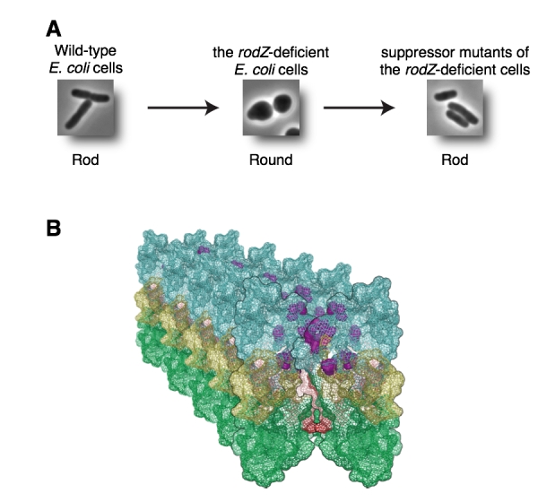
(A) Wild-type E. coli is rod (left). RodZ-deficient E. coli cell is round (middle). Suppressor mutants are rod (right). (B) Mutation sites shown by purple are clustered in domain 1A and it is a surface between MreB filaments. These mutations change properties of MreB filaments.
Division of Molecular Genetics・Fukagawa Group
The kinetochore forms a dynamic interface with microtubules from the mitotic spindle during mitosis. The Ndc80 complex acts as the key microtubule-binding complex at kinetochores. However, it is unclear how the Ndc80 complex associates with the inner kinetochore proteins that assemble upon centromeric chromatin. Here, based on a high-resolution structural analysis, we demonstrate that the N-terminal region of vertebrate CENP-T interacts with the “RWD” domain in the Spc24/25 portion of the Ndc80 complex. Phosphorylation of CENP-T strengthens a cryptic hydrophobic interaction between CENP-T and Spc25 resulting in a phospho-regulated interaction that occurs without direct recognition of the phosphorylated residue. The Ndc80 complex interacts with both CENP-T and the Mis12 complex, but we find that these interactions are mutually exclusive, supporting a model in which two distinct pathways target the Ndc80 complex to kinetochores. Our results provide a model for how the multiple protein complexes at kinetochores associate in a phospho-regulated manner.
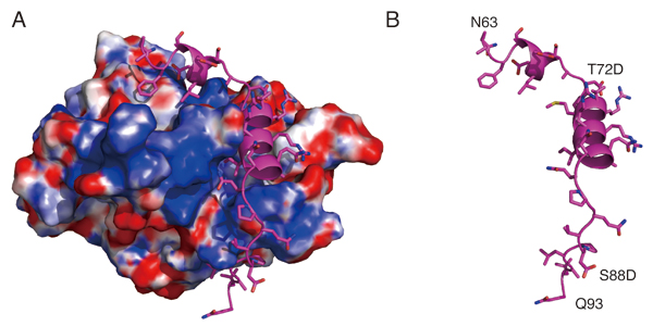
Structural model showing the surface charge of the Spc24/25 complex interacting with phospho-mimetic CENP-T peptide (Cyan). (B) Structural model showing the phospho-mimetic CENP-T peptide from the CENP-T-Spc24/25 complex structure in (A) on its own.

Mammalian Development Laboratory・Saga Group
In the mouse, testicular development is triggered in somatic cells by the function of Sry followed by the activation of fibroblast growth factor (FGF) 9, which regulates testicular differentiation in both somatic and germ cells. However, the mechanism is unknown. We show here that Nodal/Activin signaling pathway is activated in both male gem cells and somatic cells. The disruption of Nodal/Activin signaling drives male germ cells into meiosis and causes ectopic initiation of female-specific genes in somatic cells. Furthermore, we prove that Nodal/Activin-A works directly on male germ cells to induce male specific gene, Nanos2 independently of FGF9. We conclude that Nodal/Activin signaling is required for testicular development and propose a model in which Nodal/Activin-A acts downstream of FGF signaling to promote male germ cell fate and protect somatic cells from initiating female differentiation.
This study is conducted by Quan Wu who is a current student of SOKENDAI.
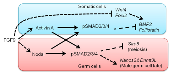
A model proposed by our study. FGF signals activate Nodal/Activin signaling pathway in both somatic cells and germ cells. Nodal/Activin-A triggers male sex differentiation by inducing male-specific genes, Nanos2 and Dnmt3L. Meanwhile, it suppresses Stra8 that is an essential gatekeeper of meiosis. In somatic cells Nodal/Activin-A thwarts the process of female differentiation by inhibiting Bmp2 and Follistatin.


