


Miyagishima Group / Symbiosis and Cell Evolution Laboratory
The closed nutrient recycling system in the Paramecium–Chlorella photosymbiosis contributes to survival under oligotrophic conditions
Kaoru Okada, Takayuki Fujiwara, Shunsuke Hirooka, Yusuke Kobayashi, Ryo Onuma, and Shin-ya Miyagishima
Science Advances 11, eadz0004 (2025) DOI:10.1126/sciadv.adz0004
Endosymbiotic relationships between a heterotrophic host and a unicellular algal endosymbiont are observed across many eukaryotic lineages. Although these relationships are prevalent in oligotrophic environments, how they function and provide an advantage under such conditions remains largely unknown. To address these issues, we examined the behaviour of the ciliate Paramecium bursaria hosting Chlorella endosymbionts under nitrogen- and prey-depleted conditions. The Paramecium host survived for up to five weeks while maintaining the number of Chlorella endosymbionts, whereas aposymbiotic Paramecium and free-living Chlorella either died or bleached, respectively, under the same conditions. In the symbiotic state, the host continuously fed on the endosymbionts without excreting nitrogenous waste into the medium, while the remaining endosymbionts continued to proliferate using heterotrophic metabolites from the host and light energy. Thus, the cyclical farming of endosymbionts by the host maintains a high concentration of nutrients within the closed system, providing a selective advantage in oligotrophic environments.
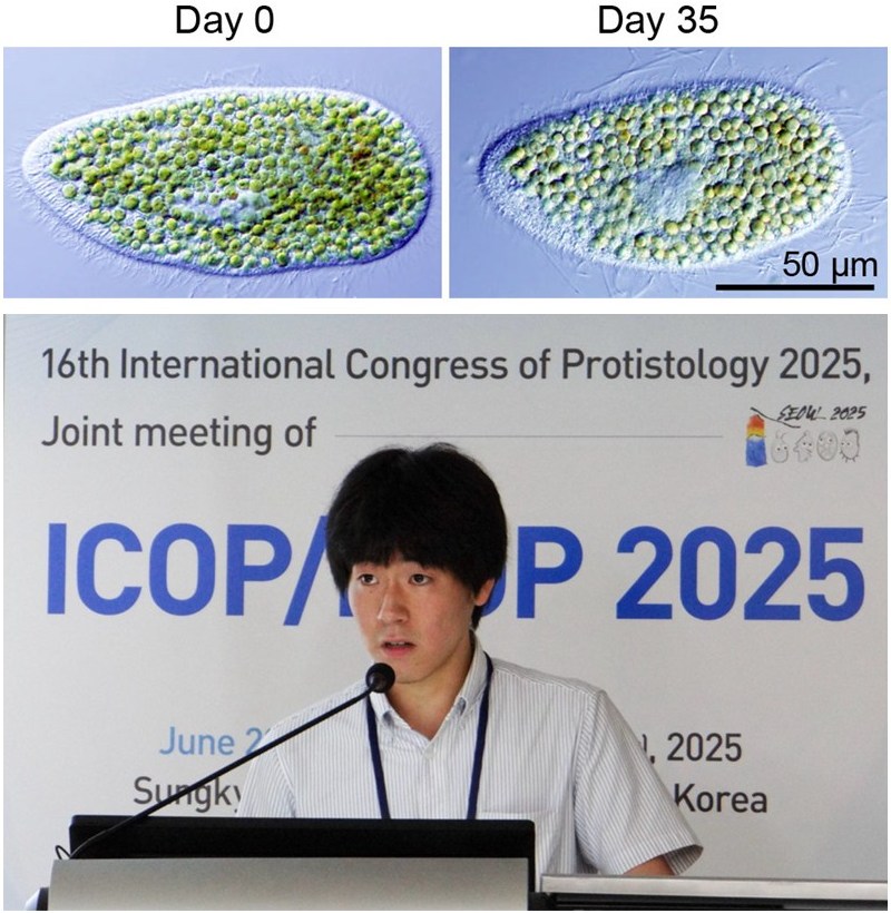
Figure. Micrographs of Paramecium bursaria and Okada-san (a graduate student) presenting the findings at an international conference
A Paramecium bursaria cell (Day 0) that had been grown with bacterial prey was further incubated for 35 days under light in a culture medium lacking prey as well as nitrogen and phosphorus sources (Day 35). The host paramecium remained viable while maintaining approximately 300 endosymbiotic Chlorella cells per host cell. These Chlorella cells were partly digested by the host but also proliferated, resulting in the apparent number of symbionts being largely maintained. Okada-san presented these findings at ICOP/ISOP 2025, an international protozoology conference held in Seoul, Korea, from June 22 to 27, 2025, and received the Best Oral Presentation Award
Press release
Intrinsically accelerated cellular degradation is amplified by TDP-43 loss in ALS-vulnerable motor neurons in a zebrafish model
Kazuhide Asakawa, Takuya Tomita, Shinobu Shioya, Hiroshi Handa, Yasushi Saeki, and Koichi Kawakami
Nature Communications DOI:10.1038/s41467-025-65097-0
![]() Press release (In Japanese only)
Press release (In Japanese only)
Amyotrophic lateral sclerosis (ALS) is a fatal disease in which motor neurons that control voluntary movement gradually degenerate, leading to paralysis. Although many studies have been conducted, the reason why motor neurons are selectively affected has remained unclear.
Using transparent zebrafish that allow live imaging of nerve cells, we found that motor neurons constantly endure a burden to keep proteins properly folded (Figure 1). Larger neurons were particularly vulnerable, and this burden was further increased by genetic manipulations mimicking ALS.
Because motor neurons connect distant organs—brain, spinal cord, and muscles—with single cells, they must grow large and synthesize vast amounts of proteins, which increases their burden. This “inevitable degradation burden” may underlie their selective vulnerability in ALS. Even in tiny zebrafish larvae, such burden was detected, suggesting that in humans, whose motor neurons can reach as long as one meter in length, the challenge is far greater.
Understanding how this degradation burden arises may open the way to new therapeutic strategies to slow ALS progression and protect motor neurons from degeneration.
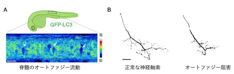
Figure: A) Large motor neurons show high protein degradation activity (autophagic flux). GFP-LC3 is degraded through autophagy. Arrows indicate large motor neurons with active GFP-LC3 degradation.
B) Inhibition of autophagy impairs axonal development of motor neurons (right). Scale bars, 20 µm.
Press release
Rad27/FEN1 prevents accumulation of Okazaki fragments and ribosomal DNA copy number changes
Tsugumi Yamaji, Yuko Katayama, Nanase Arata, and Mariko Sasaki
FEBS Letters 2025 DOI:10.1002/1873-3468.70193
![]() Press release (In Japanese only)
Press release (In Japanese only)
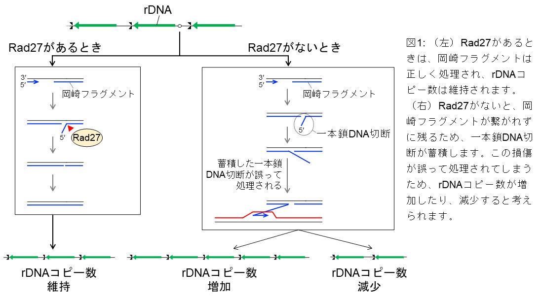
DNA copy number changes are the most frequent genomic alterations in cancer cells. Here, we 19 demonstrate that Rad27/FEN-1, a structure-specific nuclease in budding yeast, plays a crucial role in 20 maintaining the stability of the ribosomal DNA (rDNA) repeats. Severe rDNA instability is observed 21 in the rad27∆ mutant, independently of Fob1-mediated DNA replication fork arrest and DNA double-22 strand break (DSB) formation in the rDNA. The rad27Δ mutant accumulates Okazaki fragments in the 23 rDNA region, without inducing formation of detectable DSBs. Similar rDNA instability is observed in 24 DNA ligaseCdc9-deficient cells. Furthermore, Exonuclease 1 and PCNA partially compensate for the 25 loss of Rad27 in the rDNA stabilization. These findings highlight the importance of proper Okazaki 26 fragment maturation in the maintenance of rDNA stability.
The National Institute of Genetics (NIG) invites applications for one full or associate professor position in the Department of Gene Function and Phenomics. Please see the details below and apply accordingly.
Application Guidelines:PDF Application Materials:Brief summary of career(excel)
1. Division: Department of Gene Function and Phenomics
2. Recruiting: Full /Associate Professor (1 qualified person)
3. Appointment Start Date: On and after April 2026 (negotiable)
4. Term of Appointment:
Indefinite term appointment (until the mandatory retirement age of 65, based on the regulations of the Research Organization of Information and Systems)
5. Qualifications:
The candidate should be a talented researcher with outstanding achievements in microbiology or any other fields of life science related to microorganisms, have the expertise and leadership skills necessary to conduct cutting-edge research in these fields, and develop the prokaryote bioresource at the National Institute of Genetics.
6. Responsibilities:
The appointed researcher will lead an independent laboratory as a principal investigator. He or she will also participate in the education of graduate students as a faculty member of the Genetics Program of SOKENDAI.
7. Research environment and support:
・As an Inter-University Research Institute, NIG provides substantial common equipment and research infrastructure and promotes collaborative research worldwide. We expect the appointed researcher to take advantage of this research environment to organize and lead his/her research team.
・One assistant professor and one technical support staff member will be allocated to the laboratory.
8. Application Deadline: Noon (Japan Time) on December 24, 2025
9. Application Materials:
I. Curriculum Vitae (In English. Japanese applicants should also submit a Japanese version. Include your e-mail address.)
II. List of publications (If there are multiple authors, briefly describe your contribution. Indicate your most significant papers.)
III. Summary of past and present research and future directions (In English, max. 1,500 words. Include figures if necessary.)
IV. Names and contacts of references (At least two domestic and two international references.)
V. Summary of career
VI. Your key papers
*All the personal data in the application is handled in strict confidentiality, and used solely for recruitment purposes.
10. Submission:
Submit items I to VI listed above electronically as follows:
(a) The subject line should be “Application for Department of Gene Function and Phenomics Full / Associate Professor”, which should be also noted in the e-mail body.
(b) Put items I to IV together in a single file by separating each item by page. E-mail the file as an attachment. The file format should be MS Word or PDF. Also attach item V (form can be downloaded from the NIG homepage) as a separate file.
(c) Send your key papers as PDF attachments. The file format should be PDF. If your key papers are available to access online, include a list of URLs in the e-mail body.
* We will notify you via e-mail upon receipt of your electronic application within two business days.
Based on gender equality National Institute of Genetics has been actively promoting female scientists. In case of equivalent aptitude and achievements in research, education, and social contributions, preference will be given to female candidates.
National Institute of Genetics is promoting safety and healthy working, including preventing passive smoking. (Indoor smoking is banned. / Smoking is allowed only in the designated outdoor areas.)
Send your application and inquiries to:
NIG Personnel Committee (Personnel Team)
E-mail:
Mailing Address:
National Institute of Genetics
Yata 1111, Mishima, Shizuoka 411-8540 JAPAN
TEL: +81-55-981-6716
Homepage: https://www.nig.ac.jp/nig/
NIG Organization Chart: https://www.nig.ac.jp/nig/research/organization-top/organization
[Notes on Submission of Documents]
The following deficiencies have been observed:
Cases where the PDF file of the key paper was not attached
Cases where access to the key paper on the internet required an account, thereby restricting viewing
If, due to file size limitations, it is difficult to send all PDF files in a single email, they may be divided across multiple emails without issue.
In any event, please ensure that the PDF files are duly submitted.
Furthermore, if access to a paper requires an account on the relevant website, kindly indicate this in the remarks section.
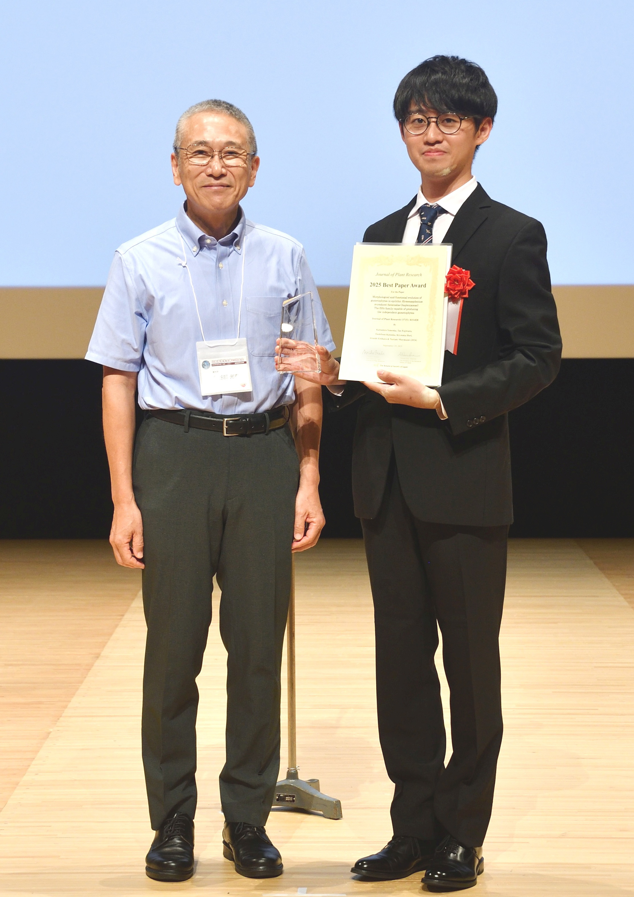
Dr. Katsuhiro Yoneoka, a postdoctoral researcher in the Plant Evolution Laboratory (Fukushima Lab), received the JPR Best Paper Award at the 89th Annual Meeting of the Botanical Society of Japan, held at the Fukuoka International Congress Center, Fukuoka, Japan, on September 17– 20, 2025.
▶ Awarded presentation title: Morphological and functional evolution of gametophytes in epilithic Hymenasplenium murakami-hatanakae (Aspleniaceae): The fifth family capable of producing the independent gametophytes
Dr. Masahito Tanaka, from the Laboratory of Physics and Cell Biology (the Shimamoto Lab) at the National Institute of Genetics, gave an invited talk at the 63rd annual meeting of the Biophysical Society of Japan and won the Early Career Presentation Award. Dr. Tanaka has been supported by a JSPS postdoctoral fellowship since 2023 and will start his new research under JST ACT-X this October.
▶ Awarded presentation title: Changes in the physical properties of early embryonic nuclei promote a transcriptional burst
▶ The 63rd Annual Meeting of the Biophysical Society of Japan
Koide Group / Mouse Genomics Resource Laboratory
Comparative Analysis of Tickling and Conspecific Play in Tame Mice and Golden Hamsters
Dagher S, DeAngelo D, Sato RY, Norimoto H, Koide T*, and Ishiyama S*
(*co-corresponding author)
Behavioural Brain Research (2026) 496 DOI: 10.1016/j.bbr.2025.115849
Social “play” is a vital behaviour through which animals strengthen bonds, acquire essential skills, and express positive emotions. Yet mice have long been regarded as creatures that simply do not play.
A research team led by Dr. Sarah Dagher (PhD student at the time of the experiment, now at the Max Planck Institute for Metabolism Research) and Dr. Shimpei Ishiyama (now at the Central Institute of Mental Health, Mannheim), and Associate Professor Tsuyoshi Koide at the National Institute of Genetics has now overturned this long-held belief. They discovered that “tame mice” — selectively bred to approach a human hand — display playful behaviours with both humans and fellow mice.
When tickled, these tame mice emit ultrasonic sounds resembling laughter and actively chase the researcher’s hand. In social settings, they engage in playful behaviours such as poking at one another and making exaggerated movements, again accompanied by ultrasonic vocalisations. By contrast, ordinary mice show little response to human tickling, interact less with peers, and often display aggression instead.
Intriguingly, the tame mice produce different types of vocalisations when playing with humans compared to when playing with other mice, indicating that they clearly distinguish their social partners.
These findings have important implications for understanding animal domestication and human–animal relationships. Breeding mice for tameness not only increased their willingness to play with humans but also enhanced social play among peers. Previous genetic analyses further revealed that regions associated with tameness in mice overlap with those linked to domestication in dogs, suggesting a shared mechanism across species.
This study shows that a tendency to “enjoy play” is not limited to certain animals but can emerge as a result of increased receptiveness to human interaction. It also challenges the long-standing notion that “rats play but mice do not,” demonstrating that mice, too, can become playful companions — both for humans and for one another.
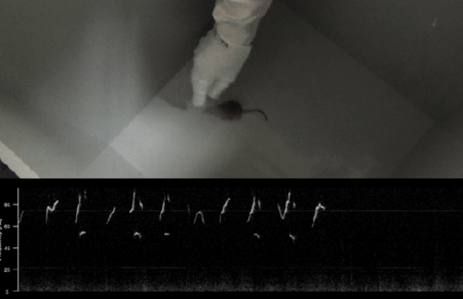
Figure: The selectively bred mice respond to tickling by emitting ultrasonic vocalisations and exhibiting play-like behaviour, such as spontaneously chasing the hand.
The video is here: https://neurogelotology.lol/2025/09/30/selective-breeding-makes-mice-playful-towards-humans-and-other-mice/
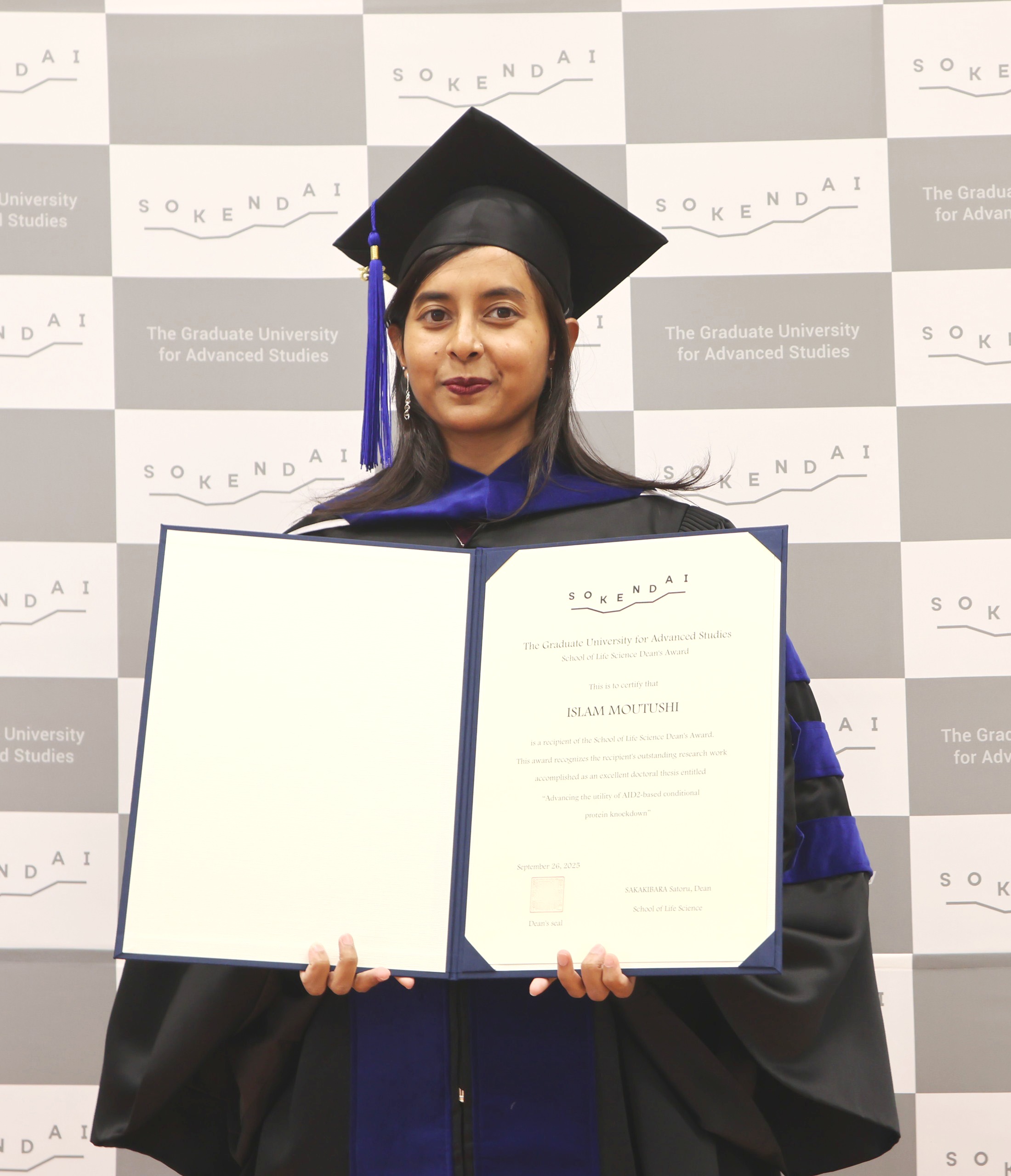
Ms. Islam, Moutushi, who graduated from SOKENDAI in September 2025 as a member of the Molecular Cell Engineering Laboratory, has been awarded the School of Life Science “Dean’s Award” for the first semester of 2025.
The Dean’s Award recognizes degree recipients who have conducted research worthy of commendation and reported their accomplishments in an outstanding doctoral thesis. The award was presented on September 26, 2025 during the graduation ceremony. In addition to the Dean’s Award, Ms. Islam has also been awarded the Genetics Program’s Morishima Award.
・Thesis title : Advancing the utility of AID2-based conditional protein knockdown
Ms. Islam has provided the following statement regarding the award.
“I am honored and delighted to receive the Dean’s Award from the School of Life Science, SOKENDAI University.
I would like to express my heartfelt gratitude to my supervisor, Professor Masato Kanemaki. Without his constant guidance and support, this achievement would not have been possible.
National Institute of Genetics, Japan is the first place where I truly learned about research, and it has provided me with invaluable opportunities to explore and grow in this field. I am also grateful to my lab members and collaborators for their contributions, and to my family for their continuous encouragement throughout this journey.
This recognition will inspire me to continue pursuing science with passion and dedication. It will remain a strong motivation as I move forward in my career.”
Ms. Islam
Kitano Group / Ecological Genetics Laboratory
Functional mutations in the thyroid-stimulating hormone receptor in natural stickleback populations at sites identical to human disease-causing mutations
Jun Kitano, Mana Sato, Hiyu Kanbe, Genta Okude, Asano Ishikawa, Yukinori Kazeto & Takashi Makino
BMC Ecology and Evolution (2025) 98 DOI:10.1186/s12862-025-02440-5
This study examined whether mutations that cause thyroid disease in humans could help identify functional mutations in natural stickleback populations. The researchers found that several Japanese stickleback populations carry non-synonymous mutations in the thyroid-stimulating hormone receptor (Tshr)2 gene. Functional assays revealed that amino acid substitutions at sites corresponding to human loss-of-function or gain-of-function mutations similarly reduced or enhanced receptor activity. Thus, we discovered natural populations carrying mutations similar to those that cause human disease. Further research is needed to determine whether these mutations are deleterious or adaptive in their habitats.
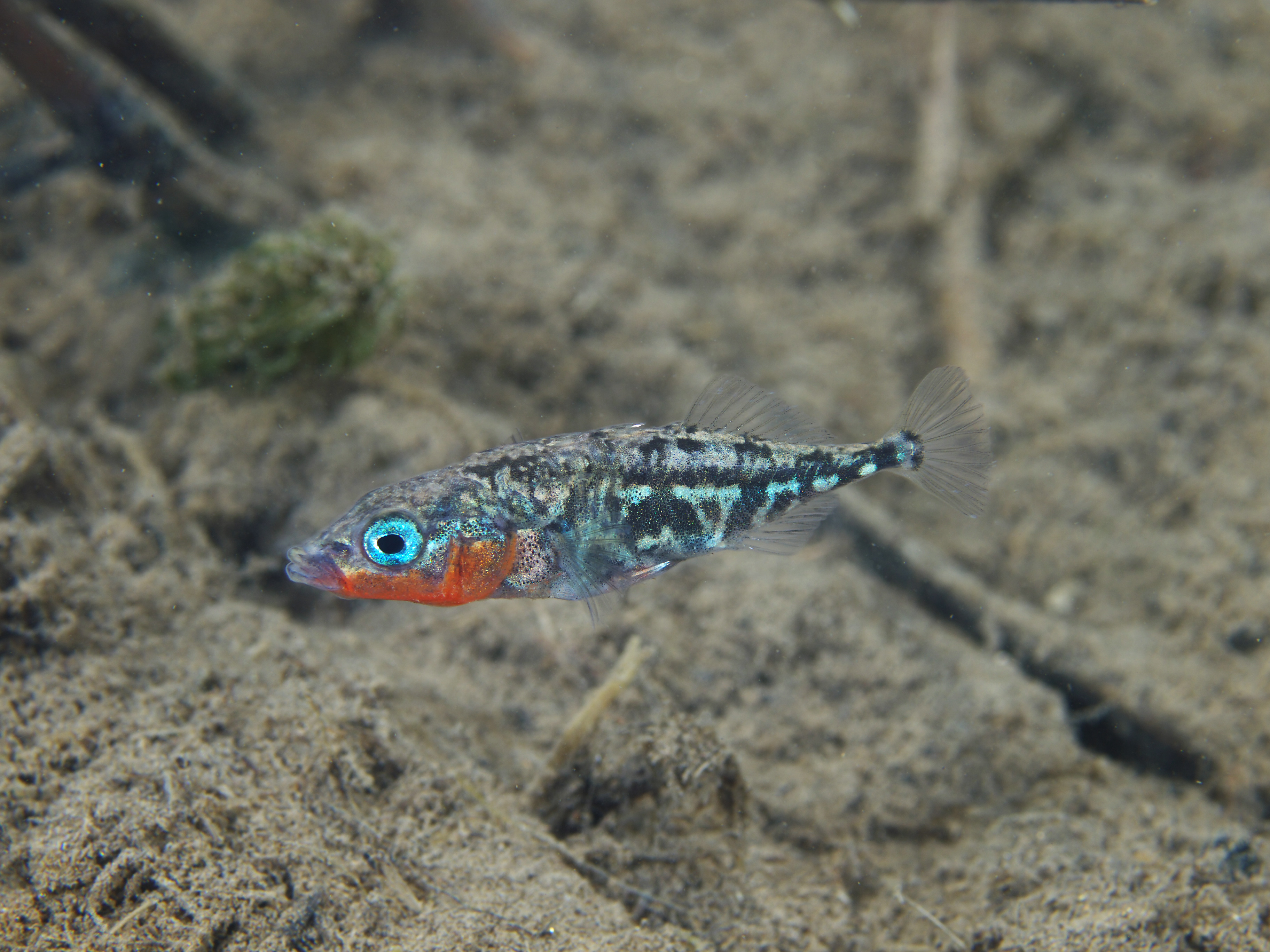
Figure: Hariyo (a freshwater population of the genus Gasterosteus, currently found in Gifu and Shiga Prefectures, and formerly inhabiting Mie Prefecture, Japan) possessed a mutation in the thyroid-stimulating hormone receptor (TSHR) gene at an identical site where mutations cause human disease.
Photo courtesy of Yasuyuki Hata.


