


Press release
Replication-dependent histone labeling dissects the physical properties of euchromatin/heterochromatin in living human cells.
Katsuhiko Minami, Kako Nakazato, Satoru Ide, Kazunari Kaizu, Koichi Higashi, Sachiko Tamura, Atsushi Toyoda, Koichi Takahashi, Ken Kurokawa, Kazuhiro Maeshima*
*corresponding author
Science Advances (2025) 11, eadu8400 DOI:10.1126/sciadv.adu8400
![]() Press release (In Japanese only)
Press release (In Japanese only)
A string of nucleosomes, where genomic DNA is wrapped around histones, is organized in the cell as chromatin, ranging from euchromatin to heterochromatin, with distinct genome functions. Understanding physical differences between euchromatin and heterochromatin is crucial, yet specific labeling methods in living cells remain limited. Here, we have developed replication-dependent histone (Repli-Histo) labeling to mark nucleosomes in euchromatin and heterochromatin based on DNA replication timing (Fig. 1). Using this approach, we investigated local nucleosome motion in the four known chromatin classes, from euchromatin to heterochromatin, of living human and mouse cells. The more euchromatic (earlier-replicated) and more heterochromatic (later-replicated) regions exhibit greater and lesser nucleosome motions, respectively. Notably, the motion profile in each chromatin class persists throughout interphase. Genome chromatin is essentially replicated from regions with greater nucleosome motions, even though the replication timing is perturbed. Our findings, combined with computational modeling, suggest that earlier-replicated regions have more accessibility, and local chromatin motion can be a major determinant of genome-wide replication timing (Fig. 2).
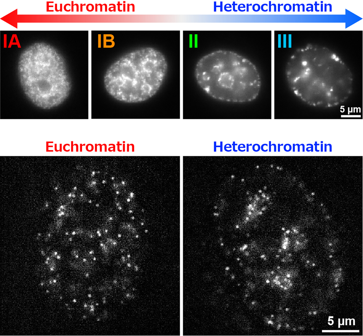
Figure1: (Top) The replication-dependent histone labeling (Repli-Histo labeling) specifically visualizes euchromatin (IA and IB), where gene expression is active, and heterochromatin (II and III), where it is repressed. (Bottom) The fluorescently labeled single-nucleosomes in euchromatin (left) and heterochromatin (right) in living HeLa cells. Each dot represents a single nucleosome.
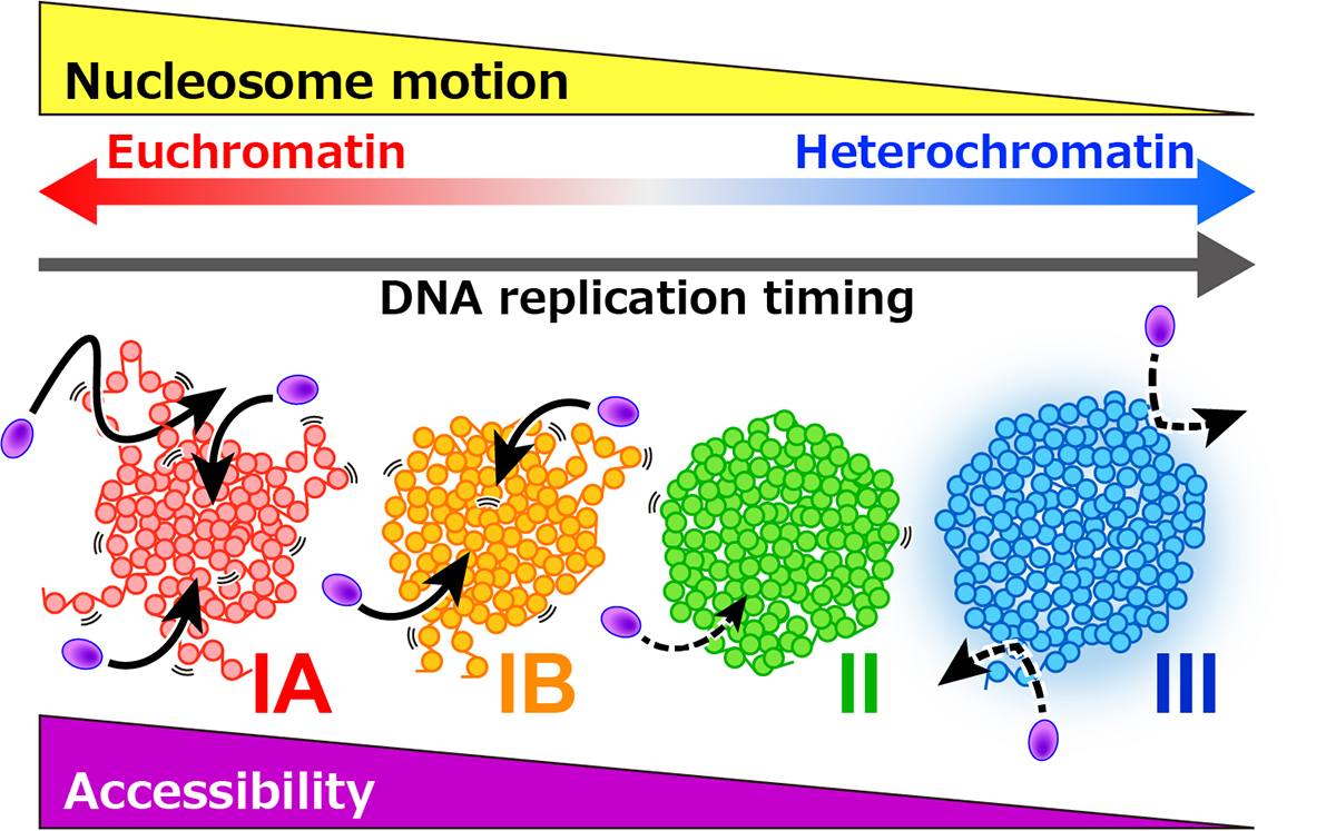
Figure2: Euchromatic regions (IA and IB) exhibit greater nucleosome motion, whereas heterochromatic regions (II and III) show more restricted motion. The nucleosome motions facilitate chromatin accessibility to large proteins (purple), which can regulate various genomic functions, including DNA replication and transcription.


