


Kawakami Group / Laboratory of Molecular and Developmental Biology
The dwarf neon rainbowfish Melanotaenia praecox, a smallspiny-rayed fish with potential as a new Acanthomorphamodel fish: II. Establishment of a microinjection procedurefor genetic engineering.
Kazuhide Miyamoto, Gembu Abe, Koichi Kawakami, Koji Tamura, Satoshi Ansai
Developmental Dynamics 2024 Feb 5 DOI:10.1002/dvdy.698
Background: Rainbowfish is a clade of colorful freshwater fish. Melanotaenia praecox is a small rainbowfish species with biological characteristics that make it potentially useful as an experimental model species. We anticipate that M. praecox could become a new model used in various fields, such as ecology, evolution, and developmental biology. However, few previous studies have described experimental set-ups needed to understand the molecular and genetic mechanisms within this species. Results: We describe detailed procedures for genetic engineering in the rainbowfish M. praecox. By using these procedures, we successfully demonstrated CRISPR/Cas-mediated knockout and Tol2 transposon-mediated transgenesis in this species. Regarding the CRISPR/Cas system, we disrupted the tyrosinase gene and then showed that injected embryos lacked pigmentation over much of their body. We also demonstrated that a Tol2 construct, including a GFP gene driven by a ubiquitous promoter, was efficiently integrated into the genome of M. praecox embryos. Conclusions: The establishment of procedures for genetic engineering in M. praecox enables investigation of the genetic mechanisms behind a broad range of biological phenomena in this species. Thus, we suggest that M. praecox can be used as a new model species in various experimental biology fields.
This study was conducted as collaboration with Tamura lab at Tohoku University.
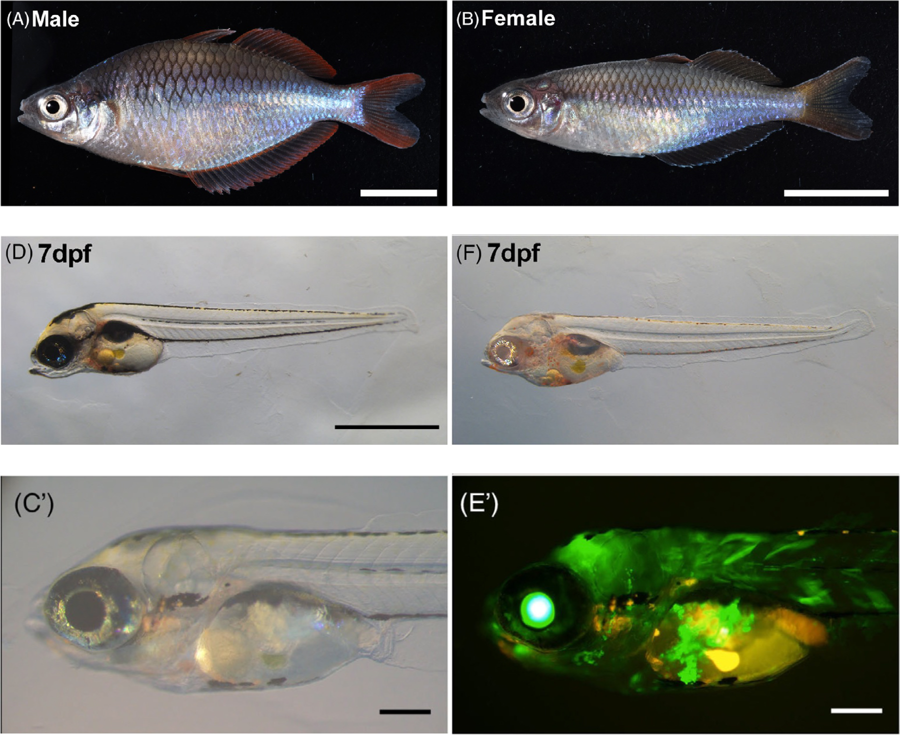
Figure: (A)(B) Male and female Melanotaenia praecox.
(D)(F) Disruption of the tyrosinase gene using CRISPR/Cas9.
(C’)(E’) Introduction of the GFP gene using Tol2.
Maeshima Group / Genome Dynamics Laboratory
Kurokawa Group / Genome Evolution Laboratory
Kuraku Group / Molecular Life History Laboratory
Chromatin organization and behavior in HRAS-transformed mouse fibroblasts
Aoi Otsuka*, Katsuhiko Minami*, Koichi Higashi, Akane Kawaguchi, Sachiko Tamura, Satoru Ide, Michael J. Hendzel, Ken Kurokawa, Kazuhiro Maeshima#
* These authors equally contributed to this work. #corresponding author
Chromosoma 2024 Feb 24 DOI:10.1007/s00412-024-00817-x
Genomic DNA is organized three-dimensionally in the nucleus as chromatin. Recent super-resolution imaging and Hi-C studies have shown that chromatin in living cells forms a number of condensed chromatin domains. Inside these domains, nucleosomes behave locally like a liquid, and such behavior is deeply related to various DNA functions. Cancer cells often show upregulated transcription and other activities, but how is the chromatin behavior in cancer cells different from non-cancer cells?
A research team led by Professor Kazuhiro Maeshima of Genome Dynamics Laboratory (NIG), including a SOKENDAI graduate student Aoi Otsuka (SOKENDAI Special Researcher), SOKENDAI graduate student Katsuhiko Minami (former SOKENDAI Special Researcher, JSPS Research Fellow DC2), Assistant Professor Satoru Ide, Technical Staff Sachiko Tamura, together with Assistant Professor Koichi Higashi and Professor Ken Kurokawa of Genome Evolution Laboratory (NIG), Assistant Professor Akane Kawaguchi of Molecular Life History Laboratory (NIG), and Professor Michael J. Hendzel of University of Alberta, have found chromatin in oncogenic RAS-transformed cells is more constrained than their parental cells.
In this work, the research team examined the chromatin behavior in mouse oncogenic HRAS-transformed cells (CIRAS-3 cells) and their parental cells (10T1/2 cells) with multiple approaches. First, they found that the CIRAS-3 cells have smaller nuclei. In addition, they investigated the individual nucleosome movements in living cells using super-resolution fluorescence microscopy and revealed that nucleosomes are locally more constrained in CIRAS-3 cells. Consistently, CIRAS-3 cells show increased heterochromatin, and in situ Hi-C revealed enriched interactions of the B-B compartments, which mainly consist of heterochromatin.
Genome chromatin in cancer cells encounters physical stress during migration through small spaces, such as metastasis. The smaller nuclei and the more constrained chromatin with enriched heterochromatin can gain elastic properties of chromatin, and may better benefit the cell metastasis (Figure).
This work was supported by JSPS Fellowship, JSPD grants (21H02453, 23K17398, 23K05798, 22H05606, 21H02535, 23KJ0998, 20H05936, 22H04925 (PAGS)), a Japan Science and Technology Agency JST SPRING(JPMJSP2104), The Naito Foundation, and the Takeda Science Foundation. Genome analysis was performed with support of Platform for Advanced Genome Science (22H04925 (PAGS)).
The journal “Chromosoma” is a historical chromosome journal first published in 1939, and is available from the first issue in the NIG Library.
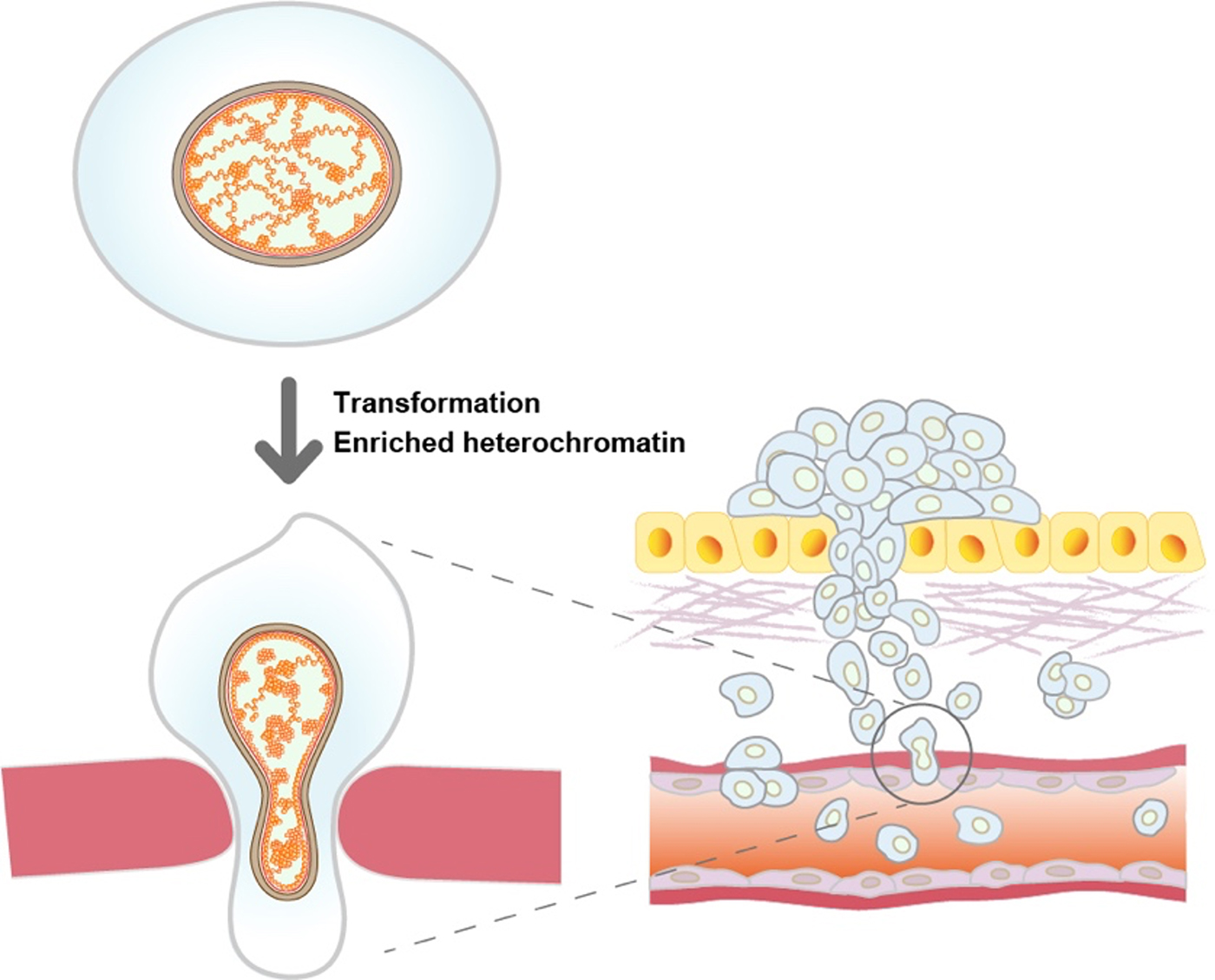
Figure: Oncogenic HRAS-transformed cells (bottom left) have more constrained chromatin with increased heterochromatin than non-transformed parental cells (top left). The more constrained chromatin may play an important role in the metastasis process.
KoideGroup / Mouse Genomics Resource Laboratory
Association of tameness and sociability but no sign of domestication syndrome in mice selectively bred for active tameness
Bharathi Venkatachalam, Bhim B. Biswa, Hiromichi Nagayama, Tsuyoshi Koide*
*責任著者
Genes, Brain and Behavior (2024) 23, e12887 DOI:10.1111/gbb.12887
Domesticated animals have been developed by selecting desirable traits following the initial unconscious selection stage, and now exhibit phenotypes desired by humans. Tameness is a common behavioural trait found in all domesticated animals. At the same time, these domesticated animals exhibit a variety of morphological, behavioural, and physiological traits that differ from their wild counterparts of their ancestral species. These traits are collectively referred to as domestication syndrome. However, whether this phenomenon exists is debatable. Previously, selective breeding has been used to enhance active tameness, a motivation to interact with humans, in wild heterogeneous stock mice derived from eight wild inbred strains. In the current study, we used tame mice to study how selective breeding for active tameness affects behavioural and morphological traits. A series of behavioural and morphological analyses on mice showed an increased preference for social stimuli and a longer duration of engagement in non-aggressive behaviour. However, no differences were observed in exploratory or anxiety-related behaviours. Similarly, selection for tameness did not affect ultrasonic vocalisations in mice, and no changes were observed in known morphological traits associated with domestication syndrome. These results suggest that there may be a link between active tameness and sociability and provide insights into the relationship between tameness and other behaviours in the context of domestication.
Source: Bharathi Venkatachalam et al., Genes, Brain and Behavior (2024) 23, e12887
Kimura Group / Cell Architecture Laboratory
Enucleation of the C. elegans embryo revealed dynein-dependent spacing between microtubule asters
Fujii, K., Kondo, T. & *Kimura
*corresponding author
Life Sci. Alliance (2023) 7, e202302427 DOI:10.26508/lsa.202302427
In psychology, personal space is known as the distance that people tend to maintain from others. A similar phenomenon can be found inside the cell. An organelle called centrosome tend to maintain certain distance from the other centrosomes. In this study, Dr. Ken Fujii and his colleagues investigated the behavior of the centrosomes in enucleated, C. elegans embryonic cells. Using genetics and computational approaches, the researchers proposed that the centrosomes compete with each other for motor proteins “dynein” distributed in the cytoplasm and at the cell cortex to maintain certain distance from the others and ensure their “personal space.” The study has implication in how cellular structures measure distances inside the cell.
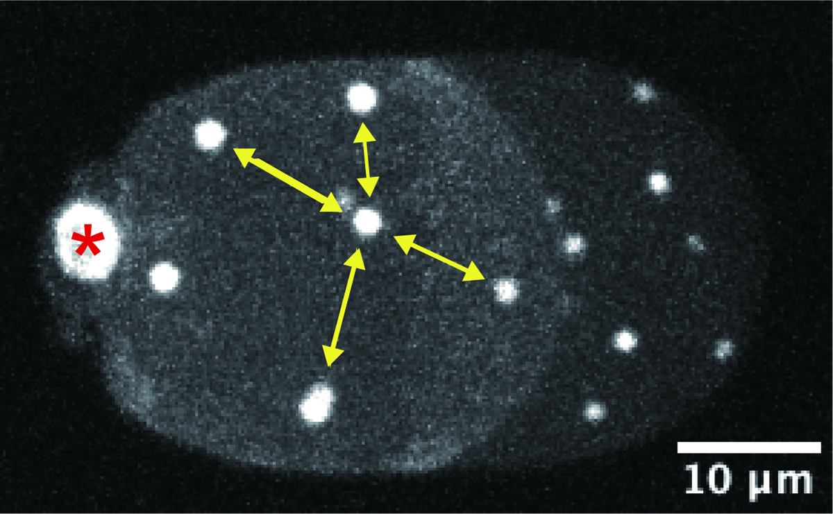
Figure: A representative microscope image of an enucleated, C. elegans embryonic cell. White signals indicate the centrosomes, which are evenly distributed inside the cell. (The image is a 2-dimensional projection of the 3-dimensional cell image. The asterisk indicates the polar body, which is not the centrosome.)
Kawakami Group / Laboratory of Molecular and Developmental Biology
Target-selective vertebrate motor axon regeneration depends on interaction with glial cells at a peripheral nerve plexus
Lauren J. Walker, Camilo Guevara, Koichi Kawakami, and Michael Granato
PLOS Biology (2023) 21, e3002223 DOI:10.1371/journal.pbio.3002223
A critical step for functional recovery from peripheral nerve injury is for regenerating axons to connect with their pre-injury targets. Reestablishing pre-injury target specificity is particularly challenging for limb-innervating axons as they encounter a plexus, a net-work where peripheral nerves converge, axons from different nerves intermingle, and then resort into target-specific bundles. Here, we examine this process at a plexus located at the base of the zebrafish pectoral fin, equivalent to tetrapod forelimbs. Using live cell imaging and sparse axon labeling, we find that regenerating motor axons from 3 nerves coalesce into the plexus. There, they intermingle and sort into distinct branches, and then navigate to their original muscle domains with high fidelity that restores functionality. We demonstrate that this regeneration process includes selective retraction of mistargeted axons, suggesting active correction mechanisms. Moreover, we find that Schwann cells are enriched and associate with axons at the plexus, and that Schwann cell ablation during regeneration causes profound axonal mistargeting. Our data provide the first real-time account of regenerating vertebrate motor axons navigating a nerve plexus and reveal a previously unappreciated role for Schwann cells to promote axon sorting at a plexus during regeneration.
This study was conducted as collaboration with Granato lab at University of Pennsylvania.
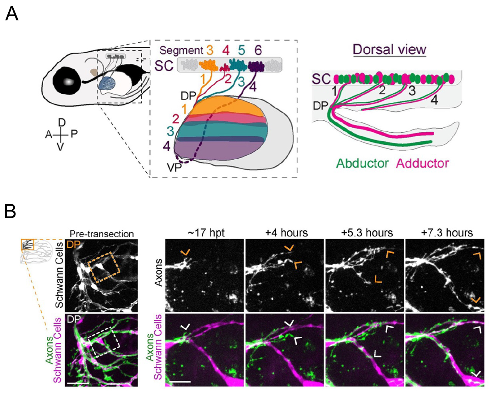
Figure: (A)Zebrafish spinal motor nerves are resorted in a plexus (DP and VP ) and connect to the muscle of the pectoral fin.
(B)Live imaging of navigation of axons along Schwann cells. Transgenic zebrafish were used to label Schwann cells.


