


![]()
Assessment of metagenomic workflows using a newly constructed human gut microbiome mock community
Hiroshi Mori*, Tamotsu Kato*, Hiroaki Ozawa, Mitsuo Sakamoto, Takumi Murakami, Todd D. Taylor, Atsushi Toyoda, Moriya Ohkuma, Ken Kurokawa, Hiroshi Ohno
*equal contribution
DNA Research 2023 May 31 DOI:10.1093/dnares/dsad010
Press release (In Japanese only)
To quantify the biases introduced during human gut microbiome studies, analyzing an artificial mock community as the reference microbiome is indispensable. However, there are still limited resources for a mock community which well represents the human gut microbiome. Here, we constructed a novel mock community comprising the type strains of 18 major bacterial species in the human gut and assessed the influence of experimental and bioinformatics procedures on the 16S rRNA gene and shotgun metagenomic sequencing. We found that DNA extraction methods greatly affected the DNA yields and taxonomic composition of sequenced reads, and that some of the commonly used primers for 16S rRNA genes were prone to underestimate the abundance of some gut commensal taxa such as Erysipelotrichia, Verrucomicrobiota and Methanobacteriota. Binning of the assembled contigs of shotgun metagenomic sequences by MetaBAT2 produced phylogenetically consistent, less-contaminated bins with varied completeness. The ensemble approach of multiple binning tools by MetaWRAP can improve completeness but sometimes increases the contamination rate. Our benchmark study provides an important foundation for the interpretation of human gut microbiome data by providing means for standardization among gut microbiome data obtained with different methodologies and will facilitate further development of analytical methods.
Source: Hiroshi Mori et al., DNA Research doi: 10.1093/dnares/dsad010
Shiori Iida, a D4 student and JSPS research fellow DC2 at Genome Dynamics Laboratory, has successfully completed the Analytical and Quantitative Light Microscopy held at Marine Biological Laboratory in Woods Hole, United States, from April 26th to May 5th, 2023. This course is a historic and educational program focused on light microscopy, held annually at the Marine Biological Laboratory.
The course fee and travel expenses were supported by Morishima Fund of the Genetics course at SOKENDAI.
Genetic Regulation of Mitosis–Meiosis Fate Decision in Plants: Is Callose an Oversighted Polysaccharide in These Processes?
Harsha Somashekar and Ken-Ichi Nonomura
Plants (2023) 12, 1936 DOI:10.3390/plants12101936
Meiosis is a special cell division that halves the chromosome number in preparation for fertilization. In this process, the genomic information of the parents is shuffled to generate a new gene combination (meiotic recombination), which is inherited to the next generation. The meiotic recombination machinery is widely conserved in eukaryotes and has been extensively studied. On the other hand, the mechanisms by which germ cells transition to meiosis are diverse among species, and little is known particularly in plants.
In this review, we mainly focused on meiosis of flowering plants, and explained how the archesporial cells, which produce meiocytes in the future, develop and eventually achieve meiosis with the latest findings. We also highlight the findings of our research group that the callose polysaccharide, a special plant cell wall components, plays an essential role in regulating the proper initiation timing and progression of male meiosis.
The image using the figures in our review paper was selected for the cover story of Plants Vol. 12, No. 10, including 143 articles.
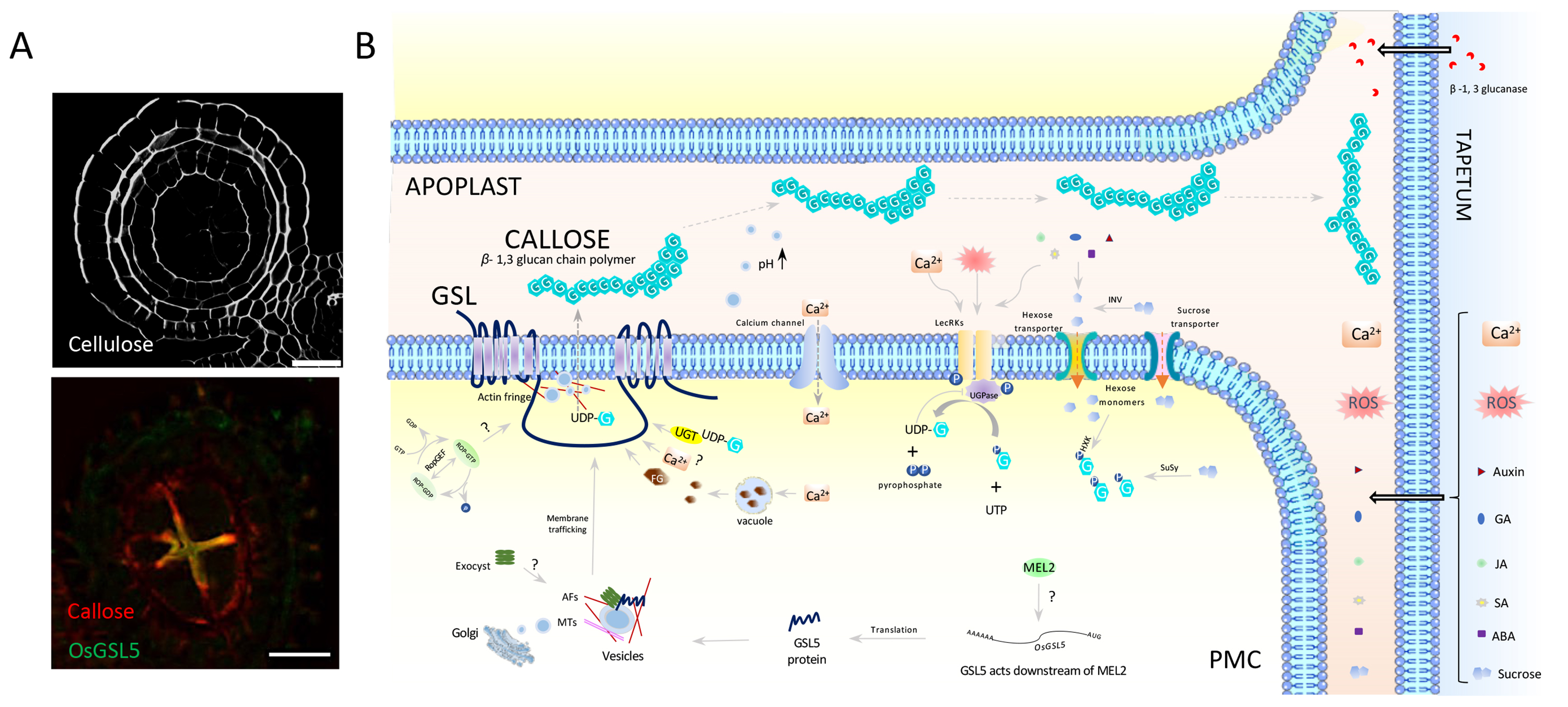
Figure: A model of callose synthesis and transport in anthers
(A) Cross sections of the anther just before meiosis. (Upper) Staining of cellulosic cell walls. (Bottom) Double immuno-staining of callose (red) and GSL5 (yellow). Bar=20µm. (B) The plasma membrane-anchored callose synthase (GSL) of male meiotic cells (PMCs) releases synthesized callose to apoplasts between PMCs (APOPLAST) just prior to meiosis transition. Callose migrates to the apoplast between PMCs and tapetum cells (TAPETUM) where GSL is absent, and eventually envelop all PMCs within the anther locule.
The assembly of the MCM2–7 hetero-hexamer and its significance in DNA replication.
Yuki Hatoyama and Masato T. Kanemaki.
Biochemical Society Transactions 2023 May 5 DOI:10.1042/BST20221465
To proliferate, cells must replicate chromosomal DNA, which encodes the genetic information. MCM2–7, the replication helicase that unwinds a double-strand DNA to generate a single-strand DNA, is essential for chromosomal DNA replication. The replicative MCM2–7 helicase forms a ring-shaped hexamer.
This review paper was published in Biochemical Society Transactions, a review journal published by the Biochemical Society. The review discusses how the MCM2–7 hexamer is efficiently assembled in cells and its physiological significance based on our recent report. In this review, we proposed a model in which MCMBP functions as a chaperone to promote the assembly of the MCM2–7 hexamer, resulting in the supply of sufficient amounts of MCM2–7 before DNA replication (Figure).
The existence of the licensing checkpoint was proposed, whereby the cell senses a lack of chromatin-bound MCM2–7 and arrests the cell cycle in the G1 phase. In this review, we proposed a new hypothesis that cells do not sense the amount of MCM2–7 directly. Instead, the cells with reduced MCM2–7 expression have DNA damage during DNA replication in the previous cell cycle, and they arrest the cell cycle by detecting this DNA damage in the next G1 phase.
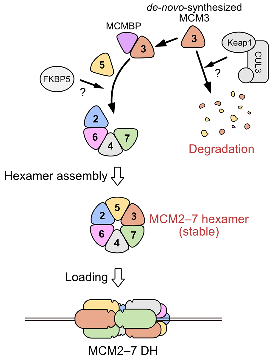
Figure: A schematic diagram of the assembling the MCM2–7 hexameric complex.
Press release
A small RNA system ensures accurate homologous pairing and unpaired silencing of meiotic chromosomes
Hiroaki Tabara*, Shohei Mitani, Megumi Mochizuki, Yuji Kohara and Kyosuke Nagata *: corresponding author
The EMBO Journal 2023 April 20 DOI:10.15252/embj.2020105002
Meiosis is a specialized mode of cell division that produces haploid gametes. During meiosis, chromosomes with homologous partners undergo synaptonemal complex (SC)-mediated pairing, while the remaining unpaired regions such as most parts of the sex chromosomes in males are heterochromatinized through unpaired silencing.
A study, published in The EMBO Journal, introduces that a small RNA system regulates meiotic pairing of homologous chromosomes in the nematode Caenorhabditis elegans. Homolog pairing initiation depends on species-specific mechanisms involving interactions between pericentric heterochromatin, centromeres, telomeres, and/or pairing center sequences. Subsequently, the SC establishes the pairing of other regions of the homologous chromosomes. In plants and mammals, DNA double-strand breaks (DSBs) are important or partly important for synapse formation between homologous chromosomes. Mysteriously, in C. elegans and fruit fly, DSBs are not necessary for synapse formation, implying the presence of an unclarified mechanism for recognizing homologous chromosomes during SC formation.
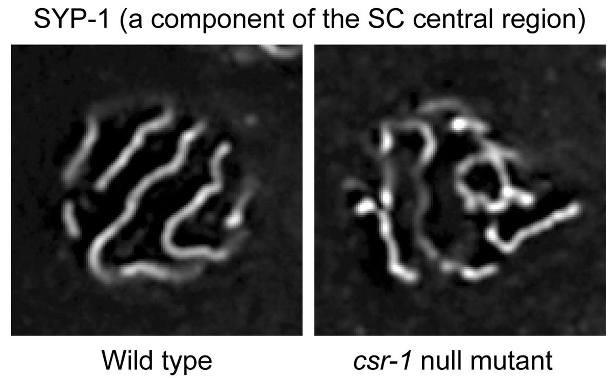
RNA interference (RNAi) is a form of RNA-mediated sequence-specific regulation of gene expression in eukaryotes. Small RNAs and Argonaute proteins play key roles in the mechanism of RNAi. Researchers at National Institute of Genetics, Tokyo Women’s Medical University, and University of Tsukuba, in Japan, found new relationships between SC formation on paired chromosomes, unpaired chromosome heterochromatinization, and an Argonaute–small RNA pathway (endogenous RNAi). The C. elegans Argonaute proteins, CSR-1 and its paralog interacting with small RNAs, are required for SC formation between chromosome pairs with accurate homology and are also involved in the condensation of unpaired chromosomes. The SC is composed of layers of strings, with two lateral elements sandwiching a central region. CSR-1 in nuclei and meiotic cohesin, constituting the SC lateral elements, were associated with non-simple DNA repeats expressing small RNAs and long non-coding RNAs (lncRNAs), and also associated weakly with coding genes. This study proposes a zipper model, in which the accurate homolog pairing is promoted via the inter-chromosomal recognition of chromosome-associated lncRNAs by the Argonaute-mediated small RNA system. The meiotic difference between paired or unpaired chromosomes is also described.
Reference
Tabara H, Mitani S, Mochizuki M, Kohara Y, Nagata K (2023) A small RNA system ensures accurate homologous pairing and unpaired silencing of meiotic chromosomes. EMBO J, in press. DOI: 10.15252/embj.2020105002.
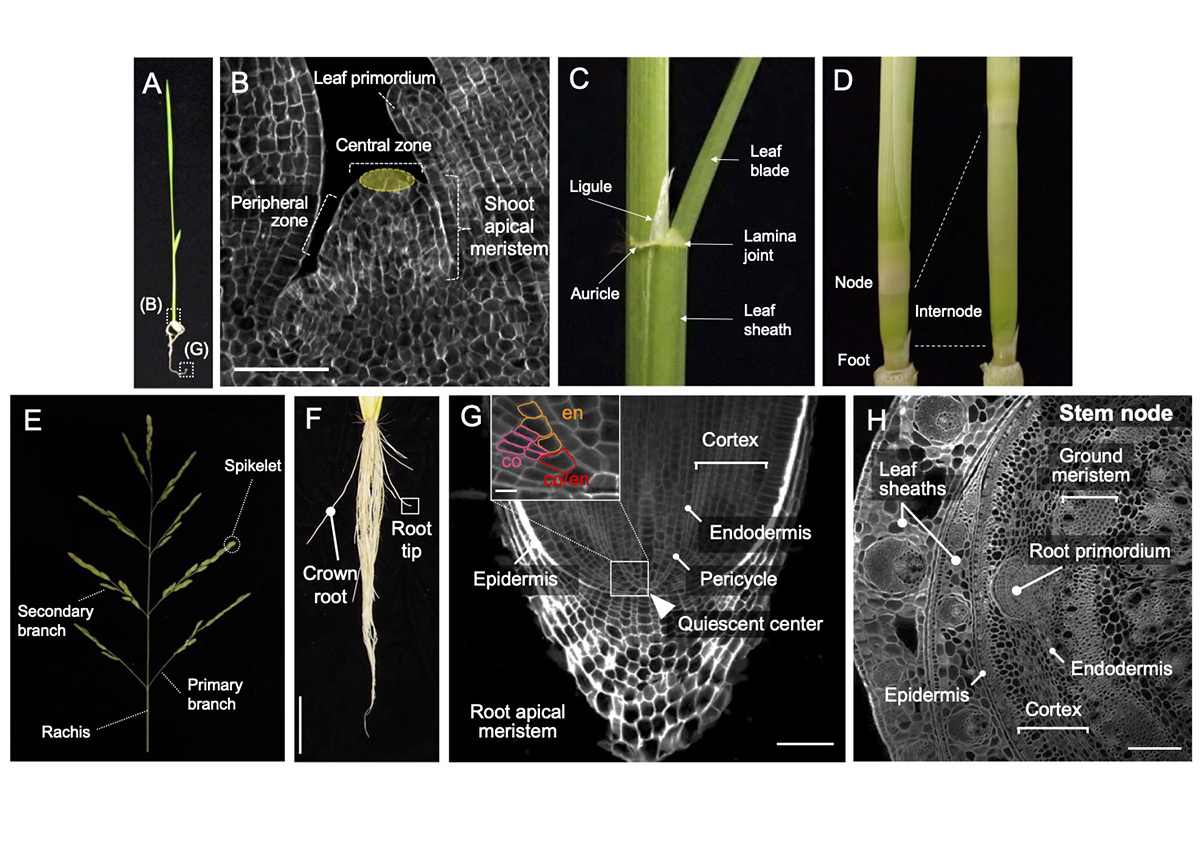
Genetic basis controlling rice plant architecture and its modification for breeding
Wakana Tanaka, Takaki Yamauchi, *Katsutoshi Tsuda *Corresponding author
Breeding Science (2023) 73, 3-45 DOI:10.1270/jsbbs.22088
Rice (Oryza sativa) is derived from the ancestral wild species Oryza rufipogon, which began to be cultivated about 10,000 years ago. In this process, the morphology of rice has undergone significant changes through human selection. For example, cultivated rice has a more upright stature for dense planting, generates shorter stems to prevent lodging (the bending over of the stem to the ground), and produces more grains in inflorescences compared to its ancestor. Recent molecular genetics has identified numerous important regulators and molecular mechanisms in development. Importantly, many regulators have been utilized as weak alleles in the process of domestication and subsequent breeding. This review aimed to comprehensively summarize a wide range of knowledge in rice developmental biology for each organ, including leaf, stem, inflorescence, and root, and to highlight examples utilized in breeding. This review will help to learn the current understanding of rice morphogenesis and provide a theoretical foundation for future crop improvement.
This work was supported by JSPS Grants (22K06267, 22H02308, 22H02319).
Press release
Condensed but liquid-like domain organization of active chromatin regions in living human cells
Tadasu Nozaki*, Soya Shinkai*, Satoru Ide*, Koichi Higashi*, Sachiko Tamura, Masa A. Shimazoe, Masaki Nakagawa, Yutaka Suzuki, Yasushi Okada, Masaki Sasai, Shuichi Onami, Ken Kurokawa, Shiori Iida, Kazuhiro Maeshima *co-first authors
Science Advances (2023) 9, eadf1488 DOI:10.1126/sciadv.adf1488
![]() Press release (In Japanese only)
Press release (In Japanese only)
The human genome chromatin can be classified into euchromatin and heterochromatin, which have high and low transcription activities, respectively. In the classical view, it was believed that euchromatin has an open and decondensed structure, while heterochromatin is highly condensed.
A research team led by Professor Kazuhiro Maeshima of Genome Dynamics Laboratory (NIG), including a graduate student (currently a Research Associate at Harvard Univ.) Tadasu Nozaki, Assistant Professor Satoru Ide, Technical Stuff Sachiko Tamura, SOKENDAI graduate student Masa A. Shimazoe, SOKENDAI graduate student (JSPS Research Fellow DC2) Shiori Iida, together with Assistant Professor Koichi Higashi and Professor Ken Kurokawa of Genome Evolution Laboratory (NIG), and Senior Research Scientist Soya Shinkai and Team Leader Shuichi Onami of RIKEN BDR, have found euchromatin forms condensed chromatin domains. This finding suggests an innovative model that condensed structure is the default state of chromatin.
In this work, motions of fluorescently labeled nucleosomes in euchromatin in living human cells were investigated with a super-resolution microscope. Two neighboring nucleosomes were observed simultaneously with different colored fluorescent dyes. The research team discovered that movements of nucleosome pairs within 150 nm in the distance were correlated. This shows that euchromatin forms irregularly condensed chromatin domains with ~150 nm diameters. Further analysis revealed that nucleosomes within a domain behave like a liquid. On the other hand, euchromatin is stable at the chromosome scale, and exhibits a solid-like behavior.
This liquid-like behavior of nucleosomes can facilitate transcription, DNA replication, and repair in condensed domains. Also, the condensed domains can protect DNA from radiation damage. Solid-like behavior of chromatin at the chromosome scale can contribute to maintaining the genome by preventing entanglements or breaks of long chromatin. Chromatin is locally dynamic and reactive, and globally stable.
This work was supported by JSPS Fellowship, JSPD grants (21H02453, 22H05606, 21H02535, 20H05550, 20H05936, 16H06279(PAGS), 22H04925 (PAGS)), a Japan Science and Technology Agency CREST grant (JPMJCR15G2), JST SPRING(JPMJSP2104), and the Uehara Memorial Foundation.
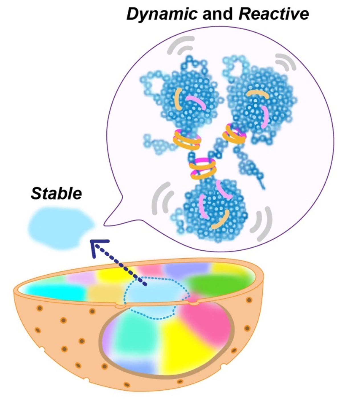
Figure: Nucleosomes form irregularly condensed chromatin domains by their intrinsic sticky nature (blue clusters in the balloon). Nucleosomes show dynamic liquid-like behavior inside a domain, which promotes DNA transactions. On the other hand, the solid-like behavior of chromatin on the chromosome scale helps to maintain the genome by preventing entanglements or breaks of long chromatin.
Video: Video of nucleosome pairs at euchromatin labeled by two different fluorescent dyes and recorded simultaneously. The upper row shows a neighboring nucleosome pair with correlated movements. The lower row depicts a distant nucleosome pair with uncorrelated movements.
Kanemaki Group / Molecular Cell Engineering Laboratory
RAD51 bypasses the CMG helicase to promoter fork reversal.
Liu W, Saito Y, Jackson J, Bhowmick R, Kanemaki MT, Vindigni A and *Cortez D.
Science (2023) 380, 382-387 DOI:10.1126/science.add7328
Replication forks are known to stall due to replication stress, and electron microscopy observation has revealed the occurrence of the fork reversal structure, also known as the chicken-foot structure. It was previously known that the RAD51 recombinase is required for the formation of the reversed forks. However, it was unclear whether CMG helicase, which was responsible for moving the replication fork, remained present in the reversed forks and, if so, where it was located. This study analyzed RAD51 mutants and found that the reversed fork structure forms behind the CMG helicase and that the requirement for RAD51 in fork reversal formation disappears when CMG helicase is absent.
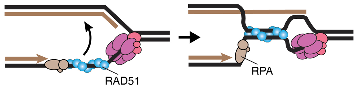
This study was conducted as collaborative research between Professor David Cortez at Vanderbilt University and the Kanemaki Laboratory at NIG. The Kanemaki Laboratory generated AID2 cells that can degrade MCM proteins, which is a main component of the CMG helicase.


