Biological Macromolecules Laboratory / Maeshima Group
Chromatin folding and DNA replication inhibition mediated by a highly antitumor-active tetrazolato-bridged dinuclear platinum(II) complex
Ryosuke Imai, Seiji Komeda, Mari Shimura, Sachiko Tamura, Satoshi Matsuyama, Kohei Nishimura, Ryan Rogge, Akihiro Matsunaga, Ichiro Hiratani, Hideaki Takata, Masako Uemura, Yutaka Iida, Yuko Yoshikawa, Jeffrey C. Hansen, Kazuto Yamauchi, Masato T. Kanemaki, and Kazuhiro Maeshima
Scientific Reports 6, Article number: 24712 (2016) DOI:10.1038/srep24712
Chromatin DNA must be read out for various cellular functions, and copied for the next cell division. These processes are targets of many anticancer agents. Platinum-based drugs, such as cisplatin, have been used extensively in cancer chemotherapy. The drug–DNA interaction causes DNA crosslinks and subsequent cytotoxicity. Recently, it was reported that an azolato-bridged dinuclear platinum (II) complex, 5-H-Y, exhibits a different anticancer spectrum from cisplatin. Here, using an interdisciplinary approach, we reveal that the cytotoxic mechanism of 5-H-Y is distinct from that of cisplatin. 5-H-Y inhibits DNA replication and also RNA transcription, arresting cells in the S/G2 phase, and are effective to cisplatin-resistant cancer cells. Moreover, it causes much less DNA crosslinking than cisplatin, and induces chromatin folding. 5-H-Y will expand the clinical applications for the treatment of chemotherapy-insensitive cancers.
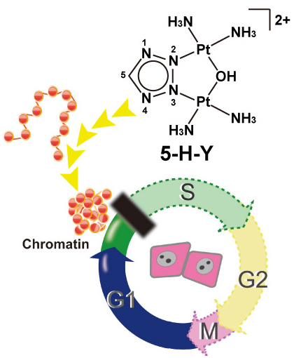
5-H-Y inhibits DNA replication and arrests the treated cells in S/G2 phase. 5-H-Y binds tightly to chromatin DNA and induces chromatin folding in vitro and in vivo.
Division of Population Genetics / Saitou Group
Genomic locations of conserved noncoding sequences and their proximal protein-coding genes in mammalian expression dynamics
Isaac Adeyemi Babarinde and Naruya Saitou
Molecular Biology and Evolution DOI:10.1093/molbev/msw058
Conserved Noncoding Sequences (CNSs) in mammals were examined with special reference to their genomic location. We extracted the CNSs conserved between chicken and four mammalian species (human, mouse, dog and cattle). Human CNSs were confirmed to be under purifying selection. The distribution pattern, ChIP-Seq and RNA-Seq data suggested that the CNSs are regulatory elements. Physical distances between CNS and their nearest protein coding genes were well conserved between human and mouse genomes. ChIP-Seq signal and gene expression patterns also suggested that CNSs regulate nearby genes. Genes with more CNSs have more evolutionarily conserved expression than those with fewer CNSs. These results suggest that the genomic locations of CNSs are important for their regulatory functions. In fact, various kinds of evolutionary constraints may be acting to maintain the genomic locations of CNSs and protein-coding genes in mammals to ensure proper regulation. First author of this paper, Dr. Babarinde, just receive Ph.D. from Department of Genetics, SOKENDAI. He also received Morishima Award from NIG.
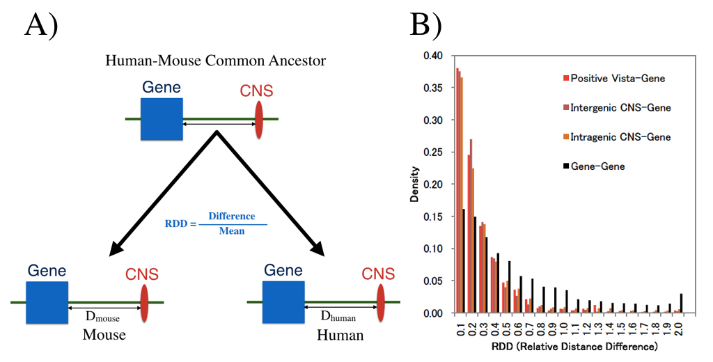
We examined whether genomic distances between genes and CNSs which already existed in the common ancestor of human and house are conserved now. (A) shows definition of distance measure RDD, and (B) shows RDD distribution. Distances between CNS and genes are much more conserved than those between genes.
Press release
Dazl is a target RNA suppressed by mammalian NANOS2 in sexually differentiating male germ cells
Yuzuru Kato, Takeo Katsuki, Hiroki Kokubo, Aki Masuda, Yumiko Saga
Nature Communications 7, Article number: 11272 DOI:10.1038/NCOMMS11272
Press Release (Only in Japanese)
Evolutionally conserved Nanos RNA-binding proteins play crucial roles in germ cell development. While a mammalian Nanos family protein, NANOS2, is required for sexual differentiation of male (XY) germ cells in mice, the underlying mechanisms and the identities of its target RNAs in vivo remain elusive. Using comprehensive microarray analysis and a bacterial artificial chromosome transgenic system, we identify Dazl, a germ cell-specific gene encoding an RNA-binding protein implicated in translation, as a crucial target of NANOS2. Importantly, removal of the Dazl 3′ UTR in XY germ cells stabilises the Dazl mRNA, resulting in elevated meiotic gene expression, abnormal resumption of the cell cycle, and impaired processing-body formation, reminiscent of Nanos2-knockout phenotypes. Furthermore, our data suggest that NANOS2 acts as an antagonist of the DAZL protein. We propose a dual system of NANOS2-mediated suppression of Dazl expression as a pivotal molecular mechanism promoting sexual differentiation of XY germ cells. This work was supported by a Grant-in-Aid for Young Scientists (B) from the Japan Society for the Promotion of Science (JSPS) to Y.K. (Grant number 25840091) and a Grant-in-Aid for Scientific Research (S) from JSPS to Y.S. (Grant number 21227008).
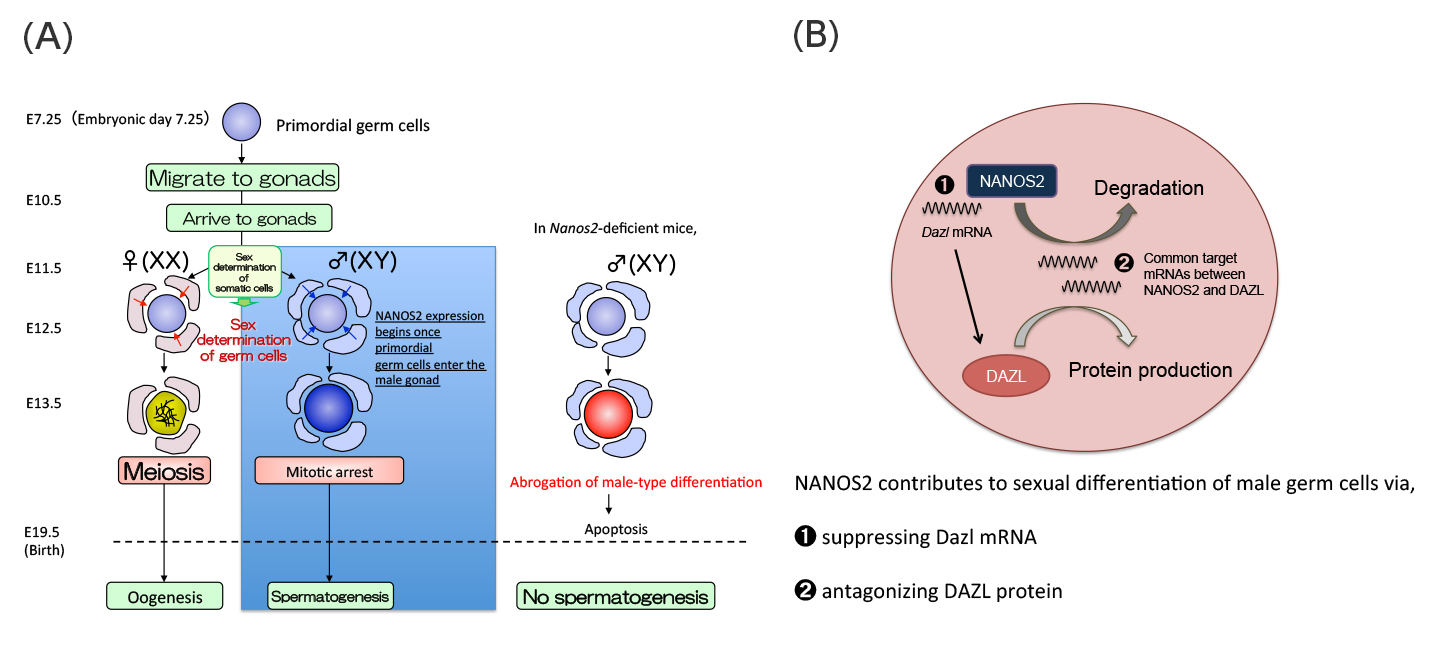
(A)Sex of mouse germ cells is determined after they are enclosed by gonadal somatic cells at E11.5. NANOS2 expression begins once primordial germ cells enter the sex-specific process of sexual differentiation in male germ cells. Germ cells lacking the Nanos2 gene die by apoptosis due to the abrogated male-type differentiation.
(B)Working model of NANOS2-dependent Dazl suppression in sexually differentiating male germ cells.
Press release
Nucleosomal arrays self-assemble into supramolecular globular structures lacking 30-nm fibers
Kazuhiro Maeshima, Ryan Rogge, Sachiko Tamura, Yasumasa Joti, Takaaki Hikima, Heather Szerlong, Christine Krause, Jake Herman, Erik Seidel, Jennifer DeLuca, Tetsuya Ishikawa, and Jeffrey C. Hansen
The EMBO Journal Published online 12.04.2016 DOI:10.15252/embj.201592660
The existence of a 30-nm fiber as a basic folding unit for DNA packaging has remained a topic of active discussion. Here we characterize the supramolecular structures formed by reversible Mg2+-dependent self-association of linear 12-mer nucleosomal arrays using microscopy and physicochemical approaches. These structures, which we call ’oligomers’, were globular throughout all stages of the cooperative assembly process, and ranged in size from ~50 nm to a maximum diameter of ~1000 nm. The nucleosomal arrays were packaged within the oligomers as interdigitated 10-nm fibers, rather than folded 30-nm structures. Linker DNA was freely accessible to micrococcal nuclease, although the oligomers remained partially intact after linker DNA digestion. The organization of chromosomal fibers in human nuclei in situ was stabilized by 1 mM MgCl2 but became disrupted in the absence of MgCl2, conditions that also dissociated the oligomers in vitro. These results indicate that a 10-nm array of nucleosomes has the intrinsic ability to self-assemble into large chromatin globules stabilized by nucleosome-nucleosome interactions, and suggest that the oligomers are good in vitro model for investigating the structure and organization of interphase chromosomes.
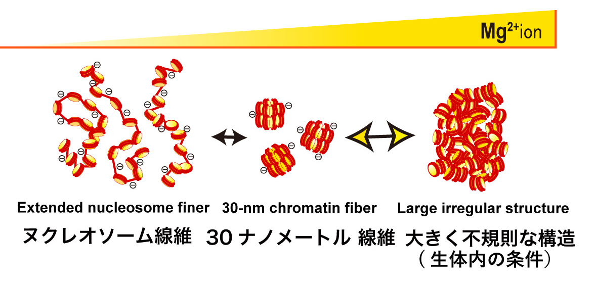
The 12-mer nucleosomal array is a well-defined model chromatin system. Without Mg2+, the nucleosomal array is extended by repulsion (Left). In 1-2 mM Mg2+, the nucleosomal array folds into a regular 30-nm chromatin fiber structure (Center). With further increases in Mg2+, the nucleosome arrays assemble into supramolecular structures (Right). The large structures are not assemblies of the 30-nm chromatin fibers, but interdigitated and melted structures of 10-nm nucleosomal arrays, reflecting on the native chromatin structure in the cell.
Press release
Allelic Imbalance in Regulation of ANRIL through Chromatin Interaction at 9p21 Endometriosis Risk Locus
Hirofumi Nakaoka, Aishwarya Gurumurthy, Takahide Hayano, Somayeh Ahmadloo, Waleed H Omer, Kosuke Yoshihara, Akihito Yamamoto, Keisuke Kurose, Takayuki Enomoto, Shigeo Akira, Kazuyoshi Hosomichi, Ituro Inoue
PLOS Genetics Published: April 7, 2016 DOI:10.1371/journal.pgen.1005893
Press Release (Only in Japanese)
A large number of variants associated with human complex diseases have been discovered by genome-wide association studies (GWASs). These discoveries have been anticipated to be translated into the definitive understanding of disease pathogeneses; however, functional characterization of the disease-associated SNPs remains a formidable challenge. Here we explored regulatory mechanism of a variant on chromosome 9p21 associated with endometriosis, a common gynecological disorder. By scrutinizing linkage disequilibrium structure and DNase I hypersensitive sites across the risk locus, we prioritized rs17761446 as a candidate causal variant. The results of our “allele-specific” functional genomic approaches sheds light on regulatory mechanisms underlying 9p21 endometriosis risk locus, in which preferential bindings of TCF7L2 and its coactivator EP300 to the protective G allele of rs17761446 lead to stronger chromatin interaction with the promoter of ANRIL, which in turn activate transcription of the non-coding RNA. Motivated by the fact that TCF7L2 was a key transcription factor of Wnt signaling pathway, we postulated that the induction of Wnt signaling activated expression levels of ANRIL and cell cycle inhibitors, CDKN2A/2B. Functional genomics on common disease will unlock functional aspect of genotype-phenotype correlations in the post-GWAS stage.
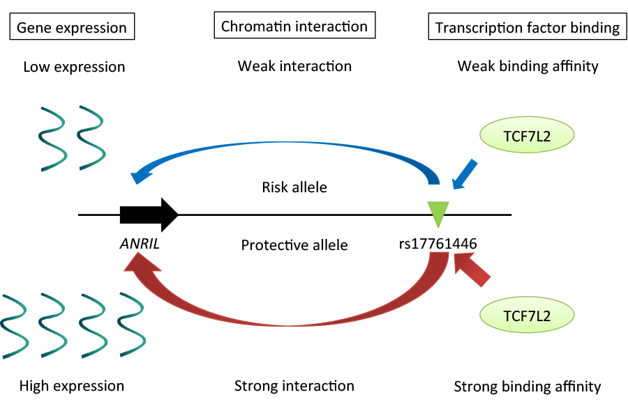
Regulatory mechanisms underlying 9p21 endometriosis risk locus. Preferential bindings of TCF7L2 and its coactivator EP300 to the protective G allele of rs17761446 lead to stronger chromatin interaction with the promoter of ANRIL, which in turn activate transcription of the non-coding RNA.
Cell Architecture Laboratory / Kimura Group
Shape–motion relationships of centering microtubule asters
Hirokazu Tanimoto, Akatsuki Kimura*, and Nicolas Minc*
J. Cell Biol. 212: 777-787, 2016. DOI:10.1083/jcb.201510064
(* corresponding authors)
A joint group of Drs. Nicolas Minc at Institut Jacques Monod (France) and Akatsuki Kimura at National Institute of Genetics/SOKENDAI analyzed the centering of microtubule asters in the sea urchin egg, and constructed a quantitative model that dictates the speed and trajectory of the centering in normal cell as well as shape-manipulated cells. The study was initiated when Prof. Kimura was a visiting researcher at the Minc lab through the SOKENDAI Young Faculty Overseas Visit Program.
In this study, Dr. Hirokazu Tanimoto et al. utilized multiple approaches such as laser ablation and pharmacological assays to demonstrate that the centering is accomplished by the forces pulling on asters from the cytoplasm. An interesting feature of the centering was to find that asters moved at a constant speed. Considering the speed was comparable to that of the growth speed of the aster, Tanimoto et al. proposed a theoretical model for the centering that explains the constant speed independent on the actual size of the aster. The model predicts well the change of the speed and trajectory of asters when the shape of the cells was manipulated in micro-chambers. Overall, this study advanced our understanding on the long-standing debate on the mechanism of the centering of microtubule asters and their associating nucleus.
See also: Ben Short “How asters find their center” J Cell Biol 212, 743 (2016) DOI: 10.1083/jcb.2127if
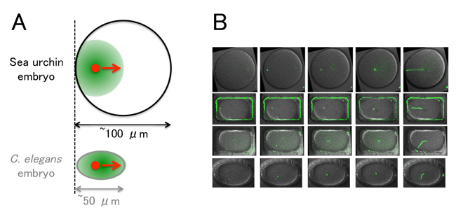
(A) The centering of microtubule asters (green) in the sea urchin embryo. The size of the embryo is large compared to C. elegans. The aster does not cover the entire cell during the centering, and the center of the aster (red) approaches the cell center with a speed comparable to the growth speed of the aster.
(B) Examples of shape manipulation using micro-chambers. The sperm pronucleus (i.e. the center of the aster, green) moves toward the center. (The entire trajectories are shown on the rightmost panels.) The theoretical model constructed in the study reproduces the speed and trajectory of aster movements even in the shape-manipulated embryos.
Time & Date
9:00 ~ 16:00, Saturday, April 2th, 2016 (No Reservations Required, Free Admission)
Access
Free Shuttle Buses Available to the National Institute of Genetics from the North Exit of Mishima Station. ( Service Time: 8:50 ~ 15:00)
Map
Map Free Shuttle Buses Available Parking around Mishima Station Available for Car Visitors.
No Pets Allowed except Service Dogs, No General Parking*
(*Disabled Parking Available on the Institute Premises)
Information
1111 Yata, Mishima, Shizuoka 411-8540, JAPAN TEL:+81-55-981-5873
Two professors and three assistant professors have joined NIG as of April 1, 2016.
professor
Yutaka SATO:Genetic Resource Center,Plant Genetics Laboratory
Ken KUROKAWA:Center for Information Biology, Genome Evolution Laboratory

assistant professor
Takayuki FUJIWARA: Division of Symbiosis and Cell Evolution, Miyagishima Group
Tomotaka MATSUMOTO: Division of Evolutionary Genetics, Akashi Group
Toshihiro KAWASAKI: Model Fish Genomics Resource Laboratory, Sakai Group
In addition, Masato KANEMAKI, tenure track associate professor at the Center for Frontier Research, has been awarded tenure and promoted to professor.
Masato KANEMAKI: Division of Molecular Cell Engineering, Department of Molecular Genetics