


Kuraku Group / Molecular Life History Laboratory
Genome assembly catalog for species in the Japanese Red List: unlocking endangered biodiversity through genomic inventory
Kryukov, K., Nakahama, N., and Kuraku, S.
F1000Research (2024) 13, 583 DOI:10.12688/f1000research.149793.2
Whole genome sequence reading, which can be regarded as a catalog of DNA information, is a prerequisite for an accurate understanding of the genetic diversity that is key to the survival of a species; DNA sequences also contain information on the molecular basis that determines the ecological, morphological, and behavioral characteristics of a species. In both ways, whole genome sequencing is essential for understanding biodiversity, yet this information is available for only a handful of species. Dr. Kirill Kryukov of Joint Support-Center for Data Science Research of Research Organization of Information and Systems (ROIS-DS), Dr. Naoyuki Nakahama of the University of Hyogo and Museum of Nature and Human Activities, Hyogo, and Dr. Shigehiro Kuraku of the National Institute of Genetics have therefore focused their attention on rare species among the wild organisms living in Japan, and have developed a database, Genome sequence data availability for the Japanese Red List, which lists the accumulation status of whole genome sequence information.
This database catalogs the registration status of NCBI whole genome sequence information for rare species based on the “Red List 2020 of the Ministry of the Environment (In Japanese only)” and the “Red List of Marine Organisms of the Ministry of the Environment”, and the summarized information is made freely accessible online. The information can be viewed as a table in a web browser. The status of accumulation can be checked at a glance not only by species or subspecies, but also by taxonomic units such as “mammals,” “insects,” and “fungi” (Fig. 1). The system is also equipped with a function to regularly update the display content to reflect new information registered daily from all over the world. In addition, by downloading the files in TSV format, it can be used for secondary data analysis.

Figure1: Status of genome sequence data availability by taxon
One of the tables from the the top page of the database. The rightmost column shows the percentage of species for which genome information has been developed among the species considered to be rare in Japan. The percentage is generally very low, although it varies from taxon to taxon.
Whole genome sequencing requires not only suitable samples for extracting high molecular weight DNA and capturing the nuclear 3D structure of DNA molecules, but also the technical capability to develop sequence information that can withstand extensive use. Although numerous technological innovations and optimizations have reduced the cost and time required for calculations, the cost sometimes runs into the millions of yen, depending on the total number of bases in the genome, and also requires the labor of specialized technical personnel. Despite these limitations, it is important to steadily accumulate information, and there are many cases overseas where national governments are taking the initiative in organizing the priorities of indigenous species and preemptively acquiring genome information. The product of this study are expected to serve as a guide for Japan to vigorously promote such efforts by identifying priorities based on the recognition of the bias in the sequence data availability, and to efficiently acquire information in a more preemptive manner.
Saga Group / Mammalian Development Laboratory
Kanemaki Group / Molecular Cell Engineering Laboratory
Establishment and characterization of mouse lines useful for endogenous protein degradation via an improved auxin-inducible degron system (AID2).
Makino-Itou H, Yamatani N, Okubo A, Kiso M, Ajima R, Kanemaki MT, Saga Y.
Development, Growth and Differentiation (2024) Sep;66(7):384-393. DOI:10.1111/dgd.12942
The development of new technologies opens new avenues in the research field. Currently, conditional gene knockout strategies are employed to examine temporal and spatial gene function. However, phenotypes are sometimes not observed because of the time required for depletion due to the long half-life of the target proteins. Protein knockdown using an improved auxin-inducible degron system, AID2, overcomes such difficulties owing to rapid and efficient target depletion. Here, we established several mouse lines useful for AID2-medicated protein knockdown, which include knock-in mouse lines in the ROSA26 locus; one expresses TIR1(F74G), and the other is the reporter expressing AID-mCherry. We also established a germ-cell-specific TIR1 line and confirmed the protein knockdown specificity. In addition, we introduced an AID tag to an endogenous protein, DCP2 via the CAS9-mediated gene editing method. We confirmed that the protein was effectively eliminated by TIR1(F74G), which resulted in the similar phenotype observed in knockout mouse within 20 h.
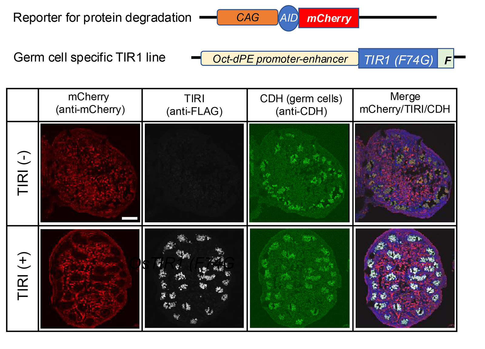
Figure: The germ-cell-specific TIR1 line was crossed with the reporter line TG-CAG-AID-mCherry. Immunofluorescence images of embryonic testes (E15.5) prepared from a pregnant CAG-AID-mCherrymother crossed with an Oct-dPE-TIR1(F74G)-FLAG male. TIR1(-) means sample without Oct-dPE-TIR1(F74G) and TIR1(+) means double transgenic sample. The mother was subjected to 5-Ph-IAA injection at E14.5. Antibodies used were anti-mCherry, anti-FLAG, and anti-E-cadherin (CDH). The scale bar indicates 100 mm.
October 24, 2024
Upon the expiration of the term of the present Director-General of the National Institute of Genetics (NIG), Research Organization of Information and Systems (ROIS) has decided to appoint Dr. Shigeru Kondo as the next Director-General of the NIG, through the deliberation of the Education and Research Council.
Dr. Kondo will arrive at his post on the 1st of December, 2024.
Term of Office: From the 1st of December, 2024 to the 30th of November, 2028 (a total of 4 years)
Mr. Bhim Bahadur Biswa, who graduated from SOKENDAI in September 2024 as a member of the Mouse Genomics Resource Laboratory, has been awarded the School of Life Science “Dean’s Award” for the first semester of 2024.
The Dean’s Award recognizes degree recipients who have conducted research worthy of commendation and reported their accomplishments in an outstanding doctoral thesis. The award was presented on September 27, 2024 during the graduation ceremony. In addition to the Dean’s Award, Mr. Biswa has also been awarded the Genetics Program’s Morishima Award.
・Thesis title : Role of gut bacteria in domestication of mice
Mr. Biswa has provided the following statement regarding the award.
“I am deeply honored to receive the Dean’s Award from the School of Life Sciences, SOKENDAI University, for my PhD research. This recognition underscores the significance of our work in understanding the role of the gut microbiome in the animal domestication process. I am immensely grateful for the guidance and support from my supervisors and lab members, as well as the collaborative environment fostered by the National Institute of Genetics, which has been crucial in advancing this project. This award motivates me to continue pursuing innovative research with the potential to make a lasting impact in the scientific community. “
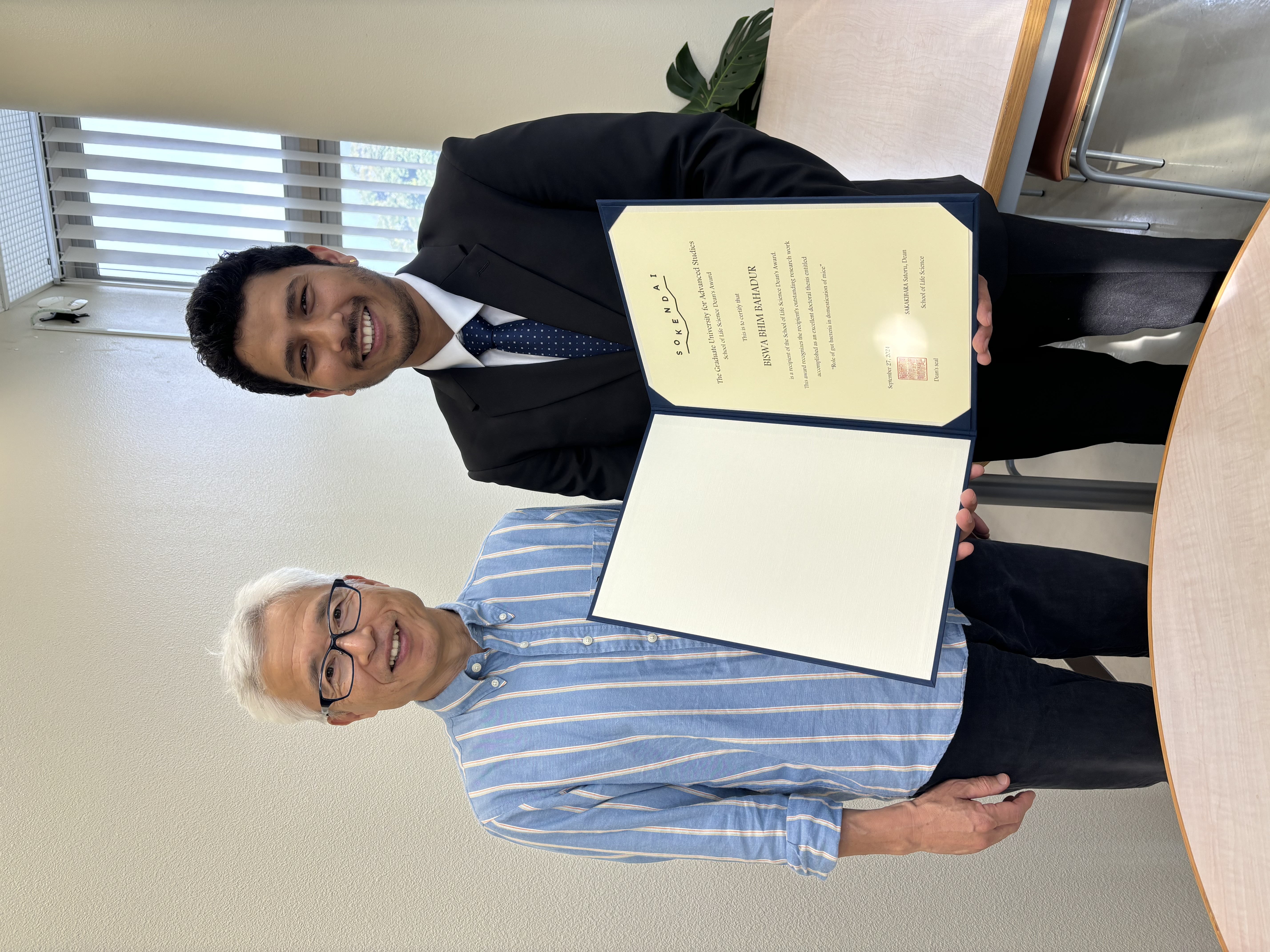
Mr. Biswa, Bhim Bahadur (right) & Associate Professor Koide
Press release
Live-cell imaging under centrifugation characterized the cellular force for nuclear centration in the Caenorhabditis elegans embryo.
Makoto Goda, Michael Shribak, Zenki Ikeda, Naobumi Okada, Tomomi Tani, Gohta Goshima, Rudolf Oldenbourg, Akatsuki Kimura
Proceedings of the National Academy of Sciences (PNAS) (2024) 121 (43), e2402759121 DOI:10.1073/pnas.2402759121
![]() Press release (In Japanese only)
Press release (In Japanese only)
Genomic DNA containing genetic information is stored in the cell nucleus. The nucleus is often located near the center of the cell. This means that there is a force inside the cell that moves and maintains the nucleus at the center. Measuring the amount of force is a challenging task. In this study, the researchers succeeded in measuring the force that maintains the cell nucleus in the center by applying centrifugal force to the cell using a special microscope called centrifuge polarizing microscope (CPM). CPM enables researchers to observe cells while rotating at a high speed. Researchers found that when a cell is rotated at a high speed, the cell nucleus is displaced from the center of the cell. The greater the centrifugal force, the greater the displacement of the nucleus from the center of the cell. Another special microscope, called an orientation-independent differential interference contrast (OI-DIC) microscope, revealed the mass density of the cell nucleus and thus enabled the researcher to calculate the centrifugal force acting on the nucleus. From this relationship between force and displacement, the researchers succeeded in quantifying the tiny force generated inside the cell to keep the nucleus centered. Cells are crowded with high concentrations of large molecules such as proteins. It has been a mystery how a large structure, such as the cell nucleus, can move inside crowded cells. This research unraveled part of this mystery using CPM and OI-DIC microscopes through the international collaboration.
Technical Section / Phenotype Research Center / Cell Architecture Laboratory
The functional roles of zebrafish HoxA– and HoxD-related clusters in the pectoral fin development
Mizuki Ishizaka, Akiteru Maeno, Hidemichi Nakazawa,, Renka Fujii, Sae Oikawa, Taisei Tani, Haruna Kanno, Rina Koita, and Akinori Kawamura
Scientific Reports (2024) 14, 23602 DOI:10.1038/s41598-024-74134-9
Although the pectoral fins of ray-finned fishes and the forelimbs of tetrapods differ in morphology and function, they share a homology with a common evolutionary origin. Comparing the developmental mechanisms of these structures is expected to provide insights into the evolution of vertebrate appendage formation. Analysis of knockout mice has shown that the deletion of HoxA and HoxD clusters, including Hox 9-13 genes results in significant shortening of the forelimbs. Similarly, deleting hox13 genes in HoxA and HoxD-related clusters of zebrafish also led to morphological abnormalities in the pectoral fins. However, the role of hox genes other than hox13 in pectoral fin formation remains unknown. In this study, we generated multiple zebrafish mutants with combinatorial deletions of HoxA and HoxD-related clusters and found that the pectoral fins in these mutants were significantly shortened, mirroring the abnormalities observed in the knockout mice. Furthermore, detailed analysis of the pectoral fins of surviving adult hox mutant zebrafish using micro-CT scans revealed morphological abnormalities in regions of the pectoral fins that are homologous to the forelimbs. These results further support the hypothesis that the formation of pectoral fins utilizing Hox genes in HoxA and HoxD clusters was established in common ancestors before the divergence of ray-finned and lobe-finned fishes.
This research was conducted by a group led by Associate Professor Akinori Kawamura of the Division of Life Science, Graduate School of Science and Engineering, Saitama University, with support from NIG-JOINT (38A2019, 7A2020, 66A2021, 18A2022, 31A2023).
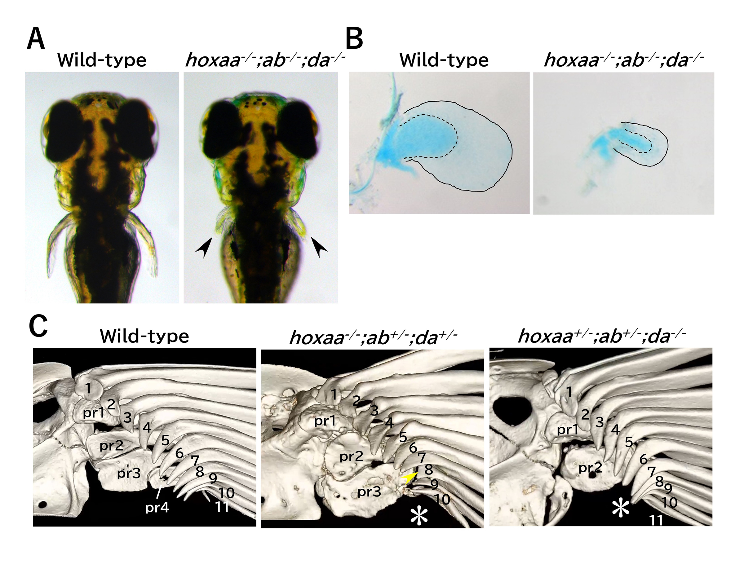
Figure: (A) Significant shortening of the pectoral fin in zebrafish hoxaa;ab;da cluster-delted mutants. 3-day post-fertilization. (B) Stained chondrocytes of the dissected pectoral fin (5-day). (C) Abnormal pectoral fin morphology in hox mutant adults revealed by micro-CT scan analysis.
Guideline for Additional Application for
2024 NIG-JOINT(Joint Researchi-(A))
(Application deadline: noon(12:00pm)
on Friday, May 31st, 2024)
Guideline for Additional Application for
2024 NIG-JOINT(Joint Researchi-(A))
(Application deadline: noon(12:00pm)
on Friday, May 31st, 2024)
Guideline for Additional Application for
2024 NIG-JOINT(Joint Researchi-(A))
(Application deadline: noon(12:00pm)
on Friday, May 31st, 2024)


