Press release
Rice GLUCAN SYNTHASE-LIKE5 promotes anther callose deposition to maintain meiosis initiation and progression
H. Somashekar, M. Mimura, K. Tsuda, K. I. Nonomura
Plant Physiology 2022 October 22 DOI:10.1093/plphys/kiac488
![]() Press release (In Japanese only)
Press release (In Japanese only)
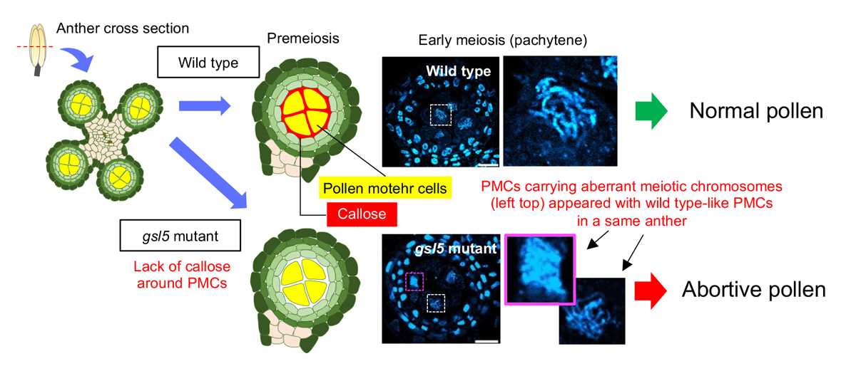
Callose is a plant cell-wall polysaccharide whose deposition is spatiotemporally regulated in various developmental processes and environmental stress responses. Appearance of callose in premeiotic anthers is a prominent histological hallmark for the onset of meiosis in flowering plants; however, the biological role of callose in meiosis remains unknown. Here we show that rice (Oryza sativa) GLUCAN SYNTHASE LIKE5 (OsGSL5), a callose synthase, localizes on the plasma membrane of pollen mother cells (PMCs) and is responsible for biogenesis of callose in anther locules through premeiotic and meiotic stages. In Osgsl5 mutant anthers mostly lacking callose deposition, aberrant PMCs accompanied by aggregated, unpaired or multivalent chromosomes were frequently observed, and furthermore, a considerable number of mutant PMCs had untimely progress into meiosis compared to that of wild-type PMCs. Immunostaining of meiosis-specific protein HOMOLOGOUS PAIRING ABERRATION IN RICE MEIOSIS2 (PAIR2) in premeiotic PMCs revealed precocious meiosis entry in Osgsl5 mutant anthers. These findings provide insights into the function of callose in controlling the timing of male meiosis initiation and progression, in addition to roles in microsporogenesis, in flowering plants.
Source: H. Somashekar et al., DOI: 10.1093/plphys/kiac488
Press release
Morphological growth dynamics, mechanical stability, and active microtubule mechanics underlying spindle self-organization
T. Fukuyama, L. Yan, M. Tanaka, M. Yamaoka, K. Saito, SC. Ti, CC. Liao, KC. Hsia, YT. Maeda, Y. Shimamoto
PNAS (2022) 119, e2209053119 DOI:10.1073/pnas.2209053119
![]() Press release (In Japanese only)
Press release (In Japanese only)
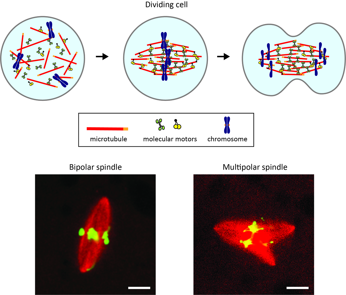
Each time a cell divides in our body, it builds a tiny micron-sized machine called the spindle. The spindle passes the copied DNA from a parental cell to the newly-created two daughter cells and should have a bipolar, football-like shape for evenly splitting the chromosomes that carry the DNA. Disrupted spindle shapes can cause errors in chromosome splitting and malfunctions in the cell offspring.
Now, a new study reveals how the shaping of this tiny segregation machine succeeds and when it ends up with failure. A team of biophysics groups in Japan (Yuta Shimamoto’s lab at the National Institute of Genetics and Yusuke Maeda’s lab at Kyushu University) and biochemistry groups abroad (Kuo-Chiang Hsia’s group at Academia Sinica and Jeff Ti’s group at the University of Hong Kong) develop a computer-based algorithm and fluorescence microscopy to track at an unprecedented resolution the way in which the machine is built from scratch. They find the unique “assembly instruction” for making the spindle with the correct shape and one for making the spindle with unfavored shapes. The team also uncovers a memory form-like material property of the spindle, which appears to be a key that separates success from failure in assembling the machine.
Abnormally shaped spindles are linked to conditions that impact human life, such as cancer and infertility. The findings of this study should be an important step toward understanding the relevant mechanisms.
Life cycle and functional genomics of the unicellular red alga Galdieria for elucidating algal and plant evolution and industrial use
S. Hirooka, T. Itabashi, T. M. Ichinose, R. Onuma, T. Fujiwara, S. Yamashita, L. W. Jong, R. Tomita, A. H. Iwane. S. Miyagishima
PNAS (2022) 119, e2210665119 DOI:10.1073/pnas.2210665119
![]() Press release (In Japanese only)
Press release (In Japanese only)
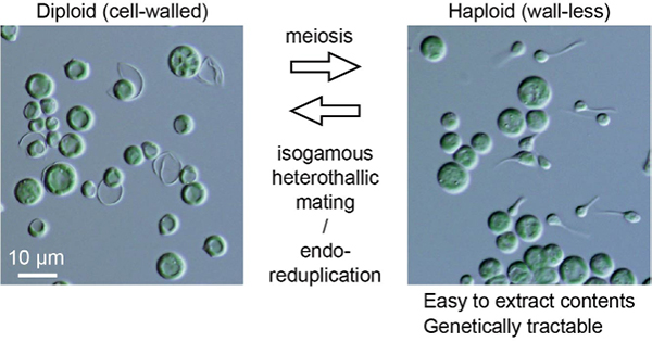
Sexual reproduction is widespread in eukaryotes. However, only asexual reproduction has been observed in unicellular red algae, including Galdieria, which branched early in Archaeplastida. Galdieria possesses a small genome; it is polyextremophile, grows either photoautotrophically, mixotrophically, or heterotrophically and is being developed as an industrial source of vitamins and pigments because of its high biomass productivity. Here, we show that Galdieria exhibits a sexual life cycle, alternating between cell-walled diploid and cell wall-less haploid and that both phases can proliferate asexually. The haploid can move over surfaces and undergo self-diploidization or generate heterozygous diploids through mating. Further, we have prepared the whole genome and comparative transcriptome dataset between the diploid and haploid and developed genetic tools for the stable gene expression, gene disruption and selectable marker recycling system using the cell wall-less haploid. The BELL/KNOX and MADS-box transcription factors, which function in haploid-diploid transition and development in plants, are specifically expressed in the haploid and diploid, respectively, and are involved in the haploid-diploid transition in Galdieria, providing information on the missing link of the sexual life cycle evolution in Archaeplastida. Four actin genes are differently involved in motility of the haploid and cytokinesis in the diploid both of which are myosin-independent and likely reflect ancestral roles of actin. We have also generated photosynthesis-deficient mutants such as blue-colored cells, which were depleted in chlorophyll and carotenoids, for industrial pigment production. These features of Galdieria facilitate understanding the evolution of algae and plants and the industrial use of microalgae.
Source: S. Hirooka et al., PNAS DOI: 10.1073/pnas.2210665119
A review by Professor Ursula Goodenough (Washington University in St. Louis) about this article will be published in this journal (PNAS) at a later day.
▶ The article of EurekAlert! is here
Iwasato Group / Laboratory of Mammalian Neural Circuits
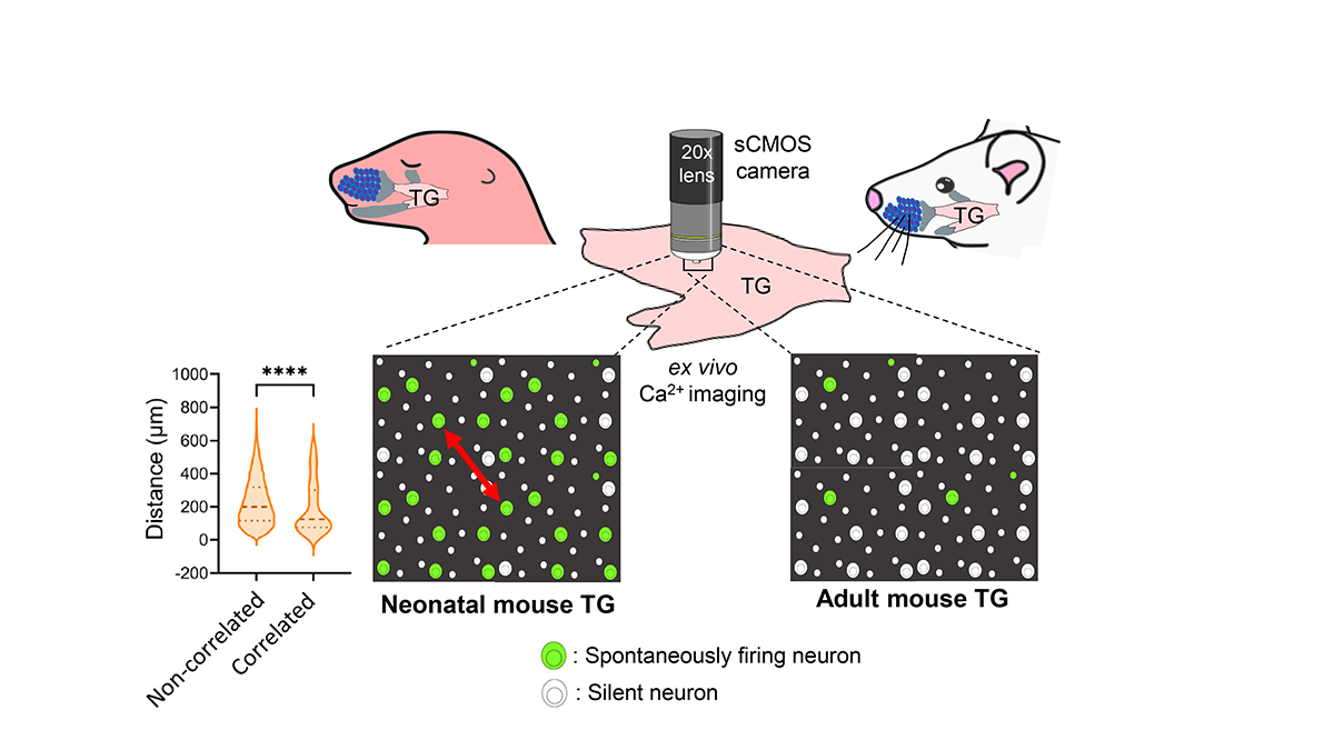
Spontaneous Activity in Whisker-Innervating Region of Neonatal Mouse Trigeminal Ganglion.
P. Banerjee, F. Kubo, H. Nakaoka, R. Ajima, T. Sato, T. Hirata, T. Iwasato.
Scientific Reports (2022) 12, 16311 DOI:10.1038/s41598-022-20068-z
Spontaneous activity is the activity that occurs without any external sensory input. During the early postnatal period in mammals, such activity is thought to be crucial for the establishment of mature neural circuits. It remains unclear if the peripheral structure of the developing somatosensory system exhibits spontaneous activity, similar to that observed in the retina and cochlea of developing visual and auditory systems, respectively. By establishing an ex vivo calcium imaging system, here we discovered that neurons in the whisker-innervating region of the trigeminal ganglion (TG) of neonatal mice generate spontaneous activity. A small percentage of neurons showed some obvious correlated activity, and these neurons were mostly located close to one another. TG spontaneous activity was majorly exhibited by medium-to-large diameter neurons, a characteristic of mechanosensory neurons, and was blocked by chelation of extracellular calcium. Moreover, this activity was diminished by the adult stage. Spontaneous activity in the TG during the first postnatal week could be a source of spontaneous activity observed in the neonatal mouse barrel cortex.
Maeshima Group / Genome Dynamics Laboratory
Chromatin organization and DNA damage.
K. Minami, S. Iida, K. Maeshima
The Enzymes “DNA Damage and Double Strand Breaks” 2022 September 27 DOI:10.1016/bs.enz.2022.08.003
Free link (until 2022.11.16)
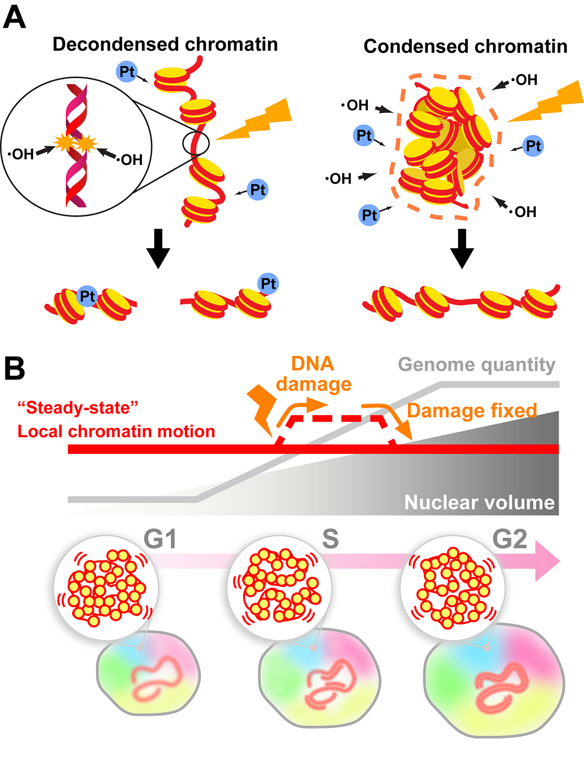
Genomic DNA is organized three-dimensionally in the nucleus as chromatin. Recent accumulating evidence has demonstrated that chromatin organizes into numerous dynamic domains in higher eukaryotic cells, which act as functional units of the genome. However, the cellular genome is constantly threatened by many sources of DNA damage (e.g., radiation). How do cells maintain their genome integrity when subjected to DNA damage?
In this review, we discuss how the compact state of chromatin safeguards the genome from radiation damage and chemical attacks. Together with recent genomics data, our finding (Takata et al. “Chromatin compaction protects genomic DNA from radiation damage”. PLOS ONE (2013). DOI: 10.1371/journal.pone.0075622) suggests that DNA compaction, such as chromatin domain formation, plays a critical role in maintaining genome integrity. But does the formation of such domains limit DNA accessibility inside the domain and hinder the recruitment of repair machinery to the damaged site(s) during DNA repair? To approach this issue, we first describe a sensitive imaging method to detect changes in chromatin states in living cells (single-nucleosome imaging). We then use this method to explain how cells can overcome potential recruiting difficulties; cells can decompact chromatin domains following DNA damage and temporarily increase chromatin motion (∼ DNA accessibility) to perform efficient DNA repair (Iida et al. “Single-nucleosome imaging reveals steady-state motion of interphase chromatin in living human cells” Science Advances (2022). DOI: 10.1126/sciadv.abn5626).
This review article will be published as the 3rd chapter in the book “The Enzymes vol.51: DNA Damage and Double Strand Breaks (Elsevier, 2022)”. This work was supported by JSPS and MEXT KAKENHI grants (19H05273 and 20H05936 to K. Maeshima), the Takeda Science Foundation (to K. Maeshima), and the Uehara Memorial Foundation (to K. Maeshima). K. Minami and S. Iida are SOKENDAI Special Researchers supported by JST SPRING JPMJSP2104.