Koide Group / Mouse Genomics Resource Laboratory
Efficient genome editing in wild strains of mice using the i-GONAD method
Yuji Imai, Akira Tanave, Makoto Matsuyama, and Tsuyoshi Koide
Scientific Reports (2022) 12, 13821 DOI:10.1038/s41598-022-17776-x
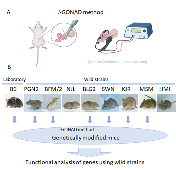
At the National Institute of Genetics, nine wild mouse strains have been established. These strains have characteristics not found in laboratory strains, such as marked genetic differences between different strains and behavior characteristic of wild mice.
These series of wild strains, named the Mishima Battery, are provided to researchers in Japan and overseas as highly unique resources, and are used for various research in the fields of cancer, immunology, development, and behavior. However, in spite of these excellent characteristics, wild strains have the problem of being difficult to apply genetic engineering technology.
In collaboration with Shigei Medical Research Institute and RIKEN, technical staff Yuji Imai and Associate Professor Tsuyoshi Koide of the Mouse Genomics Resource Laboratory have applied genome-editing using i-GONAD method, which does not involve any ex vivo manipulation of unfertilized or fertilized egg. The group showed that it is possible to efficiently modify genes in most wild strains by using this method.
First, in vitro fertilization was performed on the experimental strain C57BL/6 (B6 strain) and nine wild strains to investigate the efficiency of in vitro fertilization necessary for general genome editing experiments. As a result, it was found that only two wild strains were able to perform in vitro fertilization with the similar efficiencies as the B6 strain, and the other strains were extremely inefficient. Therefore, we applied a method called i-GONAD, developed by Professor Masato Otsuka of Tokai University, to wild strains, which performs genome editing without ex vivo manipulation of fertilized eggs. As a result, we succeeded in performing genome editing in 7 out of the 9 strains examined. This result indicates that it has become possible to efficiently perform genetic modification using wild strains in the future, and the use of wild strains in many research fields can be expected.
Kimura Group / Cell Architecture Laboratory
Sequential accumulation of dynein and its regulatory proteins at the spindle region in the Caenorhabditis elegans embryo
Takayuki Torisawa, & Akatsuki Kimura
Scientific Reports (2022) 12, 11740 DOI:10.1038/s41598-022-15042-8
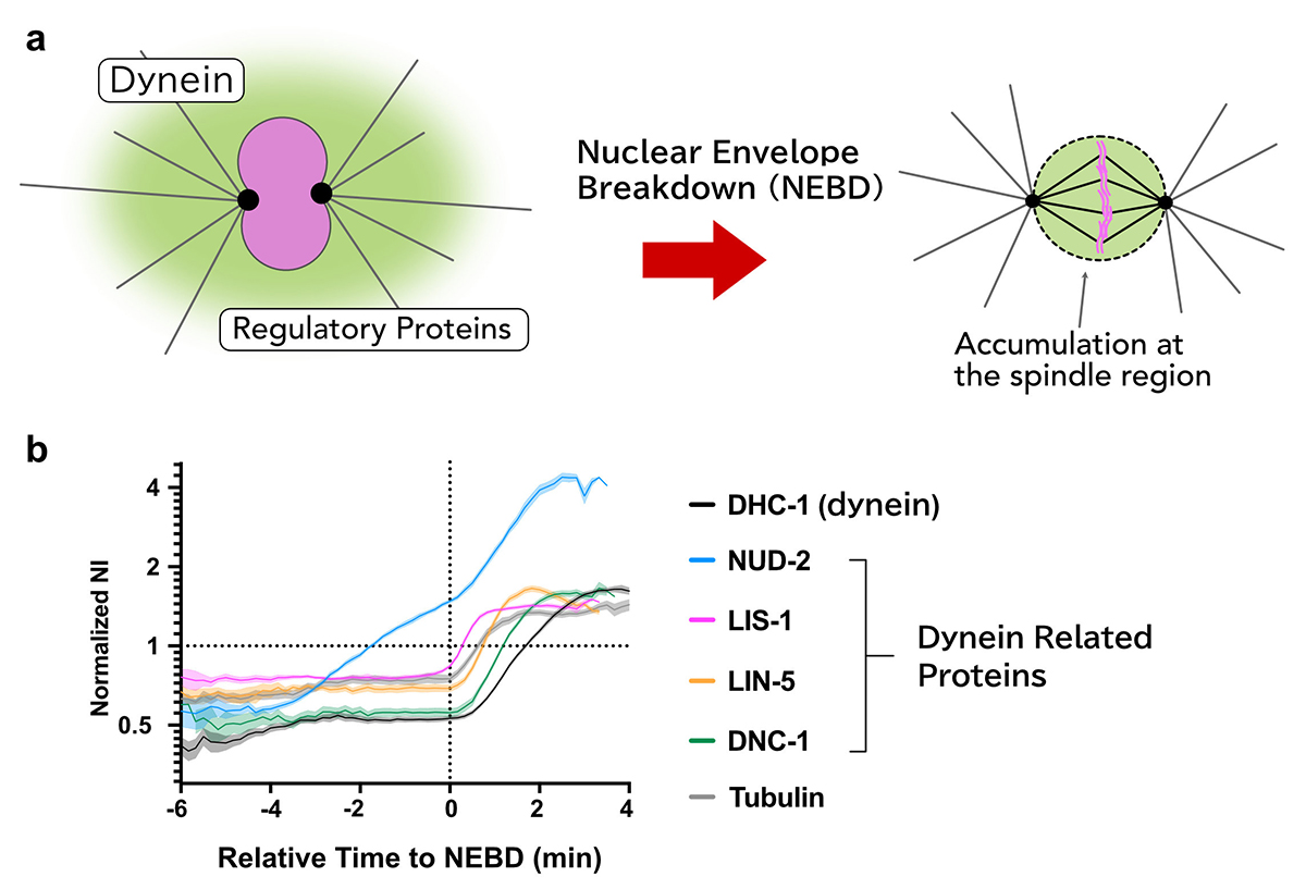
Cytoplasmic dynein is a molecular motor responsible for various cellular activities, including intracellular transport and cell division. To achieve various functions, dynein needs to be regulated by other proteins, and it has still been elusive how these regulations are achieved in many cellular contexts.
Using the early embryos of Caenorhabditis elegans, we focused on the spatiotemporal regulation of dynein during mitosis, where dynein and its regulatory proteins translocated from the cytoplasm to the spindle region, and observed the dynamics of dynein and the regulatory proteins.
We revealed that there are i) selectivity, ii) varieties in the accumulation sites, and iii) the order of accumulation in the accumulation dynamics in the spindle region.
Furthermore, we found that the accumulation of NUD-2 was unique among the dynein regulators we analyzed. NUD-2 started to accumulate before NEBD (pre-NEBD accumulation). Using a protein injection approach, we revealed that the C-terminal helix of NUD-2 was responsible for pre-NEBD accumulation. These findings suggest a fine temporal control of the subcellular localization of regulatory proteins.
This work was supported by JSPS Grants-in-Aid for Scientific Research JP19K16094, JP18H02414, JP18H05529, and JP18KK0202.
Maeshima Group / Genome Dynamics Laboratory
A phase transition for chromosome transmission when cells divide
Kazuhiro Maeshima
Nature 2022 August 03 DOI:10.1038/d41586-022-01925-3
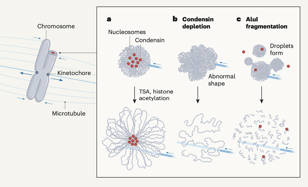
Mitotic chromosomes are the structure where DNA is tightly compacted, and they carry genetic information to be passed on to the next generation. These chromosomes are transmitted from the mother cell onto the daughter cell with physical force from microtubes and other factors. In other words, chromosomes should have mechanical resistance to endure such forces.
Recently, Daniel W. Gerlich and his colleagues have shown that chromosomes condense and gain such mechanical resistance by phase transition(Schneider et al. “A chromatin phase transition protects mitotic chromosomes against microtubule perforation” Nature 2022 doi: 10.1038/ s41586-022-05027-y). In this paper, Schneider et al. have shown that global histone deacetylation cause phase transition in mitotic chromosomes, which makes them condensed and resistant to mechanical forces. The authors have also shown that a protein complex called condensin, which was previously thought to be essential for chromosome condensation, is not involved in the condensation process itself.
Professor Kazuhiro Maeshima at Genome Dynamics Laboratory wrote a commentary on this paper in the News & Views section of Nature. Maeshima discussed how global histone deacetylation causes chromosome condensation and the role of condensin in shaping rod-like chromosomes (chromatin loop formation mechanism).