


A bidirectional network for appetite control in zebrafish. Caroline Lei Wee
Erin Yue Song, Robert Evan Johnson, Deepak Ailani, Owen Randlett, Ji-Yoon Kim, Maxim Nikitchenko, Armin Bahl, Chao-Tsung Yang, Misha B Ahrens, Koichi Kawakami, Florian Engert, and Sam Kunes.
eLife 8:e43775 (2019). DOI:10.7554/eLife.43775
Medial and lateral hypothalamic loci are known to suppress and enhance appetite, respectively, but the dynamics and functional significance of their interaction have yet to be explored. Here we report that, in larval zebrafish, primarily serotonergic neurons of the ventromedial caudal hypothalamus (cH) become increasingly active during food deprivation, whereas activity in the lateral hypothalamus (LH) is reduced. Exposure to food sensory and consummatory cues reverses the activity patterns of these two nuclei, consistent with their representation of opposing internal hunger states. Baseline activity is restored as food-deprived animals return to satiety via voracious feeding. The antagonistic relationship and functional importance of cH and LH activity patterns were confirmed by targeted stimulation and ablation of cH neurons. Collectively, the data allow us to propose a model in which these hypothalamic nuclei regulate different phases of hunger and satiety and coordinate energy balance via antagonistic control of distinct behavioral outputs.
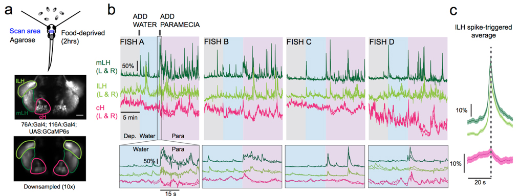
Figure: Calcium imaging of cH and LH by using transgenic zebrafish. By addition of paramecia (food), cH activity was reduced and LH activity was increased.
▶This study is based on the previous study.
Essential roles of autophagy in metabolic regulation in endosperm development during rice seed maturation
Yuri Sera, Shigeru Hanamata, Shingo Sakamoto, Seijiro Ono, Kentaro Kaneko, Yuudai Mitsui, Tomoko Koyano, Naoko Fujita, Ai Sasou, Takehiro Masumura, Hikaru Saji, Ken-Ichi Nonomura, Nobutaka Mitsuda, Toshiaki Mitsui, Takamitsu Kurusu, Kazuyuki Kuchitsu
Scientific Reports 9, 18544 (2019) DOI:10.1038/s41598-019-54361-1
Autophagy, the recycling system of metabolites in eukaryotic cells, plays crucial roles in developmental processes, reproduction and biotic/abiotic-stress responses. However, the role in plants has been largely elusive.
We found that rice autophagy-deficient mutants set smaller seeds with chalky endosperms, in which starch granules were smaller and sparser than in the wild type (Figure). The activity of α-amylases, starch degradation enzymes, was abnormally activated in the mutant endosperm, and in contrast, the level of starch synthesis-promoting enzymes was reduced. The levels of heat shock-, oxidation- or high temperature-responsible proteins were elevated in the mutant endosperm. These results suggest the importance of autophagy in starch degradation and/or stress response pathways in rice endosperm development, and will be useful for breeding of high yielding varieties tolerant to environmental changes.
This work was achieved by collaboration of Tokyo University of Science, Suwa University of Science, Niigata University, National Institute of Advanced Industrial Science and Technology, National Institute of Genetics, Akita Prefectural University, Kyoto Prefectural University and National Institute for Environmental Studies, and was supported by NIG-JOINT (84A2018).
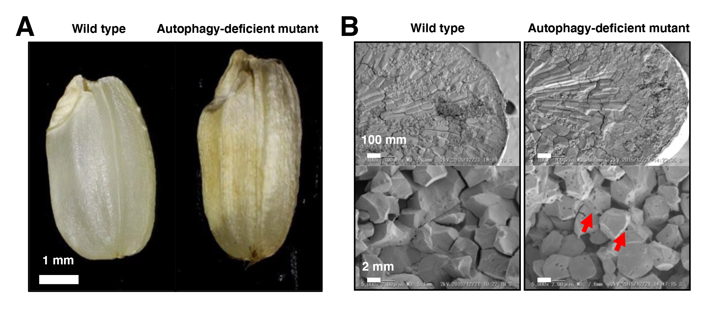
Figure: Mutant phenotypes in the endosperm of autophagy-deficient rice seeds
(A) The autophagy-deficient grain displays chalky appearance. (B) Scanning electron microscopic (top) and electron probe microscopic (bottom) images of endosperm sections. The starch granules had a lot of small pits in autophagy-deficient seeds (red arrows).
Molecular mechanism for the recognition of sequence-divergent CIF peptides by the plant receptor kinases GSO1/SGN3 and GSO2
Satohiro Okuda*, Satoshi Fujita*, Andrea Moretti, Ulrich Hohmann, Verónica G. Doblas, Yan Ma, Alexandre Pfister, Benjamin Brandt, Niko Geldner, Michael Hothorn
*These authors are equally contributed to this work
Proceedings of the National Academy of Sciences PNAS first published January 21, 2020 DOI:10.1073/pnas.1911553117
Higher plants develop Casparian strips in their roots to restrict simple diffusion of small molecules in their extracellular spaces to maintain homeostasis of whole plant bodies. Previous studies showed that a pair of receptor-kinase, GSO1(SGN3), and 21a.a.-sulfated peptides, CIF1/2, were required for Casparian strip maturation, but detail molecular mechanisms remained unclear.
Okuda and Fujita et al. presented a crystal structure of CIF2- GSO1(SGN3) peptide-receptor complex at 3 Å resolution. Quantitative analysis revealed that the GSO1(SGN3) and CIF2 pair had one of the highest affinities among known peptide-receptor pairs. Structure-based prediction uncovered Ile81 of CIF2 as a critical residue to form a tri-complex with newly identified co-receptors, SERK family proteins, to activate the downstream of the signaling pathway. Moreover, the structure-guided analysis identified homologs of CIF1/2, namely CIF3/4. CIF3/4 directly binds to GSO1(SGN3) and its homolog GSO2, but their binding properties are different from CIF1/2. Additionally, neither CIF3 nor 4 are expressing in root endodermal cells, where Casparian strips are made, implying these newly identified peptides play roles in different contexts. Our biochemical and developmental approaches provide a molecular framework to understand the recognition of diverse peptide hormones in plants.
This work was aided by grants no. 31003A_156261 and 310030E_176090 (N.G.), 31003A_176237 and 31CP30_180213 (M.H.) from the Swiss National Science Foundation, an ERC Consolidator Grant (616228-ENDOFUN) (N.G.), and an International Research Scholar Award from the Howard Hughes Medical Institute (M.H.), a Human Frontier Science Program Organization (HFSPO) postdoctoral fellowship no. LT000567/2016-L (S.O.) and a Japan Society for the Promotion of Science (JSPS) fellowship (S.F.).
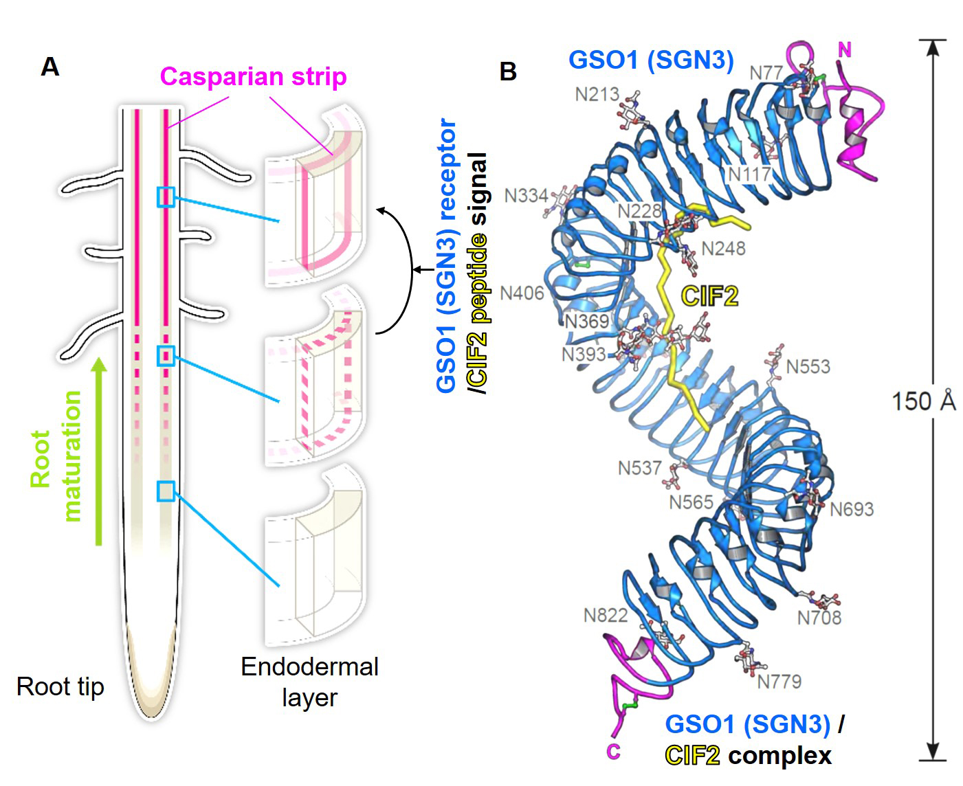
Figure: (A) Schematic model of Casparian strips (magenta). After establishing the connectivity (third row), this ring-like structure functions as an extracellular barrier. (Illustrated by Hiroko Uchida http://uchidahiroko.com/)
(B) Crystal structure of the GSO1(SGN3)/CIF2 receptor-peptide complex.
New assistant professor joins NIG as of January 1, 2020.
SASAKI, Takema : Oda Group • Cell Dynamics and Signaling Laboratory


