


Cell Architecture Laboratory / Kimura Group
Scaling relationship between intra-nuclear DNA density and chromosomal condensation in metazoan and plant.
Hara Y, Adachi K, Kagohashi S, Yamagata K, Tanabe H, Kikuchi A, Okumura S-I, Kimura A.
Chromosome Science, 19, 43-49 (2016). DOI:10.11352/scr.19.43
Because the fundamental structure of chromosomes is conserved across eukaryotes, it might be assumed that an increase in the number of DNA base-pairs in a chromosome would lead to a corresponding increase in the physical length of chromosome. This does not appear to be the case, however. We compared the lengths of mitotic chromosome from several diverse species to determine the relationship between chromosome length, number of base-pairs, and the extent of chromosome packing. We found that all species share the same relationship among these, indicating that as base-pairs are added, chromosomes become more tightly packed so that the overall length increases less than expected. Our results suggest that instead of being related to the number of DNA base-pairs, chromosome length might be proportional to the surface area of the nucleus. This may be due to the need for the chromosomes to fit within a nuclear area known as the metaphase plate during mitosis, which occurs during cellular reproduction. This study provides insight into the features that drive the evolution of genome, chromosome, nucleus, and cell size and indicates that these characteristics are shared across eukaryotes.
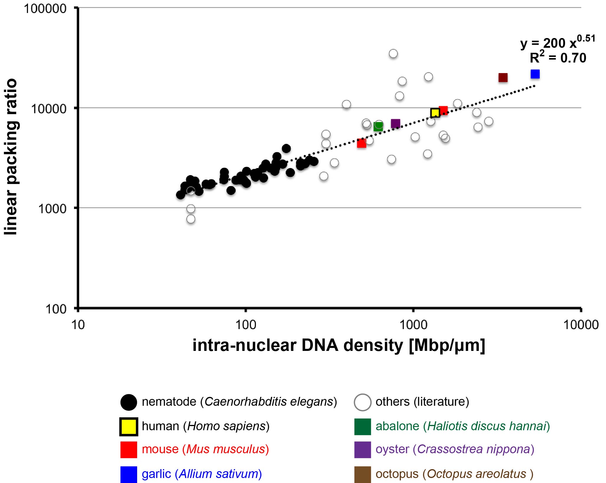
Figure. The degree of chromosome condensation (linear packing ratio) is correlated with intra-nuclear DNA density across species (Figure 1 of this paper). The double logarithmic plot of linear packing ratio against intra-nuclear DNA density is shown. The values obtained in this study are denoted with colored squares.
Cell Architecture Laboratory / Kimura Group
Reduction in chromosome mobility accompanies nuclear organization during early embryogenesis in Caenorhabditis elegans.
Arai R, Sugawara T, Sato Y, Minakuchi Y, Toyoda A, Nabeshima K, Kimura H, Kimura A.
Scientific Reports, 7, 3631 (2017). DOI:10.1038/s41598-017-03483-5
Many of us may imagine the DNA inside our cells as a jumble of noodles. However, DNA is more organized than this inside the nucleus, with chromosomes occupying distinct nuclear territories, for example. It is unclear though whether this organization is always present or whether it appears at some point during development after fertilization of the egg. We studied chromosome organization by observing the mobility of chromosomes inside in the nucleus in developing nematode embryos from the 2-cell to the 48-cell stage. We found that chromosome mobility decreases in 8-cell embryos, suggesting the initiation of chromosome organization at this point. Chromosome organization in the nucleus is important for gene expression and may have other purposes as well. For example, we found that in nematodes, the timing of chromosome organization coincides with the appearance of epigenetic marks, which regulate gene expression, and of a nuclear domain called the nucleolus. Now that we have identified the timeline of nuclear chromosome organization in nematodes, we will be able to conduct future studies to determine the factors responsible for initiating this organization.
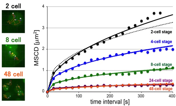
Figure. [Left images] Examples of the tracking (lines) of specific chromosomal loci (white dots) at indicated stages. The yellow dot reveals the center of the nucleus (not shown for the 48-cell stage). [Right graph] MSCD (mean squared change in distance) analyses of mobility: MSCD of the loci was plotted against the time interval (τ) for each stage. Some of the panels are identical as the panels in the paper (Arai et al., 2017)
![]()
Enhancer adoption caused by genomic insertion elicits interdigital Shh expression and syndactyly in mouse
Kousuke Mouri, Tomoko Sagai, Akiteru Maeno, Takanori Amano, Atsushi Toyoda, Toshihiko Shiroishi
PNAS Published online before print December 18, 2017 DOI:10.1073/pnas.1713339115
Pressrelease (In Japanese only)
Gene expression is regulated by tissue or organ-specific enhancer(s). Acquisition of new enhancers can alter the regulation of developmental genes, and may introduce morphological novelty in evolution. Reconfiguration between a gene and pre-existing enhancers of another gene by chromosomal rearrangement, which is referred to as “enhancer adoption”, is one possible source of new enhancers.
In this study, we re-examined an old mouse mutant named Hammer toe (Hm), which arose spontaneously over a half century ago and exhibits syndactyly with interdigital webbing. We revealed that a 150-kb non-coding genomic fragment that was originally located in chromosome 14 has been inserted into a genomic region upstream to Sonic hedgehog (Shh), located in chromosome 5. ATAC-seq and subsequent reporter assays in vivo revealed that the inserted fragment contains three interdigital enhancers to induce Shh expression in the interdigital regions in Hm. This ectopic Shh upregulates Chordin (Chrd), which in turn inhibits BMP signaling, and eventually results in syndactyly and web formation. Since the donor fragment residing in chromosome 14 has enhancer activity to induce interdigital gene expression, the Hm mutation appears to be an archetypal case of enhancer adoption. Our series of genomic deletion induced by the CRISPR-Cas9 system revealed that three enhancers in 150-kb fragment induce syndactyly in a cooperative manner. Acquisition of the combination of enhancers may have important role for the enhancer adoption.
This study was carried out as a collaboration of Kousuke Mouri, Tomoko Sagai, Akiteru Maeno, Takanori Amano and Toshihiko Shiroishi of Mammalian Genetics Laboratory, and Atsushi Toyoda of Comparative Genomics Laboratory, NIG. This study was supported by JSPS KAKENHI JP15J06985, JP17K15162 and JP17K19411.
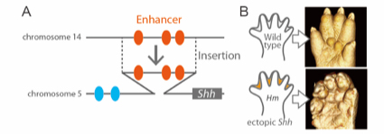
Fig. 1 (A) Hm has an insertion into the upstream region of Shh gene. Inserted fragment includes three interdigital enhancers. (B) X-ray micro-CT images of limb (P0). Shh is ectopically expressed in the interdigital region, and causes interdigital webbing in Hm.
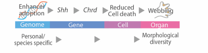
Fig. 2 The process that the morphological alteration in Hm was caused by the enhancer adoption.
You can see the several data of micro CT-scan which is one of the methods for this research below.
You can read the short review for this article.
You can read our news release in EurekAlert! about this article.
Division of Neurogenetics / Iwasato Group
Protocadherin-αC2 is required for diffuse projections of serotonergic axons
Shota Katori, Yukiko Noguchi-Katori, Atsushi Okayama, Yoshimi Kawamura, Wenshu Luo, Kenji Sakimura, Takahiro Hirabayashi, Takuji Iwasato & Takeshi Yagi
Scientific Reports, 7, Article number: 15908 (2017) DOI:10.1038/s41598-017-16120-y
Serotonergic axons extend diffuse projections throughout various brain areas, and serotonergic system disruption causes neuropsychiatric diseases. Loss of the cytoplasmic region of protocadherin-α (Pcdh-α) family proteins, products of the diverse clustered Pcdh genes, causes unbalanced distributions (densification and sparsification) of serotonergic axons in various target regions. However, which Pcdh-α member(s) are responsible for the phenotype is unknown. Here we demonstrated that Pcdh-αC2 (αC2), a Pcdh-α isoform, was highly expressed in serotonergic neurons, and was required for normal diffusion in single-axon-level analyses of serotonergic axons. The loss of αC2 from serotonergic neurons, but not from their target brain regions, led to unbalanced distributions of serotonergic axons. Our results suggest that αC2 expressed in serotonergic neurons is required for serotonergic axon diffusion in various brain areas. The αC2 extracellular domain displays homophilic binding activity, suggesting that its homophilic interaction between serotonergic axons regulates axonal density via αC2’s cytoplasmic domain.
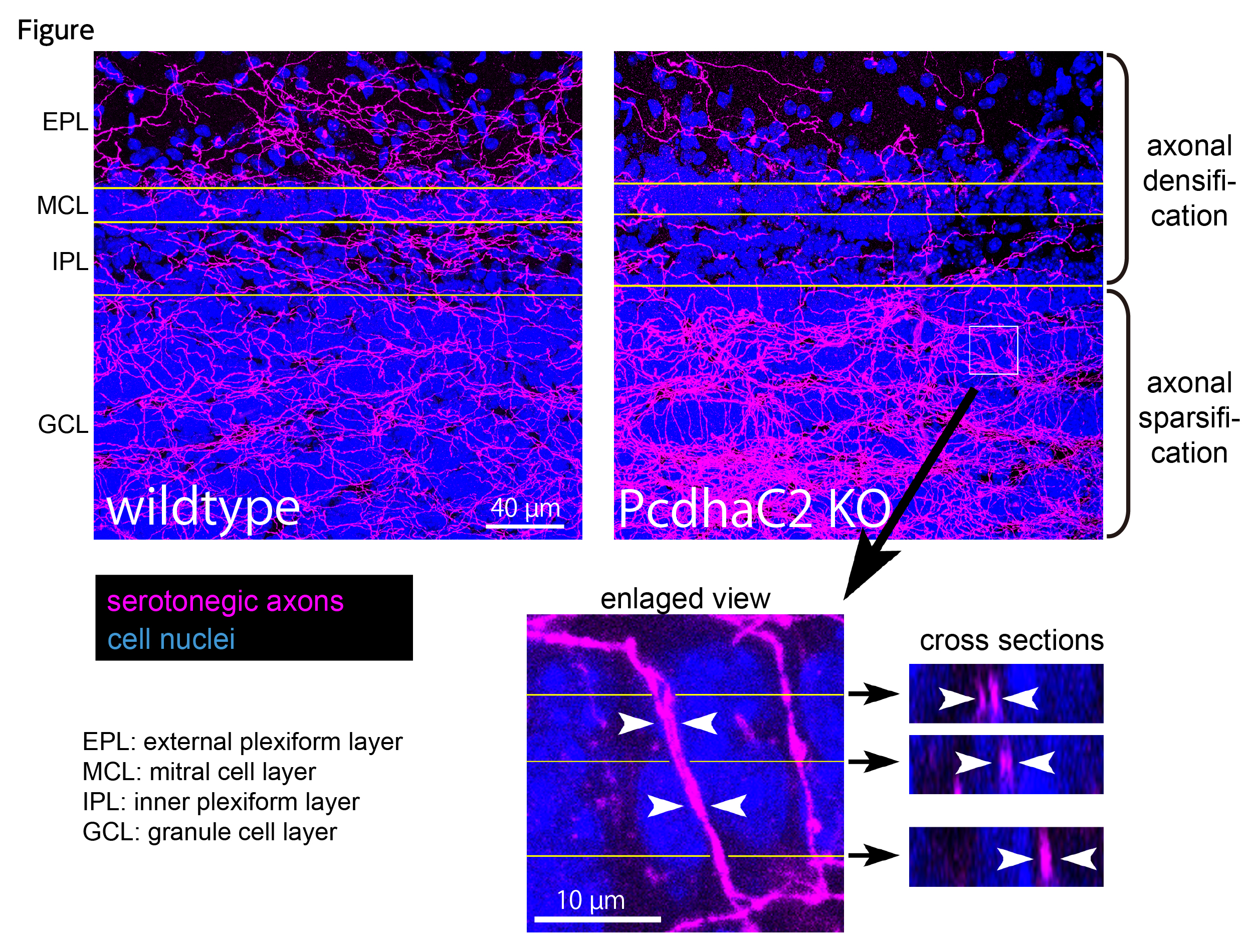
Figure. Serotonergic axons in the olfactory bulb. Wildtype mice (left) show almost uniform distributions in serotonergic axons (magenta) in all layers. In contrast, protocadherin-α C2 deficient mice (right) exhibit unbalanced serotonergic-axon distributions (densification in granule cell layer and sparsification in the other layers). In the granule cell layer (box), fasciculating serotonergic axons are observed.
Source: Scientific Reports 7 Article number: 15908(2017)
DOI: 10.1038/s41598-017-16120-y
Division of Brain Function / Hirata Group
Netrin-1 Derived from the Ventricular Zone, but not the Floor Plate, Directs Hindbrain Commissural Axons to the Ventral Midline
Kenta Yamauchi, Maya Yamazaki, Manabu Abe, Kenji Sakimura, Heiko Lickert, Takahiko Kawasaki, Fujio Murakami, and Tatsumi Hirata
Scientific Reports, DOI:10.1038/s41598-017-12269-8
In this study, we have provided evidence that Netrin-1 (Ntn1) from the ventricular zone (VZ), but not the floor plate (FP), a glial structure occupying the ventral midline (VM), guides hindbrain commissural axon (CA) to the VM. Our results are incompatible with the prevailing view that Ntn1 is a chemoattractant for CAs, rather suggest a novel mechanism that VZ-derived Ntn1 directs CAs to the VM by its local actions.
More than a century ago, based on his observations of CA growth towards the VM, Ramón y Cajal proposed the ‘chemotropic theory’, attraction of growth cones by a long-range diffusible cue emanating from their targets. Ntn1 emanating from the VM has been assumed as the chemoattractant. Consistent with this view, Ntn1 is expressed in the VM and can attract CAs at a distance in vitro. However, Ntn1 is expressed in the vicinity of the CA path, the VZ, in addition to the FP, raising an alternative possibility that Ntn1 of extra-FP origin directs CAs to the VM. To test this possibility, we generated Ntn1 FP conditional mutant (Ntn1FP-Ko) and VZ conditional mutant mice (Ntn1VZ-Ko). If Ntn1 acts CAs as a long-range diffusible chemoattractant, CA guidance to the VM should be disrupted in Ntn1FP-Ko mice. Contrary to the prediction, deletion of Ntn1 from the VZ highly disrupted CA guidance to the VM, whereas deletion of Ntn1 from the FP had little impact on it (Figure). Our results fail to support the chemotropic theory proposed by Ramón y Cajal, and rather suggest that local actions of Ntn1 from the VZ direct CAs to the VM.
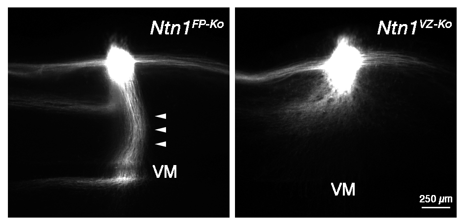
Hindbrain CA growth in Ntn1FP-Ko and Ntn1VZ-Ko mice. Deletion of Ntn1 from the VZ highly disrupts CA guidance to the VM (right), whereas deletion of Ntn1 from the FP has little impact on it (left, arrowheads). CA is labeled with a fluorescent lipophlic dye, DiI.
Fumi KUBO joined the Center for Frontier Science as of December 1, 2017.
KUBO, Fumi:Center for Frontier Research,Systems Neuroscience Laboratory

Center for Frontier Research is an incubation center to simultaneously develop two elements: human resources and new research fields. Promising young scientists conduct research as principal investigator (tenure-track associate professor) to explore new frontiers in genetics and related areas, taking advantage of NIG’s research infrastructure and various support systems.


