


Press release
Spinal RacGAP α-chimaerin is required to establish the midline barrier for proper corticospinal axon guidance
Shota Katori, Yukiko Noguchi-Katori, Shigeyoshi Itohara, Takuji Iwasato
Journal of Neuroscience 26 July 2017, 3123-16; DOI:10.1523/JNEUROSCI.3123-16.2017
Pressrelease (In Japanese only)
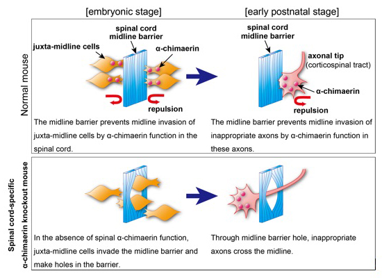
The midline barrier plays a critical role in midline axon guidance, which is fundamental to the formation of neural circuits that are responsible for proper left-right coordination of our body. Studies have revealed some of the mechanisms underlying how the midline barrier navigates axons. In contrast, the establishment of the midline barrier during embryonic development remains unclear. In this study, Katori et al. determined that α-chimaerin is required for the formation of an intact midline barrier. Spinal cord-specific α-chimaerin knockout mice had spinal midline barriers with numerous breaks (holes), through which corticospinal axons aberrantly crossed the midline. Katori et al. propose that α-chimaerin protects the midline barrier by mediating cell repulsive signaling in juxta-midline cells, which prevents these cells from invading the midline.
Source: Journal of Neuroscience 26 July 2017, 3123-16;
DOI:10.1523/JNEUROSCI.3123-16.2017
Division of Human Genetics / Inoue Group
Systematic Identification and Characterization of Regulatory Elements Derived from Human Endogenous Retroviruses.
Jumpei Ito, Ryota Sugimoto, Hirofumi Nakaoka, Shiro Yamada, Tetsuaki Kimura, Takahide Hayano, and Ituro Inoue.
PLoS Genetics. Jul 12;13(7):e1006883. 2017. DOI:10.1371/journal.pgen.1006883
Human endogenous retroviruses (HERVs) have regulatory elements that possibly influence the transcription of host genes. We systematically identified these HERV regulatory elements (HERV-REs) based on publicly available datasets of ChIP-Seq. Overall, 794,972 HERV-REs were identified. Clustering analysis showed that HERVs can be grouped according to the TF binding patterns; HERV groups bounded by pluripotent TFs (e.g., SOX2, POU5F1, and NANOG), endoderm TFs (e.g., GATA4/6, SOX17, and FOXA1/2), hematopoietic TFs (e.g., SPI1, GATA1/2, and TAL1), and CTCF were identified. Regulatory elements of HERVs tended to locate nearby genes involved in immune responses, indicating that the regulatory elements play an important role in controlling the immune regulatory network. Finally, we constructed dbHERV-REs, an interactive database of HERV regulatory elements (http://herv-tfbs.com/). This study provides fundamental information in understanding the impact of HERVs on host transcription, and offers insights into the transcriptional modulation systems of HERVs.
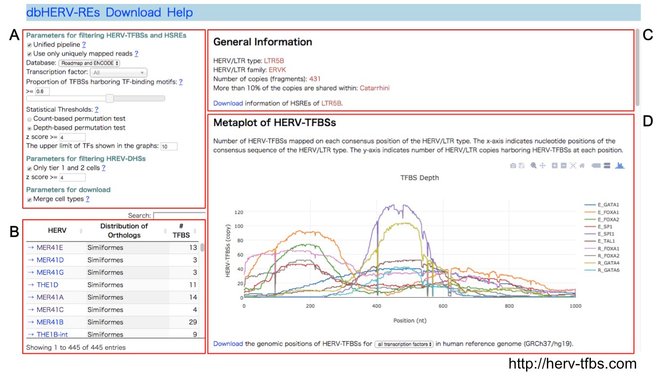
dbHERV-RE (http://herv-tfbs.com/). First, users choose a transcription factor and other parameters (A). Second, users select a type of HERVs (B). dbHERV-REs displays general information of the HERVs (phylogenetic classification, copy number, and insertion date) (C) and visualizes genetic positions of HERV-REs on the HERV sequence and the human reference genome (D).
Press release
Dynamic organization of chromatin domains revealed by super-resolution live-cell imaging
Tadasu Nozaki, Ryosuke Imai, Mai Tanbo, Ryosuke Nagashima, Sachiko Tamura, Tomomi Tani, Yasumasa Joti, Masaru Tomita, Kayo Hibino, Masato T. Kanemaki, Kerstin S. Wendt, Yasushi Okada, Takeharu Nagai, and Kazuhiro Maeshima
Molecular Cell Published: July 13, 2017 DOI:10.1016/j.molcel.2017.06.018
Press release (In Japanese only)
The eukaryotic genome is organized within cells as chromatin. For proper information output, higher-order chromatin structures can be regulated dynamically. How such structures form and behave in various cellular processes remains unclear. Here, by combining super-resolution imaging (photoactivated localization microscopy [PALM]) and single-nucleosome tracking, we developed a nuclear imaging system to visualize the higher-order structures along with their dynamics in live mammalian cells. We demonstrated that nucleosomes form compact domains with a peak diameter of ∼160 nm and move coherently in live cells. The heterochromatin-rich regions showed more domains and less movement. With cell differentiation, the domains became more apparent, with reduced dynamics. Furthermore, various perturbation experiments indicated that they are organized by a combination of factors, including cohesin and nucleosome-nucleosome interactions. Notably, we observed the domains during mitosis, suggesting that they act as building blocks of chromosomes and may serve as information units throughout the cell cycle.
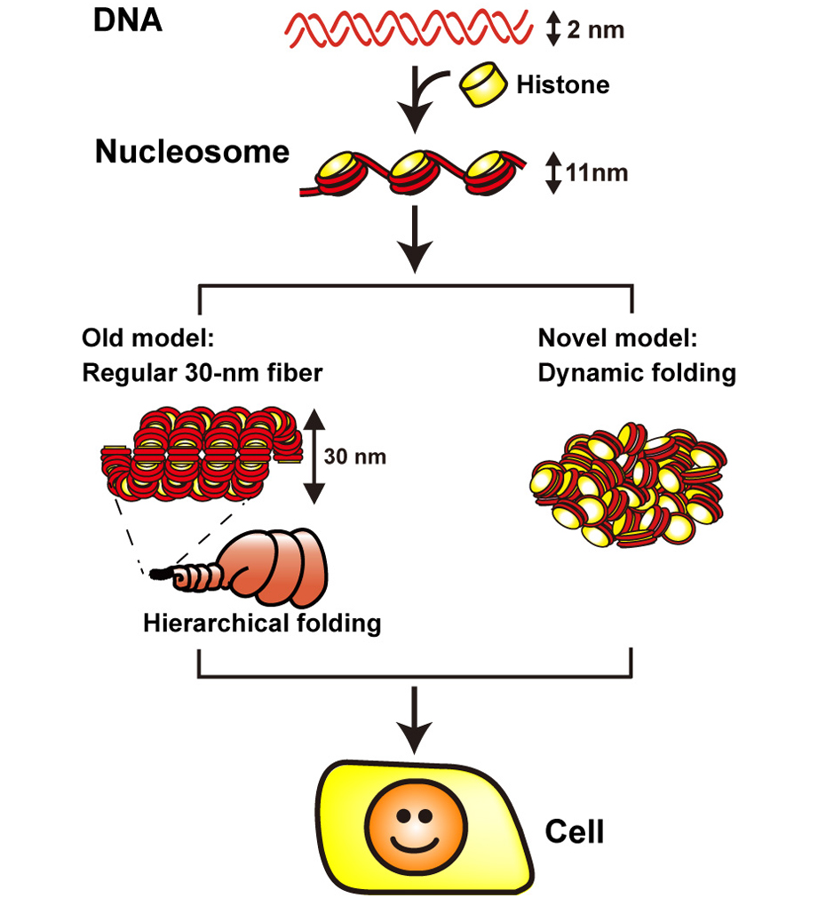
1. DNA (in red) is wrapped around core histone (in yellow) and forms a nucleosome structure (or 10-nm fiber). In the old model (left), the nucleosome fiber had long been assumed to fold into a 30-nm chromatin fiber, and subsequently into helically folded larger fibers (Hierarchical folding). In new current model (right), chromatin is composed of irregularly folded 10-nm fibers, without 30-nm chromatin fibers (dynamic folding) and stored in the cell.
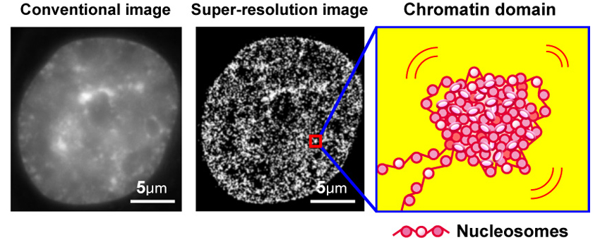
2. Left, conventional DNA staining image; center, super-resolution image of chromatin; a model of chromatin domain.
Press release
Selective breeding and selection mapping using a novel wild-derived heterogeneous stock of mice revealed two closely-linked loci for tameness
Yuki Matsumoto, Tatsuhiko Goto, Jo Nishino, Hirofumi Nakaoka, Akira Tanave, Toshiyuki Takano-Shimizu, Richard F Mott, Tsuyoshi Koide
Scientific Reports 7, Article number: 4607 (2017) DOI:10.1038/s41598-017-04869-1
Pressrelease (In Japanese only)
Tameness play important role during the process of domestication, and can be divided into two potential components: motivation to approach humans (active tameness) and reluctance to avoid them (passive tameness). To understand the genetic basis associated with active tameness in mice we applied selective breeding of a genetically heterogeneous population that we founded by crossing eight wild mouse strains. As a result of selective breeding, the level of active tameness increased over the generations, compared to an unselected control experiment. We performed two selection and two control experiments. Genetic differences between the selected and control groups, measured using a high-dense array of single-nucleotide polymorphisms, were assessed using a computer simulation experiment. In one selection experiment we found a significant increase in the occurrence of a particular genomic segment present in just one of the founder strains, compared to the control groups. This selected region contains two loci related to active tameness and is syntenic to genomic regions which are known to be a region selected during dog domestication, suggesting that responsible genes in these loci are associated with active tameness in both mouse and dog.
Yasuto MURAYAMA joined the Center for Frontier Science as of July 1, 2017.
MURAYAMA, Yasuto:Center for Frontier Research,Chromosome Biochemistry Laboratory

Center for Frontier Research is an incubation center to simultaneously develop two elements: human resources and new research fields. Promising young scientists conduct research as principal investigator (tenure-track associate professor) to explore new frontiers in genetics and related areas, taking advantage of NIG’s research infrastructure and various support systems.


