


Mammalian Development Laboratory / Saga Group
Sfrp5 identifies murine cardiac progenitors for all myocardial structures except for the right ventricle
Fujii Masayuki, Akane Sakaguchi, Ryo Kamata, Masataka Nagao, Shilvia M. Evans, Masao Yoshizumi, Akihiko Shimono, Yumiko Saga, and Hiroki Kokubo
Nature Communications. DOI:10.1038/NCOMMS1466
Upon acquirement of pulmonary circulation, the ancestral heart may have been remodelled coincidently with, or accompanied by, the production and rearrangement of progenitor cells. However, the progenitor populations that give rise to the left ventricle (LV) and sinus venosus (SV) are still ambiguous. Here we show that the expression of Secreted frizzled-related protein Sfrp5 in the mouse identifies common progenitors for the outflow tract (OFT), LV, atrium and SV but not the right ventricle (RV). Sfrp5 expression begins at the lateral sides of the cardiac crescent, excluding early differentiating regions, and continues in the venous pole, which gives rise to the SV. Lineage-tracing analysis revealed that descendants of Sfrp5-expressing cells at E7.5 contribute not only to the SV but also to the LV, atria and OFT and are found also in the dorsal splanchnic mesoderm accompanied by the expression of the secondary heart field marker, Islet1. These findings provide insight into the arrangement of cardiac progenitors for systemic circulation. This study was conducted as a collaboration research with Dr. Kokubo who used to be an assistant professor in Mammalian Development Lab and currently a lecturer of Hiroshima University.
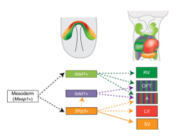
Upper panels show the expression area ofSfrp5 (orange) andIslet1 (light green) at E7.5 (left) and the distribution of descendant cells at E9.5 (right).
The lower panel shows the lineage contribution of the heart. Mesp1-positive mesodermal cells are common cardiac precursors.TransientIslet1-expressing cells that do not expressSfrp5 contribute to the RV, OFT and atrium (green). Sfrp5-positive cells contribute to the LV, atrium and OFT (orange) the SV
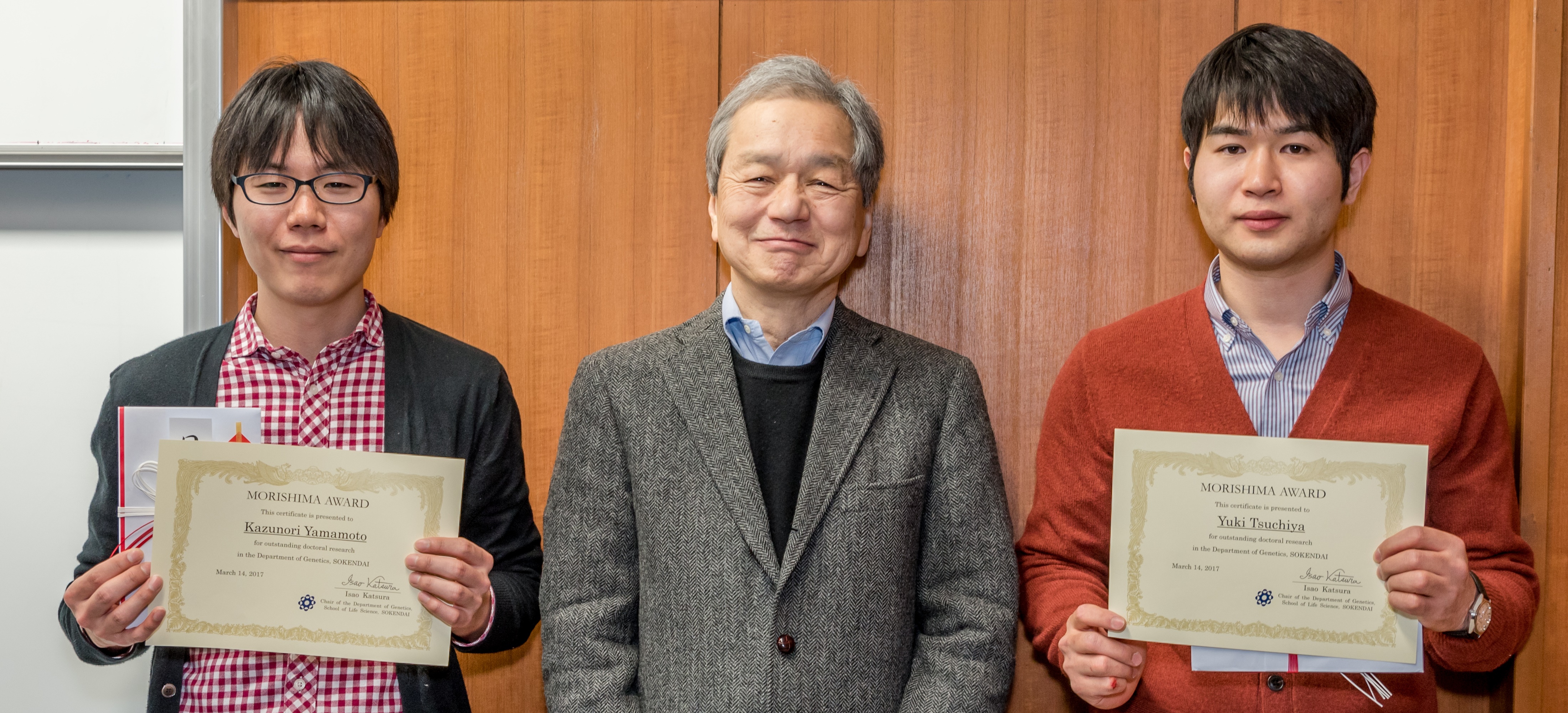
On March 14, 2017 two students who graduated in March 2016 received Morishima Awards from the Department of Genetics, SOKENDAI. The Morishima Award was established using a donation from the Morishima family honoring the late professor emeritus of NIG, Dr. Hiroko MORISHIMA. It awards students who receive a doctoral degree from the Department of Genetics to acknowledge their outstanding performances during PhD studies.
・Yuki TSUCHIYA (Division of Centrosome Biology, thesis advisor Daiju KITAGAWA)
thesis title : The role of Cep295 in centrosome biogenesis of human cells
・Kazunori YAMAMTO (Cell Architecture Laboratory, thesis advisor: Akatsuki KIMURA)
thesis title : A study on inter-cellular forces for blastomere configurations in developing embryos
Press release
Endoplasmic Reticulum-Mediated Microtubule Alignment Governs Cytoplasmic Streaming
Kenji Kimura, Alexandre Mamane, Tohru Sasaki, Kohta Sato, Jun Takagi, Ritsuya Niwayama, Lars Hufnagel, Yuta Shimamoto, Jean-François Joanny, Seiichi Uchida, and Akatsuki Kimura
Nature Cell Biology (2017) DOI:10.1038/ncb3490
Pressrelease (In Japanese only)
A research group lead by Drs. Kenji Kimura and Akatsuki Kimura at NIG revealed the self-organization mechanism of cytoplasmic streaming in Caenorhabditis elegans zygotes. Cytoplasmic streaming refers to a collective movement of cytoplasm observed in many cell types. The mechanism of meiotic cytoplasmic streaming (MeiCS) in C. elegans zygotes was puzzling as the direction of the flow is not predefined by cell polarity and occasionally reverses. The research group demonstrated that the endoplasmic reticulum (ER) network structure is required for the collective flow (Fig. 1). Using a combination of RNAi, microscopy, and image processing of C. elegans zygotes, the group devised a theoretical model, which reproduced and predicted the emergence and reversal of the flow. They proposed a positive feedback mechanism, where a local flow generated along a microtubule is transmitted to neighboring regions through the ER (Fig. 2). This, in turn, aligns microtubules over a broader area to self-organize the collective flow. The proposed model could be applicable to various cytoplasmic streaming phenomena in the absence of predefined polarity. The increased mobility of cortical granules by MeiCS correlated with the efficient exocytosis of the granules to protect the zygotes from osmotic and mechanical stresses.
The research was conducted as collaboration between NIG (Cell Architecture lab and Quantitative Mechanobiology lab), Kyushu University, Institut Curie (France) and EMBL (Germany).
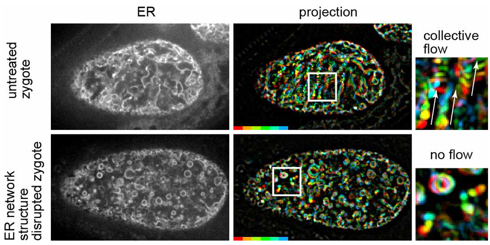
Fig. 1
Left panels show fluorescence confocal images of the ER during MeiCS in the C. elegans zygote (upper: untreated zygote, bottom: RNAi-treated zygote in which the network structure of the ER was fragmented). Middle panels show projections of sequential images of the ER. Fluorescence signals are colored to indicate their trajectories. Fragmentation of the ER network inhibited the flow. Boxed region is magnified on the right. White arrows indicate the flow direction.
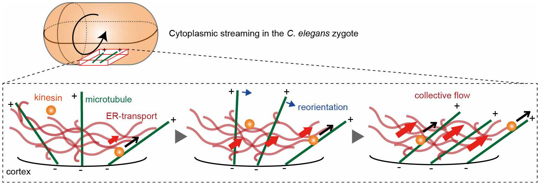
Fig. 2
ER-mediated positive feedback model. When kinesin transports the ER network along a cortical microtubule (black arrows), neighboring microtubules are biased to reorient (blue arrows) toward the ER motion, enhancing collective flow (red arrows). (−) and (+) signs indicate microtubule polarity.
Multicellular Organization Laboratory / Sawa Group
PIGN prevents protein aggregation in the endoplasmic reticulum independently of its function in the GPI synthesis
Shinji Ihara, Sohei Nakayama, Yoshiko Murakami, Emiko Suzuki, Masayo Asakawa, Taroh Kinoshita and Hitoshi Sawa
J Cell Sci 2017 130: 602-613; DOI:10.1242/jcs.196717
1. Protein aggregation is a common feature of many neurodegenerative diseases and often results from defective folding processes. We found that PIGN functions in protein quality control to prevent protein aggregation within the ER in C. elegans and in mammalian cells. Although PIGN was originally identified as an enzyme involved in glycosylphosphatidylinositol (GPI)-anchor biosynthesis, we show that the function of PIGN in protein quality control is independent of its enzymatic activities. In human, several mutations in PIGN have been shown to cause multiple congenital anomalies-hypotonia-seizures syndrome 1 (MCAHS1) that shows dysmorphic facial features and Fryns syndrome that cause lethality in the neonatal period. C. elegans with pign mutations created by CRISPR/Cas9 that correspond to MCAHS1 patients also cause protein aggregation in the ER, implying that the dysfunction of the PIGN non-canonical function might affect symptoms of MCAHS1 and potentially those of other diseases.
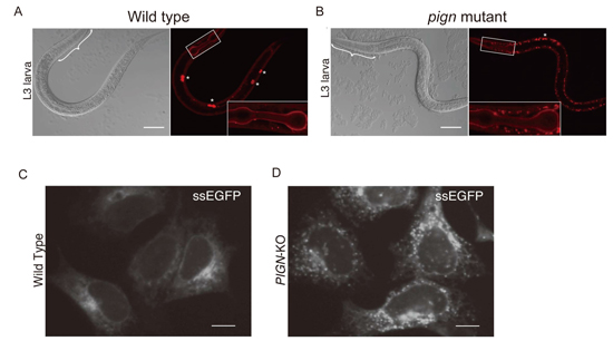
Accumulation of secretory proteins in C. elegans and mammalian cells with PIGN mutations.
(A) The localization of collagen-IV::mCherry (right) and differential interference contrast (DIC) images (left) of wild-type (A) and PIGN mutant (B) C. elegans.
The localization of secreted soluble EGFP (ssEGFP) in wild-type (C) and PIGN-knockout (D) HEK293 cells.
New assistant professors join NIG as of March 1, 2017.


