


Nonomura Group / Plant Cytogenetics Laboratory
Impact of protein domains on the MEL2 granule, a cytoplasmic ribonucleoprotein complex maintaining faithful meiosis progression in rice.
Manaki Mimura, Seijiro Ono, Harsha Somashekar, Ken-Ichi Nonomura
New Phytologist 2024 Jul 24. DOI:10.1111/nph.19968
Cytoplasmic ribonucleoprotein (RNP) granules are membraneless structures composed of various RNAs and proteins that play important roles in post-transcriptional regulation. While RNP granules are known to regulate the meiotic entry in some organisms, little is known about their roles in plants.
In this study, we observed the cytoplasmic granular structures of rice RNA-binding protein MEL2, which contributes to the control of meiotic entry timing, in leaf protoplasts and spore mother cells. Our results indicated that MEL2 granules colocalized with processing body and stress granule factors. The maintenance of granule properties modulated by LOTUS domain and the intrinsically disordered region (IDR) is essential for proper MEL2 function in meiosis progression (Figure). MEL2-like proteins widely found in plant kingdom conserved LOTUS domain followed by the IDR despite their diverse domain structures, suggesting the functional conservation of these domains among plant species.
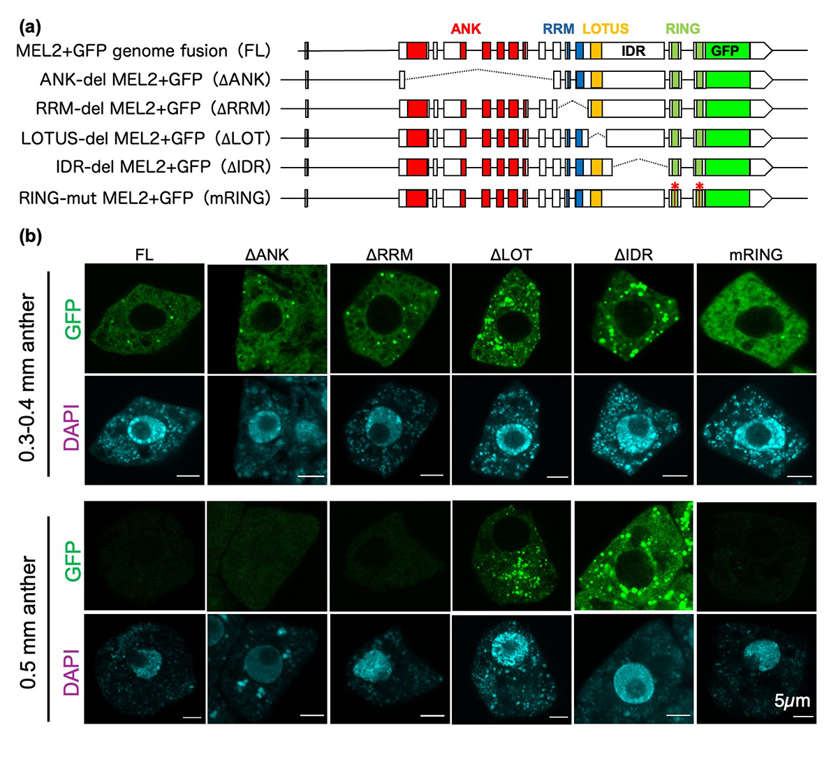
Figure: Roles of MEL2 functional domains in granule formation
(a) Genomic fusion constructs of MEL2 and GFP genes, and its derivatives with deletions (dashed lines) or mutations (asterisks) in MEL2 domains.
(b) MEL2 granule formation in spore mother cells in rice anthers transformed with the constructs in (a). The wildtype MEL2 (FL) formed granules (green) at premeiosis (0.3-0.4mm anthers), and disappeared by meiosis (0.5mm anthers). The mRING failed granule formation, and the granules of ∆LOTUS and ∆IDR were aberrantly carried over to meiocytes, all resulting in defective meiosis and pollen development.
Miyagishima Group / Symbiosis and Cell Evolution Laboratory
Development of a rapamycin-inducible protein-knockdown system in the unicellular red alga Cyanidioschyzon merolae.
Takayuki Fujiwara, Shunsuke Hirooka, Shota Yamashita, Fumi Yagisawa and Shin-ya Miyagishima
Plant Physiology (2024) kiae316 DOI:10.1093/plphys/kiae316
An inducible protein-knockdown system is highly effective for investigating the functions of proteins and mechanisms essential for the survival and growth of organisms. However, this technique is not available in photosynthetic eukaryotes. The unicellular red alga Cyanidioschyzon merolae possesses a very simple cellular and genomic architecture and is genetically tractable but lacks RNA interference machinery. In this study, we developed a protein-knockdown system in this alga. The constitutive system utilizes the destabilizing activity of the FK506-binding protein 12 (FKBP12)-rapamycin-binding (FRB) domain of human target of rapamycin kinase or its derivatives to knock down target proteins. In the inducible system, rapamycin treatment induces the heterodimerization of the human FRB domain fused to the target proteins with the human FKBP fused to S-phase kinase associated protein 1 or Cullin 1, subunits of the SCF E3 ubiquitin ligase. This results in the rapid degradation of the target proteins through the ubiquitin-proteasome pathway. With this system, we successfully degraded endogenous essential proteins such as the chloroplast division protein dynamin-related protein 5B and E2 transcription factor, a regulator of the G1/S transition, within 2 to 3 h after rapamycin administration, enabling the assessment of resulting phenotypes. This rapamycin-inducible protein-knockdown system contributes to the functional analysis of genes whose disruption leads to lethality.
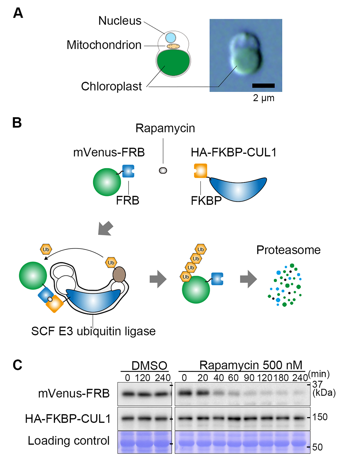
Figure: A rapamycin-inducible protein knockdown system.
(A) The cellular structure of the unicellular red alga Cyanidioschyzon merolae and a differential interference contrast microscopy image.
(B) Mechanism of the rapamycin-inducible protein knockdown system. FRB tags and FKBP proteins form a heterodimer in the presence of rapamycin. The target protein fused with an FRB tag (in this figure, the fluorescent protein mVenus) and the CUL1 subunit of ubiquitin ligase fused with FKBP bind in the presence of rapamycin, resulting in the ubiquitination of FRB-mVenus. The ubiquitinated FRB-mVenus is rapidly degraded by the proteasome.
(C) Immunoblot analysis showing the decrease in mVenus-FRB protein level after the addition of rapamycin.
NIG will be closed from August 15 to August 16, 2024 for summer holiday.
Thank you for your understanding and cooperation.
Due to the facility maintenance, the NIG website service will be stopped several times on the following time.
Thank you for your understanding and cooperation.
Time and Date: July 11 Thu, 00:00 – July 11 Thu, 08:00.
以下の期日において、遺伝研公式ウェブサイトの断続的な停止を伴う保守作業が予定されております。
皆様のご理解とご協力をお願いいたします。
停止日時: 7月11日(木) 00:00~7月11日(木) 08:00
理由: 保守作業のため
Due to the test of the NIG mail system, NIG account mails may not be able to send or receive between 12:45-13:45 on July 4.
We apologize for any inconvenience and appreciate your understanding and cooperation.
Kanemaki Group / Molecular Cell Engineering Laboratory
Replication initiation sites and zones in the mammalian genome: Where are they located and how are they defined?
Xiaoxuan Zhu, Masato T. Kanemaki*
*Corresponding author
DNA Repiar (2024) 141, 103713 DOI:10.1016/j.dnarep.2024.103713
The question of how genomic DNA is replicated is one of the key themes in molecular biology, as abnormalities in DNA replication are related to genome instability, genetic disorders, cell death, and cancer. Therefore, DNA replication has been extensively studied over the years using Escherichia coli and budding yeast as model organisms. In these unicellular models, DNA replication starts from genomic regions with consensus DNA sequences. On the other hand, in mammalian cells, including humans, consensus sequences appear to be absent, and it is still not well understood where DNA replication begins and how are they defined in the genome. Hence, this review paper compiles and compares the results of large-scale high-throughput sequencing-based explorations for mapping initiation sites/zones reported so far. Furthermore, it highlights the similarities and differences in the DNA replication mechanisms between budding yeast and mammalian cells, presents a hypothetical mechanism for how replication initiation zones are defined in mammalian cells, and discusses the future directions and challenges in DNA replication research.
The first author, Dr Xiaoxuan Zhu, has been supported as an NIG post-doctoral fellow.
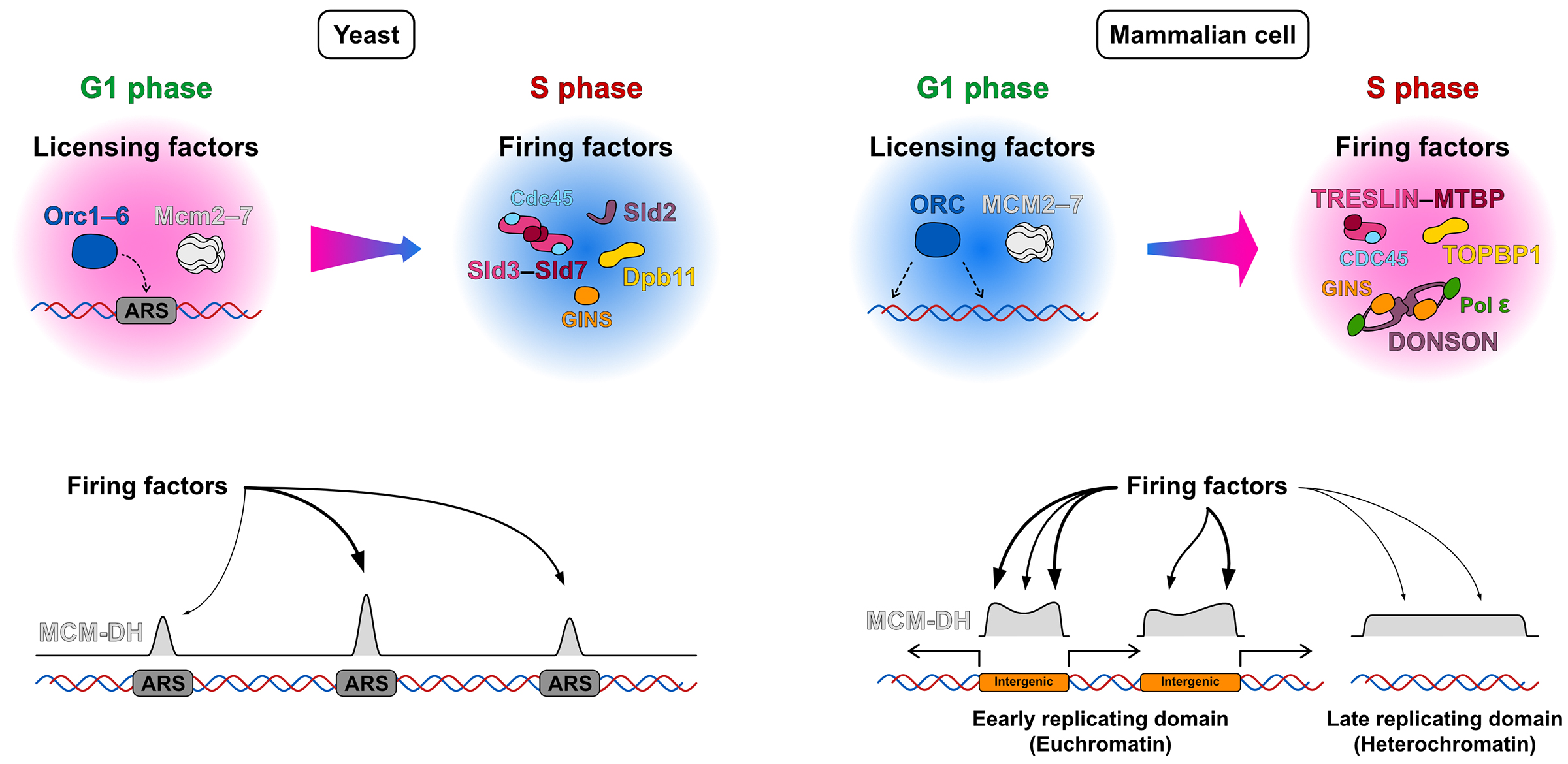
Figure: Differences in the mechanisms of replication initiation sites/zones selection between budding yeast and mammalian cells: In yeast, licensing factors accumulate at ARS (autonomously replicating sequence) sites containing a consensus sequence, leading to replication initiation at these ARS sites. In contrast, in mammalian cells, licensing factors are widely distributed across the genome. It is thought that the subsequent accumulation of firing factors at specific locations is crucial for forming initiation zones.


