


Press release
An Optical Illusion Pinpoints an Essential Circuit Node for Global Motion Processing
Yunmin Wu, Marco dal Maschio, Fumi Kubo*, Herwig Baier *Corresponding author
Neuron 2020 September 22 DOI:10.1016/j.neuron.2020.08.027
By exposing larval zebrafish to a well-known optical illusion, researchers at the Max Planck Institute of Neurobiology and National Institute of Genetics in Japan have found a clever way to isolate key clusters of neurons critical to processing the direction of motion in the zebrafish’s environment.
Details were published in the journal Neuron in September 2020. read more
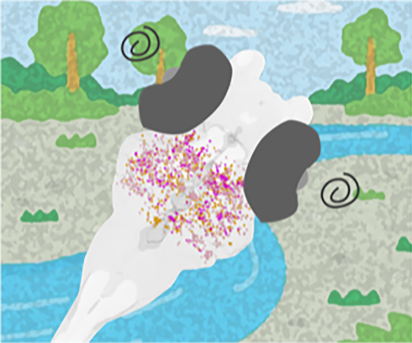
Figure: Isolating a key element in the motion processing using MAE. By inducing an optical illusion in zebrafish larvae, scientists have identified a key cluster of cells in the brain that play a crucial role in motion processing.
Press release
Identification of ancient viruses from metagenomic data of the Jomon people
Luca Nishimura, Ryota Sugimoto, Jun Inoue, Hirofumi Nakaoka, Hideaki Kanzawa-Kiriyama, Ken-ichi Shinoda, Ituro Inoue
Journal of Human Genetics 2020 September 30 DOI:10.1038/s10038-020-00841-6
Press release (In Japanese only)
Ancient DNA studies provide genomic information about the origins, population structures, and physical characteristics of ancient humans that cannot be solely examined by archeological studies. The DNAs extracted from ancient human samples such as teeth contain not only ancient human genomes but also microbial ones infecting those of ancient humans. Information on ancient viral genomes is useful in making inferences about the viral evolution. Here, we have utilized metagenomic sequencing data from the dental pulp of five Jomon individuals, who lived on the Japanese archipelago more than 3000 years ago; this is to detect ancient viral genomes. We were able to obtain eleven putative ancient viral genomes. Among them, we reconstructed the complete sequence of the Siphovirus contig89 (CT89) viral genome. As a result of the genomic comparison between the Jomon CT89 and the modern CT89 sequences, the Jomon CT89 may reflect the ancestral states. Our results suggest that metagenomic information from the dental pulp of the Jomon people is essential in retrieving ancient viral genomes used to examine their evolution.
Press release
Transcriptional suppression of ribosomal DNA with phase separation
Satoru Ide, Ryosuke Imai, Hiroko Ochi, and Kazuhiro Maeshima
Science Advances 6,eabb5953(2020) DOI:10.1126/sciadv.abb5953
Press release (In Japanese only)
The nucleolus is a nuclear body with multiphase liquid droplets for ribosomal RNA (rRNA) transcription. How rRNA transcription is regulated in the droplets remains unclear. Here, using single-molecule tracking of RNA polymerase I (Pol I) and chromatin-bound upstream binding factor (UBF), we reveal suppression of transcription with phase separation. For transcription, active Pol I formed small clusters/condensates that constrained rDNA chromatin in the nucleolus fibrillar center (FC). Treatment with a transcription inhibitor induced Pol I to dissociate from rDNA chromatin and to move like a liquid within the nucleolar cap that transformed from the FC (Fig. 1). Expression of a Pol I mutant associated with a craniofacial disorder inhibited transcription by competing with wild-type Pol I clusters and transforming the FC into the nucleolar cap (Fig. 2). The cap droplet excluded an initiation factor, ensuring robust silencing. Our findings suggest a mechanism of rRNA transcription suppression via phase separation of intranucleolar molecules governed by Pol I.
This work was supported by the Japan Society for the Promotion of Science KAKENHI grants (15K18580, 15H01361, and 18K06187 to S.I.; 16H04746 and 19H05273 to K.M.), a Japan Science and Technology Agency CREST grant (JPMJCR15G2 to K.M.), and the Takeda Science Foundation (to K.M.).
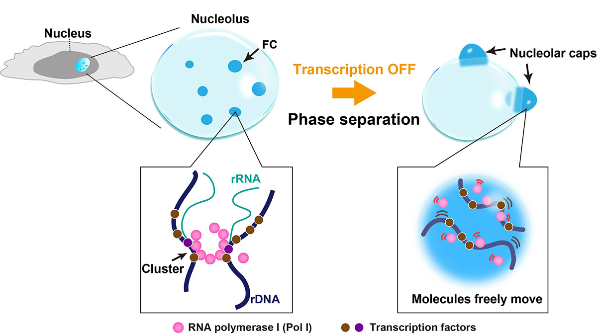
Fig. 1. Diagram of nucleolar reorganization in response to transcription inhibition through phase separation. Left: In the nucleolus fibrillar center (FC, dark blue), active Pol I molecules (pink) form a stable cluster/condensate to transcribe rRNA genes (rDNA), constraining rDNA chromatin. Right: Once transcription is inhibited by a drug, the FC components segregate to the nucleolar periphery, where they coalesce to form large bodies called nucleolar caps. The Pol I cluster/condensate detaches from chromatin, thus releasing the chromatin constraint. Pol I and rDNA behave like a liquid in the nucleolar cap through further phase separation.
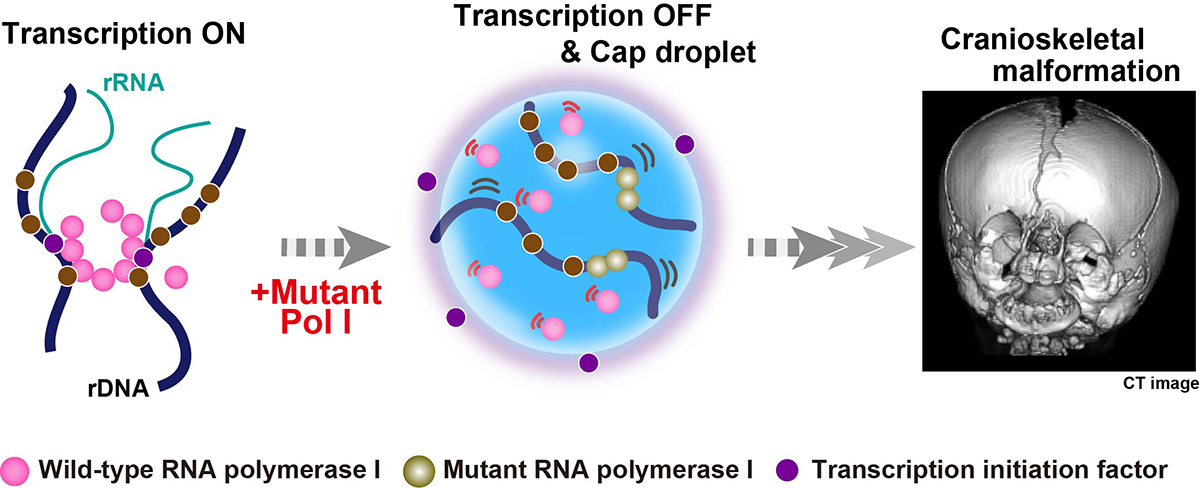
Fig. 2. Suppression of transcription with phase separation by a mutant Pol I associated with a craniofacial disorder. Mutant Pol I stably binds to rDNA chromatin and inhibits wild-type Pol I cluster/condensate formation (middle). The whole Pol I population becomes mobile in the nucleolar cap. A transcription initiation factor (violet) is excluded from the cap droplet, ensuring robust transcription suppression within the cap (middle). This process can cause the failure of ribosome biogenesis on craniofacial skeletal development in a patient (right, the image given by Dr. K. Nicole Weaver in Cincinnati Children’s Hospital Medical Center).
Transient and lineage-restricted requirement of Ebf3 for sternum ossification
Mao Kuriki, Fuminori Sato, Hiroyuki N Arai, Maina Sogabe, Mari Kaneko, Hiroshi Kiyonari, Koichi Kawakami, Yuki Yoshimoto, Chisa Shukunami, Atsuko Sehara-Fujisawa
Development 147, dev186239 (2020). DOI:10.1242/dev.186239
Osteoblasts arise from bone-surrounding connective tissue containing tenocytes and fibroblasts. Lineages of these cell populations and mechanisms of their differentiation are not well understood. Screening enhancer-trap lines of zebrafish allowed us to identify Ebf3 as a transcription factor marking tenocytes and connective tissue cells in skeletal muscle of embryos. Knockout of Ebf3 in mice had no effect on chondrogenesis but led to sternum ossification defects as a result of defective generation of Runx2+ pre-osteoblasts. Conditional and temporal Ebf3 knockout mice revealed requirements of Ebf3 in the lateral plate mesenchyme cells (LPMs), especially in tendon/muscle connective tissue cells, and a stage-specific Ebf3 requirement at embryonic day 9.5-10.5. Upregulated expression of connective tissue markers, such as Egr1/2 and Osr1, increased number of Islet1+ mesenchyme cells, and downregulation of gene expression of the Runx2 regulator Shox2 in Ebf3-deleted thoracic LPMs suggest crucial roles of Ebf3 in the onset of lateral plate mesoderm differentiation towards osteoblasts forming sternum tissues.
This study was conducted as collaboration with Professor Atsuko Sehara Laboratory at Institute for Frontier Life and Medical Sciences, Kyoto University. This study was partly supported by NBRP.
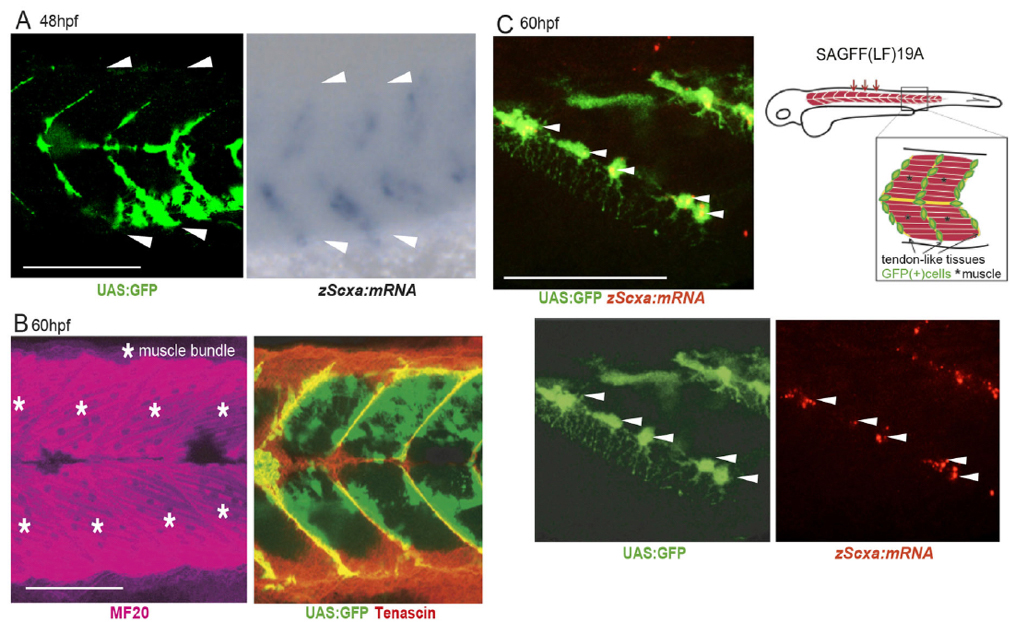
Figure1: Expression of GFP (Gal4) in tenocytes and muscle connective tissues in the zebrafish ebf3 gene trap line (48 and 60 hours post fertilization).
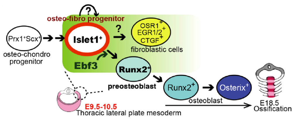
Figure2: A role of Ebf3 in pre-osteoblast formation.
Press release
The “COVID-19 Data Portal JAPAN ” has been launched through collaboration between the Research Center for Open Science and Data Platform (RCOS, Center Director: YAMAJI Kazutsuna, Professor, NII Digital Content and Media Sciences Research Division) of the National Institute of Informatics (NII, Director General: KITSUREGAWA Masaru, Tokyo, Japan) and the Bioinformation and DDBJ Center (Head of DDBJ Center: ARITA Masanori, Professor, NIG Biological Networks Laboratory) of the National Institute of Genetics (NIG, Director General: HANAOKA Fumio, Shizuoka, JAPAN), both belonging to the Research Organization of Information and Systems (ROIS, President: FUJII Ryoichi, Tokyo, Japan).
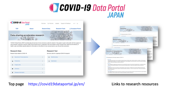
Fig: The portal site lists the collected research data and tools related to COVID-19 by field. You can access resources promptly by referring to the description of each resource.


