


HLA-B*39:01:01 is a novel risk factor for antithyroid drug-induced agranulocytosis in Japanese population
Saya Nakakura, Kazuyoshi Hosomichi, Shinya Uchino, Akiko Murakami, Akira Oka, Ituro Inoue, Hirofumi Nakaoka
The Pharmacogenomics Journal 2020 September 22 DOI:10.1038/s41397-020-00187-4
Anti-thyroid drug (ATD) is a mainstay of Graves’ disease. About 0.1-0.5% of patients with Graves’ disease treated with ATD exhibit agranulocytosis, which is characterized by severe reduction of circulating neutrophils. Although it has been reported that the HLA class II allele (HLA-DRB1*08:03) was associated with ATD-induced agranulocytosis, the entire HLA region have not been explored in Japanese. Therefore, we performed HLA sequencing for 10 class I and 11 class II genes in 87 patients with ATD-induced agranulocytosis and 384 patients with Graves’ disease who did not develop ATD-induced agranulocytosis. By conducting case-control association studies at the HLA allele and haplotype levels, we identified HLA-B*39:01:01 as an independent risk factor. To verify the reproducibility of the association of HLA-B*39:01:01, we retrieved allele frequency data for HLA-B*39:01:01 from previous case-control association studies. The association of HLA-B*39:01:01 was significantly replicated in Chinese, Taiwanese, and European populations. A meta-analysis combining results from the previous and current studies reinforced evidence of association between HLA-B*39:01:01 and ATD-induced agranulocytosis. The results of this study will provide a better understanding of the pathogenesis of ATD-induced agranulocytosis in the context of HLA-mediated hypersensitivity reaction.
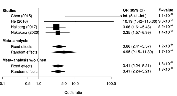
Figure: Meta-analysis for association between ATD-induced agranulocytosis and HLA-B*39:01:01 by combining previous three studies and current study. For single studies, odds ratio and 95% confidence interval are represented by box and whisker. For meta-analyses, odds ratio and 95% confidence interval are represented by a diamond. In all the single studies, HLA-B*39:01:01 was significantly associated with the risk for ATD-induced agranulocytosis. A meta-analysis combining results from the previous and current studies reinforced evidence of the association.
Developmental Phase Transitions in Spatial Organization of Spontaneous Activity in Postnatal Barrel Cortex Layer 4
Shingo Nakazawa, Yumiko Yoshimura, Masahiro Takagi, Hidenobu Mizuno, Takuji Iwasato.
Journal of Neuroscience 2020 September 4 DOI:10.1523/JNEUROSCI.1116-20.2020
Developing sensory cortices exhibit spatially-organized spontaneous activity, which is critical for the cortical circuit maturation. We previously reported “patchwork-type” spatial organization of spontaneous activity in the mouse barrel cortex at postnatal day 5 (P5) (Press release at 2018)
In the present study, we analyzed in detail how the spatial organization of barrel cortex spontaneous activity changes during the first two postnatal weeks. We found that spontaneous activity between P1 to P5 exhibited a patchwork-type pattern (Phase I). While around P9, a new type of pattern, showing wide area synchronization (Phase II), was observed, and at P11, neurons fired sparsely as observed in the adult brain (Phase III).
When thalamus was genetically silenced, Phase I cortical activity was abolished but Phase II and III activity remained intact, suggesting that the Phase I to II transition is associated with the shift of the activity source. On the other hand, the Phase II to III transition was impaired by cortical Rac1 inhibition. Phase II to III transition may be facilitated by Rac1-mediated synapse maturation.
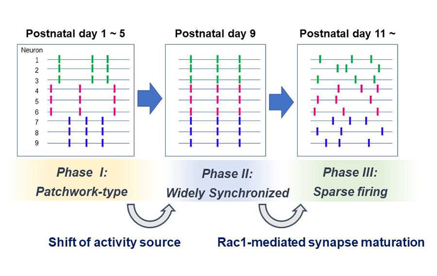
Figure: Three phases of spontaneous network activity were found in the barrel cortex layer 4 during the first two postnatal weeks. The Phase I to II transition is associated with the loss of thalamocortical input dependency. The Phase II to III transition may rely on cortical Rac1-dependent synapse maturation.
Zebrafish can regenerate endoskeleton in larval pectoral fin but the regenerative ability declines
Keigo Yoshida, Koichi Kawakami, Gembu Abe, Koji Tamura
Developmental Biology 463, 110-123 (2020). DOI:10.1016/j.ydbio.2020.04.010
We show for the first time endoskeletal regeneration in the developing pectoral fin of zebrafish. The developing pectoral fin contains an aggregation plate of differentiated chondrocytes (endochondral disc; primordium for endoskeletal components, proximal radials). The endochondral disc can be regenerated after amputation in the middle of the disc. The regenerated disc sufficiently forms endoskeletal patterns. Early in the process of regenerating the endochondral disc, epithelium with apical ectodermal ridge (AER) marker expression rapidly covers the amputation plane, and mesenchymal cells start to actively proliferate. Taken together with re-expression of a blastema marker gene, msxb, and other developmental genes, it is likely that regeneration of the endochondral disc recaptures fin development as epimorphic limb regeneration does. The ability of endoskeletal regeneration declines during larval growth, and adult zebrafish eventually lose the ability to regenerate endoskeletal components such that amputated endoskeletons become enlarged. Endoskeletal regeneration in the zebrafish pectoral fin will serve as a new model system for successful appendage regeneration in mammals.
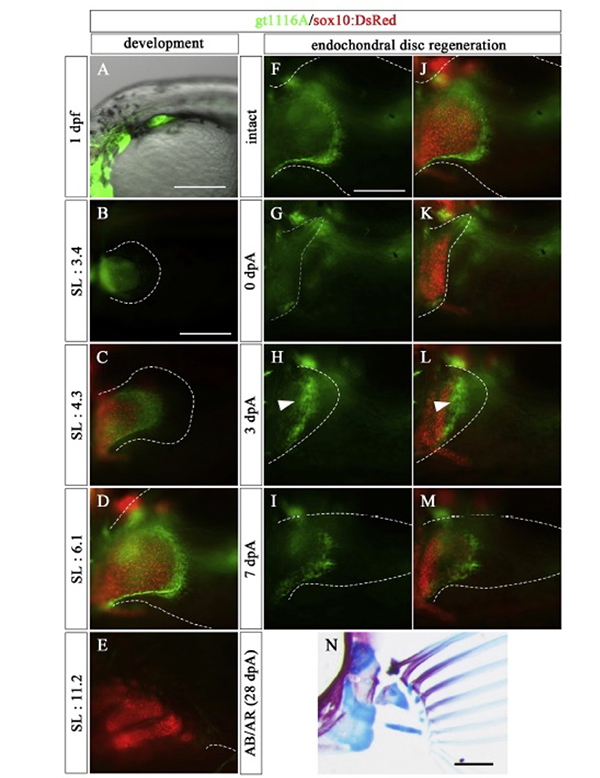
Figure: In the prdm16 gene trap line, the mesenchymal cells of the endoskeletal part of the pectoral fin expressed GFP (green) during the embryonic development. In this line, GFP was also expressed in the mesenchymal cells of the endoskeletal part during endochondrial disc regeneration. The red color in the figure indicates the expression of the sox10 gene, and labeled chondrocytes.
Press release
The dynamics, causes and impacts of mammalian evolutionary rates revealed by the analyses of capybara draft genome sequences
Isaac Adeyemi Babarinde and Naruya Saitou
Genome Biology and Evolution (2020) evaa157 DOI:10.1093/gbe/evaa157
Capybara (Hydrochoerus hydrochaeri) is the largest species among the extant rodents. The draft genome of capybara was sequenced with the estimated genome size of 2.6 Gbp. Although capybara is about 60 times larger than guinea pig, comparative analyses revealed that the neutral evolutionary rates of the two species were not substantially different. However, analyses of 39 mammalian genomes revealed very heterogeneous evolutionary rates. The highest evolutionary rate, 8.5 times higher than the human rate, was found in the Cricetidae-Muridae common ancestor after the divergence of Spalacidae. Muridae, the family with the highest number of species among mammals, emerged after the rate acceleration. Factors responsible for the evolutionary rate heterogeneity were investigated through correlations between the evolutionary rate and longevity, gestation length, litter frequency, litter size, body weight, generation interval, age at maturity, and taxonomic order. The regression analysis of these factors showed that the model with three factors (taxonomic order, generation interval and litter size) had the highest predictive power (R2 = 0. 74). These three factors determine the number of meiosis per unit time. We also conducted transcriptome analysis, and found that the evolutionary rate dynamics affects the evolution of gene expression patterns.
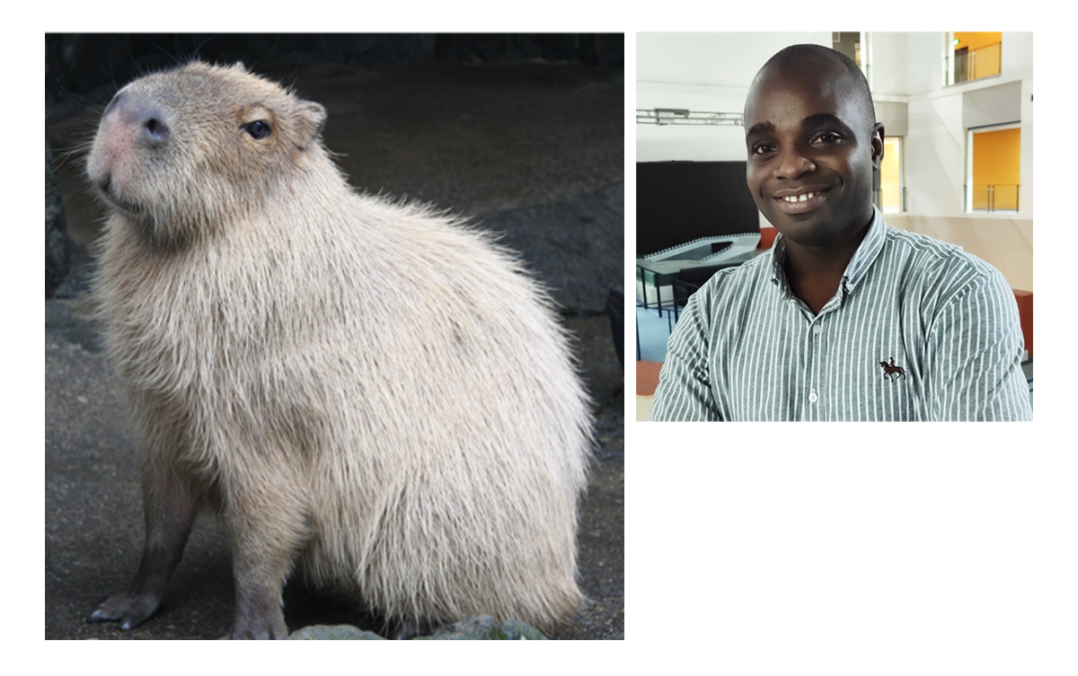
Left Panel: The memory of capybara “Rai-chan” (Photo: Izu Shaboten Zoo)
right Panel:Dr. Babarinde (from Nigeria, Africa), who received his Ph.D. from the Graduate University for Advanced Studies, SOKENDAI, in Saito’s lab and is currently a post-doctoral fellow in China.


