


Choice between 1- and 2-furrow cytokinesis in Caenorhabditis elegans embryos with tripolar spindles
Tomo Kondo and Akatsuki Kimura
Molecular Biology of the Cell Published Online:20 Feb 2019 DOI:10.1091/mbc.E19-01-0075
Excessive centrosomes often lead to multipolar spindles, and thus probably to multipolar mitosis and aneuploidy. In Caenorhabditis elegans, approximately 70% of the paternal emb-27APC6 mutant embryonic cells contained more than two centrosomes and formed multipolar spindles. However, only 30% of the cells with tripolar spindles formed two cytokinetic furrows (Figure A, right). The rest formed one furrow, like normal cells (Figure A, left). To investigate the mechanism underlying this inconsistency, we conducted live-cell imaging in emb-27APC6 mutant cells. We observed that the chromatids were aligned only on two of the three sides of the tripolar spindle, and the angle of the tripolar spindle relative to the long axis of the cell correlated with the number of cytokinetic furrows (Figure B). Our numerical modeling showed that the combination of cell shape, cortical pulling forces, and heterogeneity of centrosome size determines whether cells with tripolar spindle form one or two cytokinetic furrows.
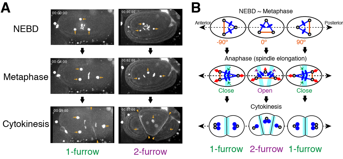
Figure:
(A) Images of a living C. elegans embryo with three centrosomes (arrows). In 1-furrow cytokinesis (left), a cleavage furrow (arrowheads) was observed between separated chromatids. In 2-furrow cytokinesis (right), two cleavage furrows (arrowheads) were observed.
(B) The proposed model showing how 1-furrow and 2-furrow cytokinesis are determined depending on the angle of tripolar spindle.
Six6 and Six7 coordinately regulate expression of middle-wavelength opsins in zebrafish
Yohey Ogawa, Tomoya Shiraki, Yoshimasa Asano, Akira Muto, Koichi Kawakami, Yutaka Suzuki, Daisuke Kojima and Yoshitaka Fukada
PNAS (2019) 116 (10) 4651-4660 DOI:10.1073/pnas.1812884116
Color discrimination in the vertebrate retina is mediated by a combination of cone cell types expressing UV- (SWS1), blue- (SWS2), green- (RH2), and red- (LWS) sensitive photoreceptive molecules (opsins). Although the tetrachromatic cone system is retained in most non-mammalian vertebrate lineages, the transcriptional mechanism underlying gene expression of cone opsins remains elusive.
In this study, we found that the retinal transcription factors, sine oculis homeobox 6 (Six6) and Six7, synergistically and positively regulate gene expression of zebrafish SWS2 and RH2 opsins. Larvae deficient for both of these transcription factors showed heavily impaired visually driven foraging behavior and were unable to compete for food when reared in a group with normal siblings. The results suggest that six6 and six7 play a pivotal role in blue- and green-light sensitivity and daylight vision.
This research was done by a team composed of Prof. Yoshitaka Fukada, Dr. Daisuke Kojima, and Dr. Yohey Ogawa at the University of Tokyo, and the Laboratory of Molecular and Developmental Biology at the National Institute of Genetics (Prof. Koichi Kawakami, Dr. Akira Muto, and Dr. Tomoya Shiraki).
This work was supported by Japan Society for the Promotion of Science (JSPS) Grants-in-Aid for Scientific Research (KAKENHI) Grants (JP16J01681, JP16K20983, JP15K07144, JP18H04988, JP24227001, and JP17H06096).
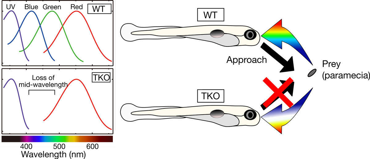
Figure: (Left) Zebrafish deficient for both of the Six7 and Six6 (TKO) lost gene expression of zebrafish SWS2 (blue) and RH2 (green) opsins, indicating that the TKO showed lower middle-wavelength sensitivity. (Right) The TKO showed heavily impaired visually driven foraging behavior and were unable to compete for food when reared in a group with normal siblings (WT).
Time & Date
Saturday, April 6th, from 9:00am to 4:00pm (No Reservations Required, Free Admission)
Access
Free Shuttle Buses Available to the National Institute of Genetics from the North Exit of Mishima Station. (Service Time: 8:50am – 3:00pm)
Map
Map Free Shuttle Buses Available Parking around Mishima Station Available for Car Visitors.
No Pets Allowed except Service Dogs, No General Parking*
(*Disabled Parking Available on the Institute Premises)
Information
1111 Yata, Mishima, Shizuoka 411-8540, JAPAN TEL: +81-55-981-5873
NIG is proud to announce that an associate professor in the Center for Frontier Research has been awarded tenure and promoted to professor as of April 1, 2019.
ODA, Yoshihisa: Cell Dynamics and Signaling Laboratory
Center for Frontier Research is an incubation center to simultaneously develop two elements: human resources and new research fields. Promising young scientists conduct research as principal investigator (tenure-track associate professor) to explore new frontiers in genetics and related areas, taking advantage of NIG’s research infrastructure and various support systems. Those who obtained tenure will establish new research divisions in NIG to lead the new fields that they contribute in creating.
New assistant professor joins NIG as of April 1, 2019.
NEGISHI, Takefumi: Multicellular Organization Laboratory • Sawa Group


