![]()
Establishment of DNA-DNA Interactions by the Cohesin Ring
Yasuto Murayama, Catarina P. Samora, Yumiko Kurokawa, Hiroshi Iwasaki and Frank Uhlmann
CellPublished Online January 18, 2018 DOI:10.1016/j.cell.2017.12.021
Pressrelease (In Japanese only)
DNA, which encodes a blueprint of life, is a very long molecular thread and is packed into a cellular nuclear by forming the protein-DNA supramolecular complex called chromosome. During cell proliferation, chromosomes are doubled up by copying DNA molecules before equal segregation to two daughter cells. To achieve this process smoothly without any mistakes, chromosome contains several special molecular structures. One of such essential chromosomal structures is sister chromatid cohesion. Literally, this is a physical connection which is formed between two duplicated chromosomes. Without cohesion, cell fails to segregate chromosomes properly and this can result in plethora of abnormal events including cell death and cancers. Cohesin is the ring-shaped protein complex and is vital for establishment of sister chromatid cohesion. How cohesin achieves cohesion establishment is one of open questions in chromosome biology.
In the current study, Murayama and colleagues purified the fission yeast cohesin and reconstituted its DNA binding reaction in a test tube. This revealed cohesin topologically entraps DNA inside of its ring. Moreover, cohesin tethers two DNA strands by topological embrace. If DNA-cohesin-DNA tethering is formed between two replicated chromosomes, this can establish chromosome cohesions.
Cohesin is a member of Structural Maintenance of Chromosome (SMC) protein complex family. SMC complexes play vital roles in chromosome organizations. Deficiencies in the complexes have been reported to cause human diseases and malfunctions including developmental disorders, cancers and infertility. The current study provides the first molecular glimpse at how an SMC complex, cohesin, tethers DNA strands.
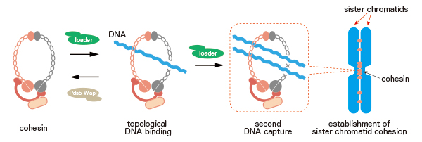
Figure: a model of establishment of sister chromatid cohesion by the cohesin ring.
Cohesin tethers two DNA strands by topological embrace. DNA-cohesin-DNA tethering can provide a molecular bridge to establish cohesion between the replicated sister chromatids.
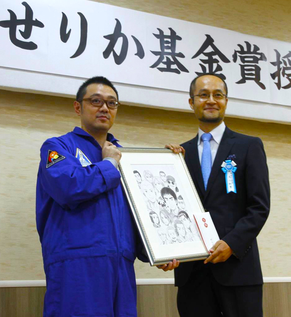
Assistant Professor Kazuhide Asakawa, Division of Molecular and Developmental Biology won the 1st “SERIKA FUND” award established for ALS (Amyotrophic Lateral Sclerosis) research.
This award is given to scientists who are expected for their innovative and developing research.
The fund is named after heroin of Japanese comic “Space Brothers” who lost her father suffering from ALS. In the story, the heroin Serika becomes an astronaut and succeeds gravity- free-space experiments to develop medicines for ALS treatments. It is hoped to make her wish come true in real life.
New assistant professor joins NIG as of January 1, 2018.
Keita MIYOSHI: Invertebrate Genetics Laboratory, Saito Group
Mammalian Genetics Laboratory / Shiroishi Group
Genetic Informatics Laboratory / Kawamoto Group
Commonly used laboratory mouse strains have mosaic genomes, which are derived predominantly from the west European subspecies with the remaining sequences derived from the Japanese subspecies. National Institute of Genetics (NIG) has established a series of experimental mouse strains named “Mishima Battery”. It consists of ten inbred strains originated from four mouse subspecies including the Japanese subspecies, which are widely distributed in the world (Fig. 1). Most of these strains have very remote genetic status from the commonly used laboratory mouse strains such as C57BL/6J (B6), and show very unique phenotypes. Genomic polymorphisms between the laboratory strains and the Japanese subspecies-derived strains are widely used in studies of epigenetics as allele-specific markers. The mouse strains in the “Mishima Battery” are now distributed to the research community via RIKEN BioResource center and NIG, and contribute to broad range of life science. NIG has also established B6-ChrNMSM consomic strains, in which every chromosome of B6 is substituted by corresponding chromosome of Japanese wild mouse-derived MSM/Ms strain, a member of “Mishima Battery”. Using a full panel of the consomic strains, researchers are able to identify chromosomes that control complex traits and diseases. NIG provides these consomic strains to the research community.
Our recent genome-resequencing project of “Mishima Battery” revealed over forty millions of SNPs due to large inter-subspecific genetic distance. Here, we release an upgraded version of the NIG Mouse Genome database named “NIG_MoG2” (http://molossinus.lab.nig.ac.jp/mog2/). NIG_MoG2 primarily comprises the whole genome sequence data of the “Mishima Battery” (Fig. 2), providing visualized genome polymorphism information with single-nucleotide polymorphisms and short insertions/deletions in the genomes of the “Mishima Battery”. It allows users, especially for wet-lab biologists, to intuitively recognize inter-subspecific genome divergence on the browser. NIG_MoG2 is thus a valuable navigator for exploring mouse genome polymorphisms.
This project was supported by NIG Advanced Genomics Project (Next Generation Genome-Research-Hub Project), “Genome Information Upgrading Program” of National BioResource Project (NBRP) from Japan Agency for Medical Research and Development (AMED). The project was performed as a mission of the Genetic Resource Center of NIG.
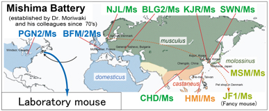
Fig. 1. Geographic distribution of mouse subspecificies, and origins of the “Mishima Battery”. Names of the strains are indicated in colored and bold prints.
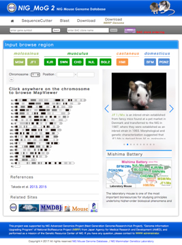
Fig. 2. Top page of the NIG_MoG2 (http://molossinus.lab.nig.ac.jp/mog2).
![]()
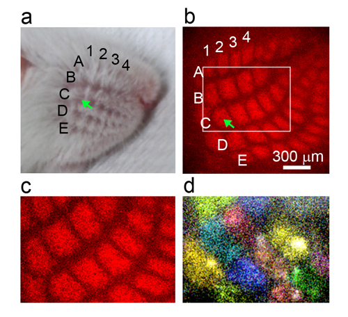
Patchwork-type spontaneous activity in neonatal barrel cortex layer 4 transmitted via thalamocortical projections
Hidenobu Mizuno, Koji Ikezoe, Shingo Nakazawa, Takuya Sato, Kazuo Kitamura, Takuji Iwasato
Cell Reports Volume 22, Issue 1, p123–135, 2 January 2018 DOI:10.1016/j.celrep.2017.12.012
Pressrelease(In Japanese only)
Establishment of precise neuronal connectivity in the mammalian neocortex relies on activity-dependent circuit reorganization during postnatal development; however, the nature of cortical activity during this period remains largely unknown.
Using two-photon calcium imaging of the mouse somatosensory cortex (barrel cortex) in vivo during the first postnatal week, we revealed that layer 4 (L4) cortical neurons within the same barrel fire synchronously in the absence of peripheral stimulation, creating a “patchwork” pattern of spontaneous activity corresponding to the barrel map. By generating transgenic mice expressing a genetically-coded calcium indicator GCaMP6s in thalamocortical axons, we showed that thalamocortical axons also demonstrated the spontaneous patchwork activity pattern. Patchwork activity was diminished by peripheral anesthesia but was mostly independent of self-generated whisker movements. The patchwork activity pattern largely disappeared during postnatal week 2, as even L4 neurons within the same barrel tended to fire asynchronously. This spontaneous L4 activity pattern has features suitable for circuit refinement in the neonatal barrel cortex.
This work was supported by JSPS KAKENHI grant numbers JP15K14322 and JP16H06143, the Takeda Science Foundation, and the Collaborative Research Project (2017-2923) of Brain Research Institute, Niigata University to H.M., and JSPS KAKENHI grant numbers JP16K14559, JP15H01454, and JP15H04263, and Grant-in Scientific Research on Innovation Areas “Dynamic Regulation of Brain Function by Scrap & Build System” (JP16H06459) from MEXT to T.I.