


Press release
Telomere Visualization in Tissue Sections using Pyrrole–Imidazole Polyamide Probes
Asuka Sasaki, Satoru Ide, Yusuke Kawamoto, Toshikazu Bando, Yukinori Murata, Mari Shimura, Kazuhiko Yamada, Akiyoshi Hirata, Kiyoshi Nokihara, Tatsumi Hirata, Hiroshi Sugiyama, Kazuhiro Maeshima
Scientific Reports 6: 29261 (2016) DOI:10.1038/srep29261
Press Release (Only in Japanese)
Pyrrole–Imidazole (PI) polyamides bind to specific DNA sequences in the minor groove with high affinity. Specific DNA labeling by PI polyamides does not require DNA denaturation with harsh treatments of heat and formamide and has the advantages of rapid and less disruptive processing. Previously, we developed tandem hairpin PI polyamide probes (TH59 series), which label telomeres in cultured cell lines more efficiently than conventional methods, such as fluorescence in situ hybridization (FISH). Here, we demonstrate that a TH59 derivative, HPTH59-b, along with immunostaining for specifying cell types in the tissues, visualizes telomeres in mouse and human tissue sections. Quantitative measurements of telomere length with single-cell resolution suggested shorter telomeres in the proliferating cell fractions of tumor than in non-tumor tissues. Thus, PI polyamides are a promising alternative for telomere labeling in clinical research, as well as in cell biology.
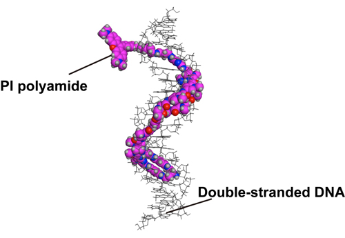
Figure1. A structural model of HPTH59-b binding to DNA.
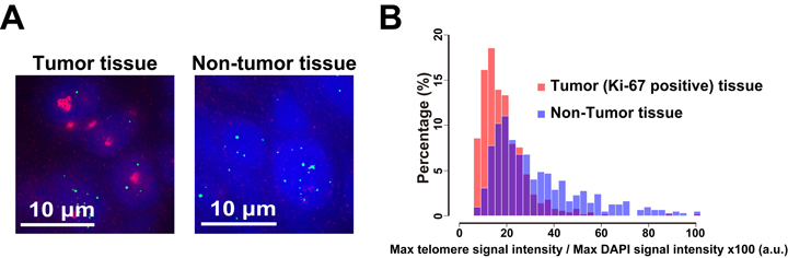
Figure2. Different telomere lengths between human tumor and non-tumor tissue sections. (A) Frozen sections of esophageal tumor/non-tumor tissue stained with DAPI (blue), anti-Ki-67 (growth marker; green) antibody, and HPTH59-b (red). Distribution histograms of telomere signal intensities in tumor and non-tumor tissue sections.


