


Division of Agricultural Genetics • Kakutani Group
Transposable elements (TEs) have a major impact on genome evolution, but they are potentially deleterious, and most of them are silenced by epigenetic mechanisms such as DNA methylation. Here we report the characterization of a TE encoding an activity to counteract epigenetic silencing by the host. In Arabidopsis thaliana, we identified a mobile copy of the Mutator-like element (MULE) with degenerated terminal inverted repeats (TIRs). This TE, named Hiun (Hi), is silent in wild type plants, but it transposes when DNA methylation is abolished. When a Hi transgene was introduced into the wild type background, it induced excision of the endogenous Hi copy, suggesting that Hi is the autonomously mobile copy. In addition, the transgene induced loss of DNA methylation and transcriptional activation of the endogenous Hi. Most importantly, the trans-activation of Hi depends on a Hi-encoded protein different from the conserved transposase. Proteins related to this anti-silencing factor, which we named VANC, are widespread in the non-TIR MULEs and may have contributed to the recent success of these TEs in natural Arabidopsis populations.
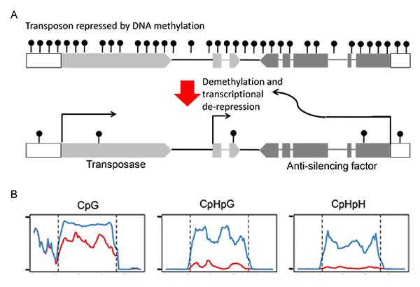
(A) Structure of the plant transposon Hiun (Hi). This transposon encodes for the transposase and the anti-silencing factor.
The antisilencing factor induces demethylation and transcriptional de-repression.
(B) DNA methylation of endogenous Hi compared between non-transgenic plant (blue) and transgenic plant expressing Hi transgene (red).
The transgene induced demethylaton of endogenous copy, especially at non-CpG sites.
Division of Molecular and Developmental Biology • Kawakami Group
Correct organ size must involve a balance between promotion and inhibition of cell proliferation. A mathematical model has been proposed, where an organ is assumed to produce its own growth activator, as well as a growth inhibitor. But, there is yet no molecular evidence to support this model. The mechanosensory organs of the fish lateral line system (neuromasts) are composed of a core of sensory hair cells surrounded by non-sensory support cells. Sensory cells are constantly replaced, and are regenerated by surrounding non-sensory cell, while each organ retains the same size throughout life. Moreover, neuromasts also bud off new neuromasts, which stop growing when they reach the same size. In this study, we show that the size of neuromasts is controlled by a balance between growth-promoting Wnt signaling activity in proliferation-competent cells, and Wnt-inhibiting Dkk activity produced by differentiated sensory cells. This negative feedback loop from Dkk (secreted by differentiated cells) on Wnt-dependent cell proliferation (in surrounding cells) also acts during regeneration to achieve size constancy. This study establishes Wnt/Dkk as a novel mechanism to determine the final size of an organ.
This study was funded by the PRESTO of the Japan Science and Technology Agency.

A) Schematic drawing of a neuromast.
(B) Schematic representation of Wnt signaling and of its inhibition by Dkk signaling.
(C) Wnt reporter activity (green) gradually subsides as hair cells (red) are formed.
(D) Dkk2 expression coincides with neuromast maturation.
Mouse Genomics Resource Laboratory (MGRL) • Koide Group
Copy number variation (CNV) of genomic segments is a common phenomenon that affects more than 10% of the human and mouse genomes, and is an important source of diversity in genomic structure. CNVs are frequently found in clusters called CNV regions (CNVRs), which are strongly associated with large segmental duplications (large SDs). However, the composition of these complex repetitive structures remains unclear. In the present study, we established new method for analyzing on complex repetitive structures of CNVRs by collaborating with National Institute of Informatics.
At first, we conducted self-comparative-plot analysis of all mouse chromosomes using the high-speed and large-scale-homology search algorithm, Similarity/Homology Efficient Analyze Procedure (SHEAP) developed by Dr. Takeaki Uno in National Institute of Informatics. By using this method, large SDs were visualized as unique tartan-checked patterns with complex arrangement of diagonal split lines (Figure 1). We focused on one SD on chromosome 13 (SD13M), which is one of the causative regions for genetic incompatibility (papers in preparation), and applied blast-based Systematic analysis of HErPlot to Extract Regional Distinction (SHEPHERD), a stepwise ab initio method, to extract core elements of repetitive sequences in SD13M (Figure 2). Then, comparative genome hybridization array analysis was empirically conducted on MSM, BLG2 (strains derived from wild mice in Mishima in Japan, and Toshevo in Bulgaria, respectively), and C57BL/6J (an experimental strain). This analysis showed that core elements are categorized to ones with CNVs and the others with constant copy number among strains, which have distinctive characters and divergences. The present study seems to be helpful for elucidating evolutional processes and functions of the CNVRs (Fig. 3).
This study was funded by the “Cultivation of integrated project” of Transdisciplinary Research Integration Center in Research Organization of Informatio and Systems (Japan).
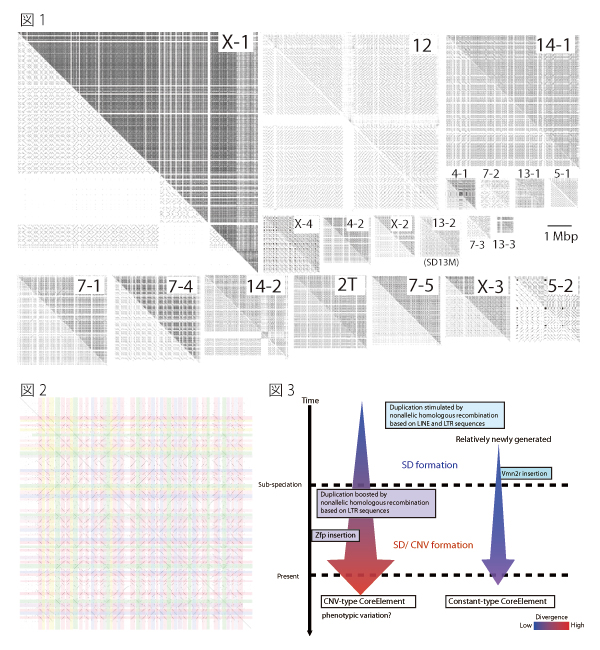
Fig.1. Tartan-checked structure of SDs visualized using SHEAP.
The lower left triangle of each panel shows a self-plot of the sequence after known repeat sequences have been masked using RepeatMasker. Each of the upper right triangles shows a self-plot of the intact sequence. Diagonal lines aligned in the same column or row represent repetitive sequences.
Fig.2. Extraction of repeat units from the self-plot of the large SD
Diagonal lines were extracted from a self-comparative-plot of SD13M that consisted of a dot-plot matrix. Then we selected one sequence and removed the other sequences represented by diagonal lines that were located in the same column or in the same row.
Fig.3 Model for the formation of SDs and CNVs
The average divergence of CNV-type core elements was greater than that of the constant type, and the CNV-type core elements contained significantly larger proportions of long terminal repeat (LTR) types of retrotransposon than the constant-type core elements. These results suggest that constant-type core elements emerged more recently than CNV-type core elements, and that retrotransposition of LTRs promotes nonallelic homologous recombination and caused CNV in SD13M.
Division of Microbial Genetics • Araki Group
Chromosomal replication origins, where DNA replication is initiated, are determined in eukaryotic cells by specific binding of a six-subunit origin recognition complex (ORC). Many biochemical analyses have revealed the detailed properties of the ORC–DNA interaction. However, because of the lack of in vitro analysis, the molecular architecture of the ORC–chromatin interaction is unclear. Recently, mainly from in vivo analyses, a role of chromatin in the ORC–origin interaction has been reported, including the existence of a specific pattern of nucleosome positioning around origins and of a specific interaction between chromatin—or core histones—and Orc1, a subunit of ORC. Therefore, to understand how ORC establishes its interaction with origin in vivo, it is essential to know the molecular mechanisms of the ORC–chromatin interaction. Here, we show that ORC purified from yeast binds more stably to origin-containing reconstituted chromatin than to naked DNA, and forms a nucleosome-free region at origins. Molecular imaging using atomic force microscopy (AFM) reveals that ORC associates with the adjacent nucleosomes and forms a larger complex. Moreover, stable binding of ORC to chromatin requires linker DNA. Thus, ORC establishes its interaction with origin by binding to both nucleosome-free origin DNA and neighboring nucleosomes.
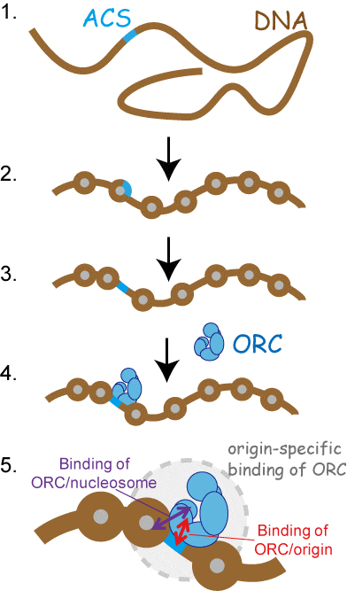
(1) DNA fragment containing origin-specific DNA motif (ACS).
(2) By chromatin reconstitution, nucleosome is formed on ACS.
(3) By the addition of ORC, ACS becomes nucleosome-free-region.
(4) ORC interacted to nucleosome-free ACS.
(5) ORC establishes its interaction with origin by binding to both nucleosome-free ACS and neighboring nucleosomes.
Microbial Genetics Laboratory • Niki Group
Three types of mitosis, which are open, closed, or semi-open mitosis, function in eukaryotic cells, respectively. The open mitosis involves breakage of the nuclear envelope before nuclear division, whereas the closed mitosis proceeds with an intact nuclear envelope. To understand the mechanism and significance of three types of mitotic division in eukaryotes, we investigated the process of semi-open mitosis, in which the nuclear envelope is only partially broken, in the fission yeast Schizosaccharomyces japonicus. In anaphase-promoting complex/cyclosome (APC/C) mutants of Sz. japonicus, the nuclear envelope remained relatively intact during anaphase, resulting in impaired semi-open mitosis. As a suppressor of apc2 mutant, a mutation of Oar2 which was a 3-oxoacyl-[acyl-carrier-protein] reductase was obtained. The level of the Oar2, which had two destruction-box motifs recognized by APC/C, was increased in APC/C mutants. Furthermore, the defective semi-open mitosis observed in an apc2 mutant was restored by mutated oar2+. Based on these findings, we propose that APC/C regulates the dynamics of the nuclear envelope through degradation of Oar2 dependent on APC/C during the metaphase-to-anaphase transition of semi-open mitosis in Sz. japonicus.
This study has been carried out as collaboration with Dr. Ogura at Tohoku University.

Observations of nuclear envelope dynamics in WT, apc2 mutant, and apc2∆oar2 mutant. The semi-open mitosis appeared in WT was defective in apc2 mutant, but partially restored in apc2∆oar2 mutant. Green: nuclear envelope marker (Cut11-GFP), magenta: chromosomal marker (H2A-mCherry). Scale bar: 5µm.


