


Miyagishima Group • Symbiosis and Cell Evolution Laboratory
IPTG- and estradiol-inducible gene expression systems in the unicellular red alga Cyanidioschyzon merolae.
Takayuki Fujiwara, Shunsuke Hirooka, Shota Yamashita, and Shin-ya Miyagishima.
Plant Physiology(2025) DOI:10.1093/plphys/kiaf575
The genetically tractable unicellular red alga Cyanidioschyzon merolae has a remarkably simple genome (4,775 nucleus-encoded proteins) and cellular architecture. It contains only a single set of most membranous organelles, making it a valuable tool for elucidating the fundamental mechanisms of photosynthetic eukaryotes. However, as in other genetically tractable eukaryotic algae, previously developed systems for inducible gene expression rely on environmental stimuli such as heat shock or ammonium depletion, which impact cellular physiology and thus limit their usage. To overcome this issue, we developed IPTG- and estradiol-inducible gene expression systems in C. merolae in which the addition of these chemicals itself has no impact on cellular growth or the transcriptome. Additionally, we established IPTG- and estradiol-inducible protein knockdown systems and successfully degraded the endogenous chloroplast division protein DRP5B using the estradiol-inducible system. These systems facilitate functional genomic analyses in C. merolae, especially for understanding physiological mechanisms and their interactions in photosynthetic eukaryotes.
IPTG-inducible gene expression system.
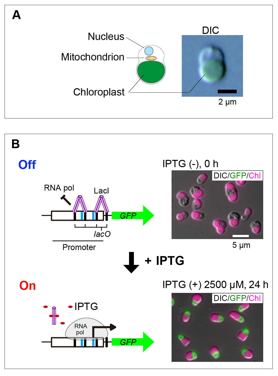
(A) Schematic illustration of the cellular structure and differential interference contrast (DIC) image of Cyanidioschyzon merolae, a species of microalgae.
(B) Schematic overview of the IPTG-inducible gene expression system and fluorescence microscopy images of cells expressing the reporter protein GFP (green) in an IPTG-dependent manner. Four lac operator (lacO) sequences were inserted into the promoter region, and an Escherichia coli–derived LacI repressor was expressed separately to suppress transcription by binding to these sites. Upon addition of IPTG, LacI dissociates from the lacO sequences, allowing transcription to initiate. GFP protein expression was detected from 4 hours after IPTG addition. DIC images show cell outlines, GFP (green) indicates cytosolic fluorescence, and Chl (magenta) represents chloroplast autofluorescence. Scale bars are indicated in the images.


