


Maeshima Group / Genome Dynamics Laboratory
The shifting paradigm of chromatin structure: from the 30-nm chromatin fiber to liquid-like organization
Kazuhiro Maeshima
Proceedings of the Japan Academy, Series B (2025) DOI:10.2183/pjab.101.020
The organization and dynamics of chromatin are critical for genome functions such as transcription and DNA replication/repair. Historically, chromatin was assumed to fold into the 30-nm fiber and progressively arrange into larger helical structures, as described in the textbook model. However, over the past 15 years, extensive evidence, including studies by Prof. Kazuhiro Maeshima at National Institute of Genetics, has dramatically transformed the view of chromatin from a static, regular structure to one that is more variable and dynamic. In higher eukaryotic cells, chromatin forms condensed yet liquid-like domains, which appear to be the basic unit of chromatin structure, replacing the 30-nm fiber. These domains maintain proper accessibility, ensuring the regulation of DNA reaction processes. Based on the available evidence, Prof. Maeshima discusses the physical properties of chromatin in live cells, emphasizing its viscoelastic nature—balancing local fluidity with global stability to support genome functions.
This work was supported by the Japan Society for the Promotion of Science (JSPS) and MEXT KAKENHI grants (JP23K17398, JP22H04925 (PAGS), and JP24H00061) and the Takeda Science Foundation.
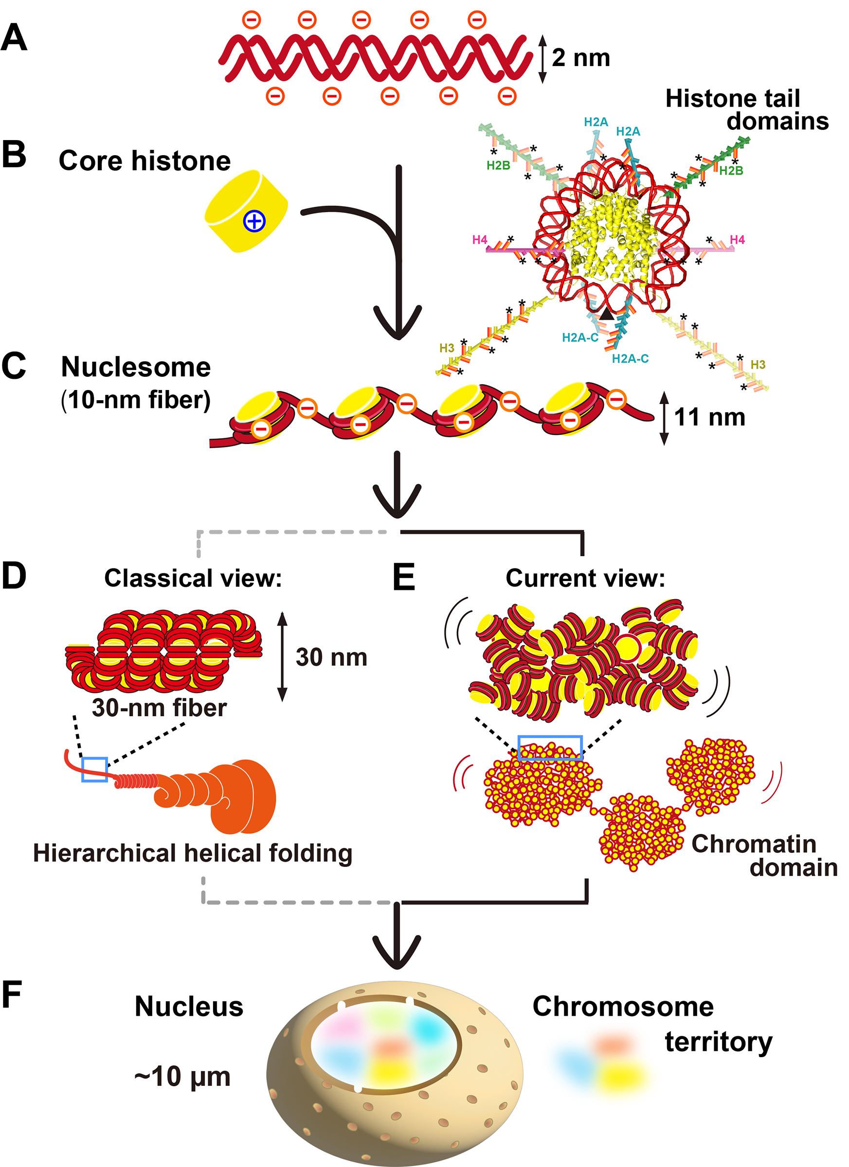
Figure: DNA, histones, nucleosomes, and chromatin.
(A) Negatively charged DNA (red). (B, left) The positively charged core histone octamers (yellow). (B, right) Nucleosome structure at 1.9 Å resolution;10) totally 10 histone tail domains (intrinsically disordered regions [IDRs]) are extended away from the nucleosome core. The dyad position was indicated with an arrowhead. Basic residues (lysines and arginines) in the tail domains are colored in orange. Asterisks indicate sites of lysine acetylation. (C) 10-nm fiber. Note that the fiber remains negatively charged. (D) A classical view of 30-nm chromatin fibers folded into a hierarchical structure. (E) A current view of irregularly folded 10-nm fibers in the chromatin domain. (F) The nucleus has chromosomes in different colors based on their territories. This figure was reproduced from Ide et al. (BioEssays 2022) with modifications.


