


Guideline for Additional Application for
2024 NIG-JOINT(Joint Researchi-(A))
(Application deadline: noon(12:00pm)
on Friday, May 31st, 2024)
Guideline for Additional Application for
2024 NIG-JOINT(Joint Researchi-(A))
(Application deadline: noon(12:00pm)
on Friday, May 31st, 2024)
Guideline for Additional Application for
2024 NIG-JOINT(Joint Researchi-(A))
(Application deadline: noon(12:00pm)
on Friday, May 31st, 2024)
Kanemaki Group / Molecular Cell Engineering Laboratory
Ran-GTP assembles a specialized spindle structure for accurate chromosome segregation in medaka early embryos
Ai Kiyomitsu, Toshiya Nishimura, Shiang Jyi Hwang, Satoshi Ansai, Masato T. Kanemaki, Minoru Tanaka & Tomomi Kiyomitsu
Nature Communications (2024) 15, 981 DOI:10.1038/s41467-024-45251
With the help of medaka fish, CRISPR and new imaging techniques, researchers have set a new standard for studying cell division at the very earliest stages of life.
The beginning of life is shrouded in mystery. While the intricate dynamics of mitosis is well-studied in the so-called somatic cells – the cells that have a specialized function, like skin and muscle cells – they remain elusive in the first cells of our bodies, the embryonic cells. Embryonic mitosis is notoriously difficult to study in vertebrates, as live functional analyses and -imaging of experimental embryos are technically limited, which makes it hard to track cells during embryogenesis.
However, researchers from the Cell Division Dynamics Unit at the Okinawa Institute of Science and Technology (OIST) have recently published a paper in Nature Communications, together with Professors Toshiya Nishimura from Hokkaido University (previously at Nagoya University), Minoru Tanaka from Nagoya University, Satoshi Ansai from Tohoku University (currently at Kyoto University), and Masato T. Kanemaki from the National Institute of Genetics. The study takes the first major steps towards answering questions about embryonic mitosis, thanks to a combination of novel imaging techniques, CRISPR/Cas9 genome editing technology, a modern protein-knockdown system, and medaka, or Japanese rice fish (Oryzias latipes). The timelapses that they have produced help answer fundamental questions about the intricate process of equally dividing chromosomes during embryonic mitosis, and simultaneously chart the next frontier of scientific exploration. As Professor Tomomi Kiyomitsu, senior author of the study, describes the timelapses: “they are beautiful, both on their own and because they lay a new foundation for elucidating embryonic mitosis.”
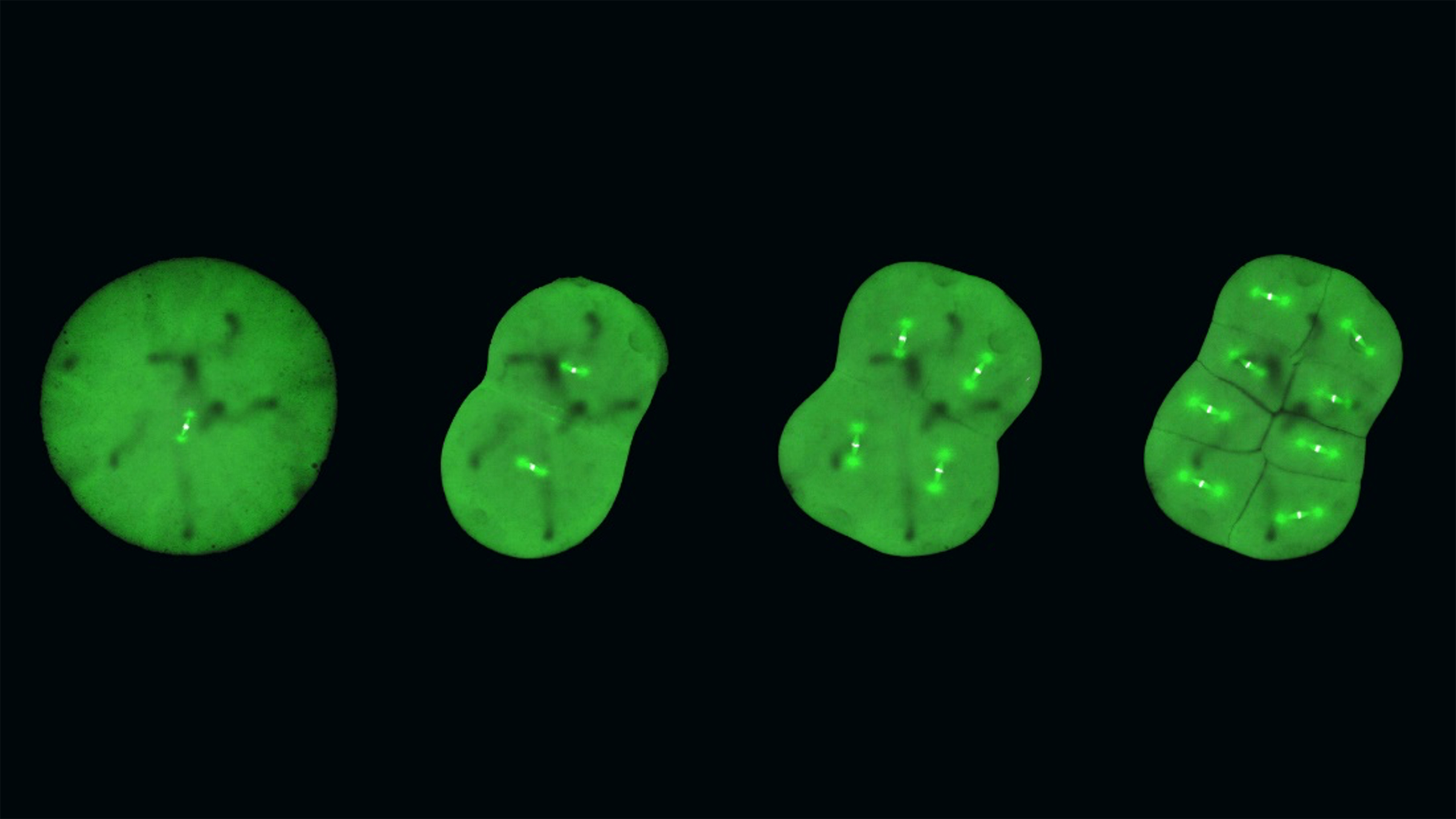
Central to the mystery of embryonic mitosis is the crucial step when the chromosomes, which contain all the genetic information of the cell, are aligned and segregated equally into daughter cells. A key player in this process is the mitotic spindle, which is made of microtubules – long protein fibers used for intra-cellular structure and transport – that radiates from opposite poles of the spindle and attaches to the chromosomes in the middle. The spindle captures duplicated chromosomes properly and segregates them equally into the daughter cells during division. There are many factors determining spindle formation, and one of these is the protein Ran-GTP, which plays an essential role in cell division of female reproductive cells, which lack centrosomes – cell organelles responsible for assembling microtubules – but not in small somatic cells, which do have centrosomes. However, it has long been unclear whether Ran-GTP is required for spindle assembly in vertebrate early embryos, which contain centrosomes but have unique features, like a larger cell size.
In contrast to mammalian early embryos, embryonic cells in fish are transparent and develop synchronously in a uniform, single-cell layer sheet, which makes them significantly easier to track. The medaka turned out to be particularly well-suited for the researchers, as these fish tolerate a wide range of temperatures, produce eggs daily, and have a relatively small genome. Being temperature-tolerant means that the medaka embryonic cells could survive at room temperature , making them particularly suited for long, live timelapse photography.
The fact that medaka produce eggs frequently and have a relatively small genome size makes them good candidates for CRISPR/Cas9-mediated genome editing. With this technology, the researchers have created genetically modified, or transgenic, medaka whose embryonic cells literally highlight the dynamics of certain proteins involved in mitosis.
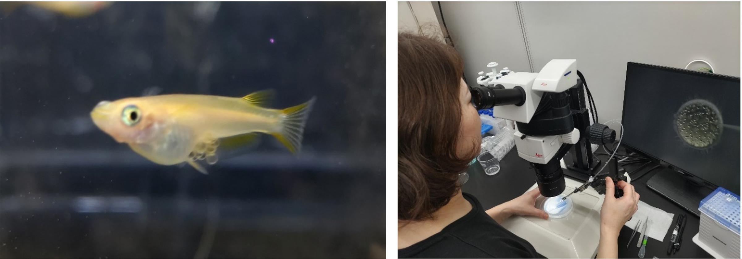
Figure: Left: Egg-carrying medaka fish, used in the study. Right: Dr. Ai Kiyomitsu from the Cell Division Dynamics Unit, first author of the paper, injecting RNA into a medaka embryo as part of the CRISPR/Cas9-mediated genome editing process. Photos: Tomomi Kiyomitsu (OIST).
In studying the timelapses of the developing mitotic spindle in live, transgenic medaka embryos, the researchers discovered that large early embryos assemble unique spindles different from somatic spindles. In addition, Ran-GTP plays a decisive role in spindle formation in early embryonic divisions, but the importance diminishes in later stage embryos. This is possibly because the spindle structure is remodeled as cells get smaller during development, though the exact reason is a subject for future research.
The researchers also discovered that the early embryonic cells do not have a dedicated spindle assembly checkpoint, which characterizes most somatic cells, and which serves to ensure that the chromosomes are properly aligned before segregation. As Professor Kiyomitsu surmises, “the checkpoint is not active, and yet the chromosome segregations are still very accurate. This could be explained by the fact that embryonic cells need to divide very quickly, but it is something that we want to study further.”
While genetically modifying the medaka fish and studying the early embryos have led to new key insights into embryonic mitosis, this is just the beginning for Professor Kiyomitsu and the team. In addition to questions related to the diminishing role of Ran-GTP in later stages and the missing spindle assembly checkpoint, he points to the satisfying symmetry of cell divisions in the timelapses: “The spindle formation is characterized by a high degree of symmetry, as the cells appear to be dividing in the sizes and defined directions, and the spindle is consistently in the center of the cells. How can the spindle orient itself so regularly across the cells, and how is it able to find the center every time?”
Moving beyond the timelapses, the team also hopes to further solidify this new foundation with additional medaka gene-lines to serve as models for research in embryonic cells, and at the same time optimize the genome editing process. Eventually, the team wants to test for generalizability of their findings by studying embryonic mitosis in other organisms, and at a later stage, they want to explore the evolution of spindle assembly and embryonic divisions, which would also contribute to a better understanding of human embryogenesis and to developing diagnosis and treatment of human infertility.
“With this paper, we have created a solid foundation,” summarizes Professor Kiyomitsu, “but we have also opened a new frontier. Embryonic mitosis is beautiful, mysterious, and challenging to study, and we hope that with our work, we can eventually get a little closer to understanding the intricate processes at the beginning of life.”
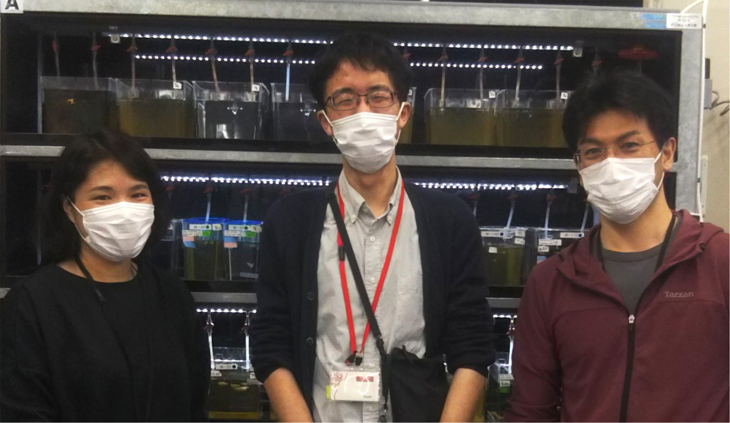
Three of the researchers involved in the study, in front of fish-tanks of the transgenic medaka fish used in the study. From left to right: Dr. Ai Kiyomitsu from OIST, Professor Satoshi Ansai from Kyoto University (previously Tohoku University), and Professor Tomomi Kiyomitsu from OIST. Photo: Tomomi Kiyomitsu.
KanemakiGroup / Molecular Cell Engineering Laboratory
Non-canonical functions of UHRF1 maintain DNA methylation homeostasis in cancer cells
Kosuke Yamaguchi, Xiaoying Chen, Brianna Rodgers, Fumihito Miura, Pavel Bashtrykov, Frédéric Bonhomme, Catalina Salinas-Luypaert, Deis Haxholli, Nicole Gutekunst, Bihter Özdemir Aygenli, Laure Ferry, Olivier Kirsh, Marthe Laisné, Andrea Scelfo, Enes Ugur, Paola B. Arimondo, Heinrich Leonhardt, Masato T. Kanemaki, Till Bartke, Daniele Fachinetti, Albert Jeltsch, Takashi Ito & Pierre-Antoine Defossez
Nature Communications (2024) 15, 2960 DOI:10.1038/s41467-024-47314-4
Replication of the genetic material DNA is essential for cell proliferation. During this process, not only DNA but also DNA methylation, which is essential epigenetic mark, is maintained from parent to daughter cells. It has been known that DNA methyltransferase 1 (DNMT1) and its activator, UHRF1, are important to maintain DNA methylation. Although it has been recognized primarily as an activator of DNMT1, UHRF1 is also implicated in cancer development, unlike DNMT1, suggesting that it may have functions through other pathways, independent of DNMT1.
To delineate the individual functions of UHRF1 and DNMT1, we utilized the auxin-inducible degron (AID)(In Japanese only) and AID2 system that can induce the total and synchronous depletion of endogenous UHRF1 or DNMT1 proteins in human cancer cells. Interestingly, UHRF1 depletion resulted in a more severe DNA methylation loss than DNMT1 removal. With the combination of whole genome DNA methylation analysis and genetic knock-out techniques, we revealed that UHRF1 regulated not only DNMT1 but also DNA methyltransferases DNMT3A/DNMT3B and demethylase TET2.
Assistant Prof. Kosuke Yamaguchi (formally a postdoctoral researcher at Paris-Cite University) led the study as the first and co-corresponding author in the Pierre-Antoine Defossez laboratory, and Prof. Masato Kanemaki was also involved in this study.
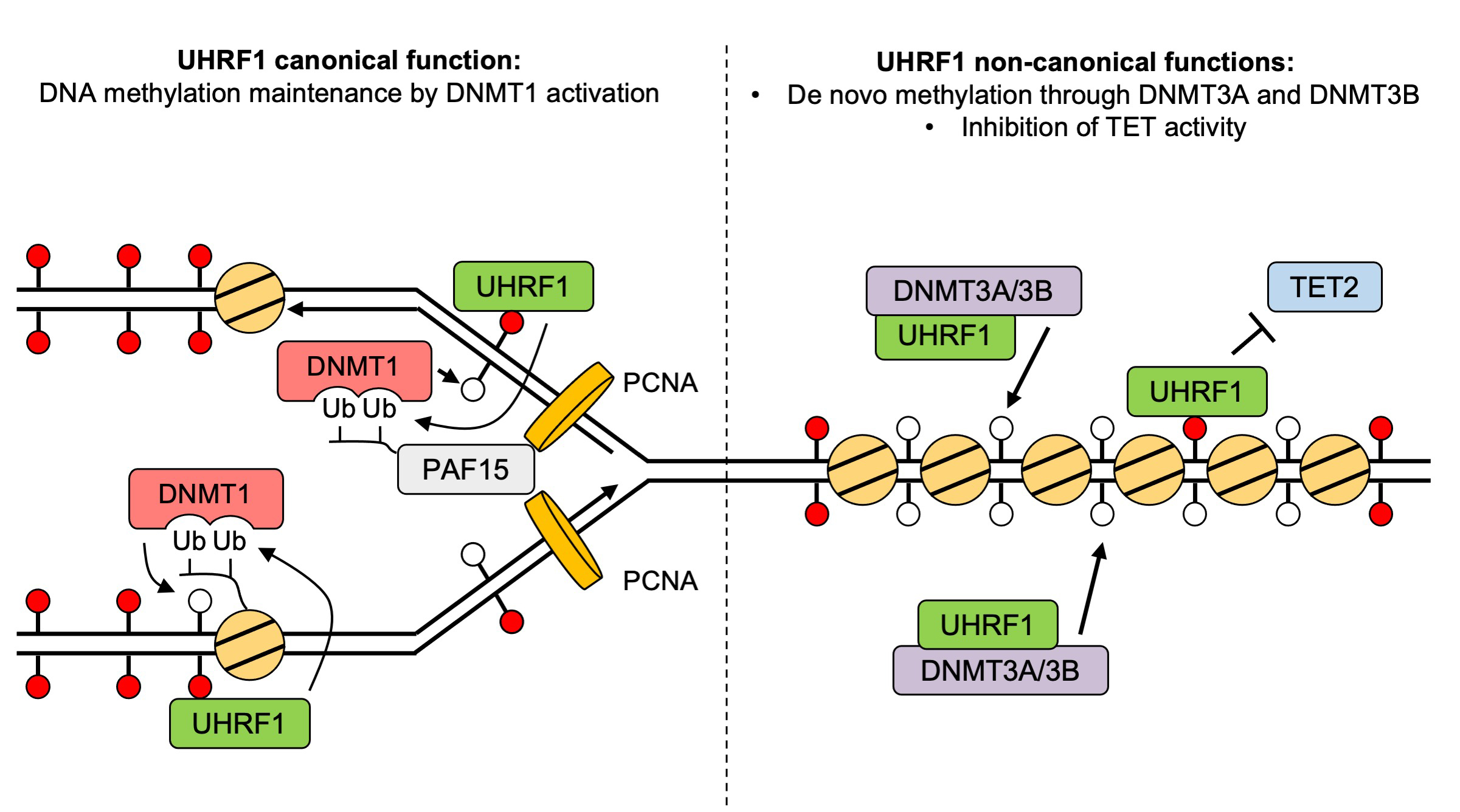
Figure: A revised and expanded model for UHRF1 functions in DNA methylation homeostasis.
Press release
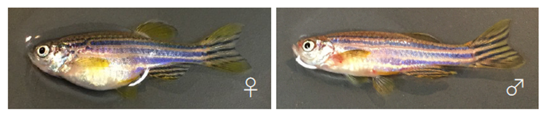
Establishment of a zebrafish inbred strain, M-AB, capable of regular breeding and genetic manipulation
Kenichiro Sadamitsu, Fabien Velilla, Minori Shinya, Makoto Kashima, Yukiko Imai, Toshihiro Kawasaki, Kenta Watai, Miho Hosaka, Hiromi Hirata and Noriyoshi Sakai.
Scientific Reports (2024) 14, 7455 DOI:10.1038/s41598-024-57699-
![]() Press release (In Japanese only)
Press release (In Japanese only)
Inbred strains of organisms are genetically highly uniform and thus useful for life science research. We have previously reported the ongoing generation of the zebrafish IM strain from the India (IND) strain through full sib-pair mating for 16 generations. However, the IM fish laid a small number of offspring and had a short lifespan, implying the need for discreet care in breeding. Here, we report the subsequent establishment of IM strain as well as the generation of a new inbred zebrafish strain, Mishima-AB (M-AB). M-AB was derived from the *AB strain by full sib-pair mating for over 20 generations, which fulfills the general criterion for the establishment of an inbred strain. In contrast to the IM case, maintenance of the M-AB strain by sib-pair mating required almost no special handling. Genome sequencing of IM individuals from the 47th generation and M-AB individuals from the 27th generation revealed that SNP-based genomic heterogeneity across whole-genome nucleotides was 0.008% and 0.011%, respectively. These percentages were much lower than those of the parental IND (0.197%) and *AB (0.086%) strains. These results indicate that the genomes of these inbred strains were highly homogenous. We also demonstrated the successful microinjection of antisense morpholinos, CRISPR/Cas9, and foreign genes into M-AB embryos at the 1-cell stage. Overall, we report the establishment of a zebrafish inbred strain, M-AB, which is capable of regular breeding and genetic manipulation. This strain will be useful for the analysis of genetically susceptible phenotypes such as behaviors, microbiome features and drug susceptibility.
Source: Kenichiro Sadamitsu et al., Scientific Reports (2024) 14, 7455
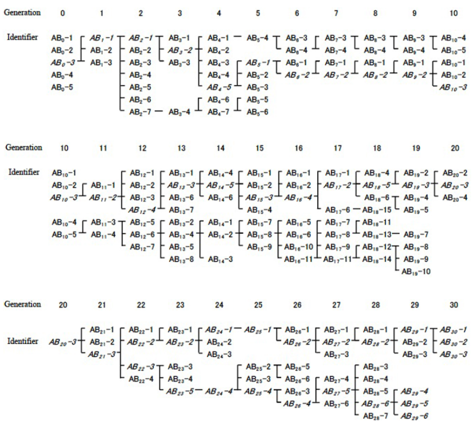
Figure: Crossbreeding of inbred M-AB strain
Dr. Kenji FUKUSHIMA joined the Center for Frontier Science as of April 1, 2024.
▶ FUKUSHIMA, Kenji : Center for Frontier Research Plant Evolution Laboratory
Center for Frontier Research is an incubation center to simultaneously develop two elements: human resources and new research fields. Promising young scientists conduct research as principal investigator (tenure-track associate professor) to explore new frontiers in genetics and related areas, taking advantage of NIG’s research infrastructure and various support systems.
Dr. Kazuhide ASAKAWA joined NIG as a Associate Professor and opened a new laboratory on April 1, 2024.
▶ ASAKAWA, Kazuhide : Neurobiology and Pathology Laboratory

ASAKAWA, Kazuhide
Associate Professor
Dr. Yasuhiro GOTO joined the Advanced Genomics Center of NIG as a Associate Professor on April 1, 2024.
▶ GOTO, Yasuhiro : Advanced Genomics Center Sequencing Division
GOTO, Yasuhiro
Associate Professor
New Assistant Professor joins NIG as of April 1, 2024.
▶ YAMAGUCHI, Kosuke : Kanemaki Group, Molecular Cell Engineering Laboratory


