


Press release
NeuroGT: A brain atlas of neurogenic tagging CreER drivers for birthdate-based classification and manipulation of mouse neurons
T Hirata*, Y Tohsato, H Itoga, G Shioi, H Kiyonari, S Oka, T Fujimori, S Onami *Corresponding author
Cell Reports Methods 1, 100012 (2021) DOI:10.1016/j.crmeth.2021.100012
Press release (In Japanese only)
Neuronal birthdate is one of the major determinants of neuronal phenotypes. However, most birthdating methods are retrospective in nature, allowing very little experimental access to the classified neuronal subsets. Hirata and her collaborators have developed four neurogenic tagging mouse lines that can assign CreER-loxP recombination to neuron subsets that share the same differentiation timing in living animals. Because this genetic tag is irreversible, the classified neuronal subset can be subsequently subjected to various experimental manipulations.
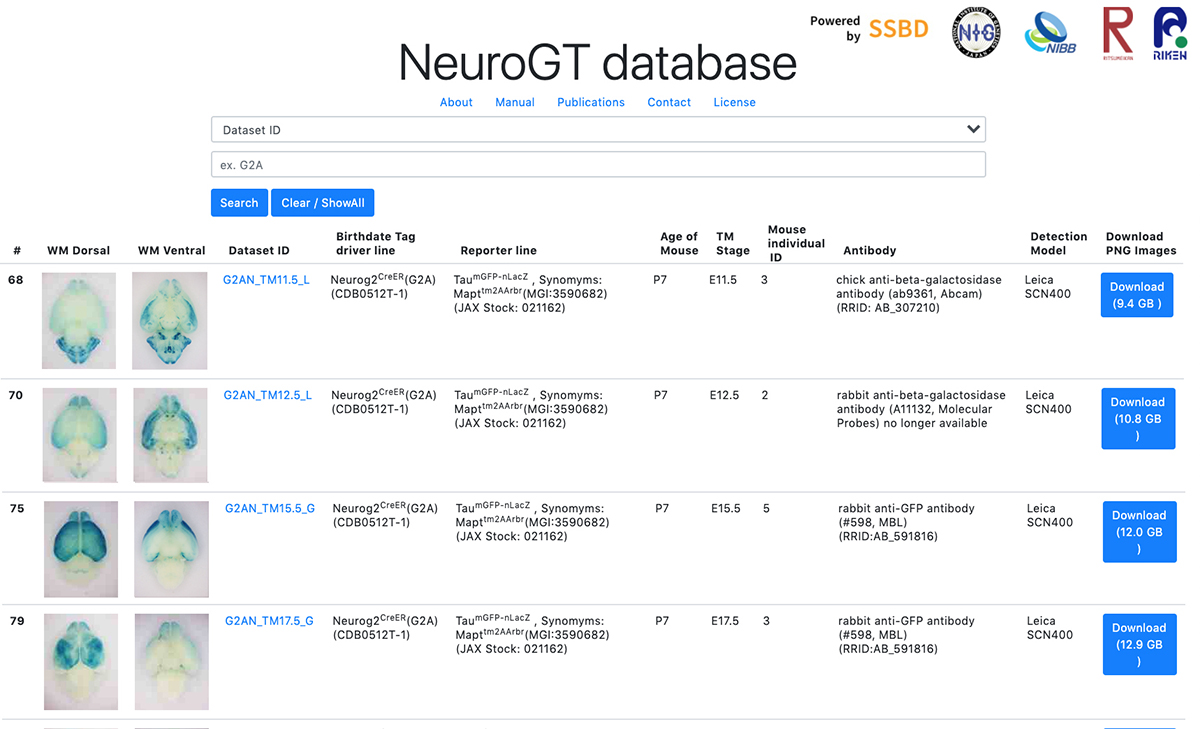
To encourage the use of this resource, Hirata et al. have launched “NeuroGT database”, a brain atlas of neurogenic tagging mouse lines, which includes holistic image data of the loxP-recombined neurons and their processes across the entire brain that were tagged at each single day during the neurodevelopmental period. Users can search for the datasets from a web browser using the terns in the meta-information, interactively view thumbnail images of the sections, and download the high-resolution section images.
The NeuroGT is now open to public, offering people the opportunity to find specific neurogenic tagging driver lines and the stages of tagging appropriate for their own research purposes. The driver mouse lines can be obtained from Riken Bioresource Center.
The NeuroGT database was constructed by joint efforts of different research groups; mouse engineering (RIKEN Center for Biosystems Dynamics Research), neuroscience (National Institute of Genetics), bioimaging (National Institute for Basic Biology), and image informatics (Ritsumeikan University, RIKEN Center for Biosystems Dynamics Research)
This research was supported by ROIS Challenging Exploratory Research Projects for the Future Grant and Grant-in-Aid for Publication of Scientific Research Results (Database, 19HP7002). High resolution digitization of section images was supported by Advanced Bioimaging Support (JP16H06280) and Grants-in-Aid for Scientific Research (20H03345, JP18H05412).
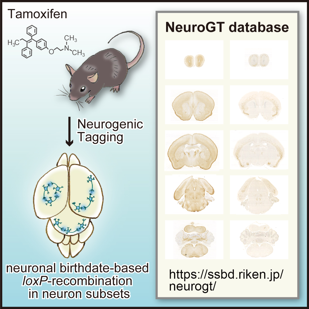
Figure: Graphical abstract of “NeuroGT Database”
Video: Thumbnail images of coronal sections can be sequentially viewed along the antero-posterior brain axis by dragging the slider. The images stained for the two different reporters, membrane-localized GFP and nucleus-localized-βGAL are stacked separately. The sync button automatically matches the section level of the two reporter images.


