


Press release
Genome-wide SNP data of Izumo and Makurazaki populations support inner-dual structure model for origin of Yamato people
Timothy Jinam, Yosuke Kawai, Yoichiro Kamatani, Shunro Sonoda, Kanro Makisumi, Hideya Sameshima, Katsushi Tokunaga, and Naruya Saitou
Journal of Human Genetics 2021 January 25. DOI:10.1038/s10038-020-00898-3
Press release (In Japanese only)
The “Dual Structure” model on the formation of the modern Japanese population assumes that the indigenous hunter- gathering population (symbolized as Jomon people) admixed with rice-farming population (symbolized as Yayoi people) who migrated from the Asian continent after the Yayoi period started. The Jomon component remained high both in Ainu and Okinawa people who mainly reside in northern and southern Japan, respectively, while the Yayoi component is higher in the mainland Japanese (Yamato people). The model has been well supported by genetic data, but the Yamato population was mostly represented by people from Tokyo area. We generated new genome-wide SNP data using Japonica Array for 45 individuals in Izumo City of Shimane Prefecture and for 72 individuals in Makurazaki City of Kagoshima Prefecture in Southern Kyushu, and compared these data with those of other human populations in East Asia, including BioBank Japan data. Using principal component analysis, phylogenetic network, and f4 tests, we found that Izumo, Makurazaki, and Tohoku populations are slightly differentiated from Kanto (including Tokyo), Tokai, and Kinki regions. These results suggest the substructure within Mainland Japanese maybe caused by multiple migration events from the Asian continent following the Jomon period, and we propose a modified version of “Dual Structure” model called the “Inner-Dual Structure” model.
This study was supported by SOKENDAI Cooperative Research grant on modern human evolution, MEXT Grant-in-aid for Scientific Research on Innovative Areas “Yaponesian Genome” (grant number 18H05505), cooperative research of the Inter-University Research Institutions (grant number I- URIC18P01), and a cooperative research with Genesis Healthcare.
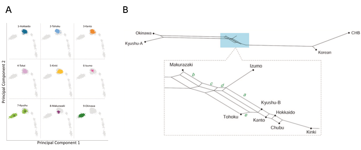
Figure: Genetic relationship of nine Yaponesian populations and some continental populations.
(A) Principal Component Analysis (PCA). DNA data for Izumo (6) and Makurazaki (8) are newly determined, and those for other seven regions are from BioBank Japan. Five continental populations are located at lower right side. Kyushu (7) population is divided into Okinawa cluster A and Yamato cluster B. (B) Phylogenetic network analysis using the Neighbor-Net method. Okinawa and Kyushu-A are located at left and Korean and CHB (Chinese Han in Beijing) are located at right. Makurazaki is the closest to the left cluster followed by Izumo, while Kinki is the closest to the right cluster.
Non-thalamic origin of zebrafish sensory nuclei implies convergent evolution of visual pathways in amniotes and teleosts
Solal Bloch, Hanako Hagio, Manon Thomas, Aure´ lie Heuze´, Jean-Michel Hermel, Elodie Lasserre, Ingrid Colin, Kimiko Saka, Pierre Affaticati, Arnim Jenett, Koichi Kawakami, Naoyuki Yamamoto, Kei Yamamoto
Elife 9, e54945 (2020) DOI:10.7554/eLife.54945
Ascending visual projections similar to the mammalian thalamocortical pathway are found in a wide range of vertebrate species, but their homology is debated. To get better insights into their evolutionary origin, we examined the developmental origin of a thalamic-like sensory structure of teleosts, the preglomerular complex (PG), focusing on the visual projection neurons. Similarly to the tectofugal thalamic nuclei in amniotes, the lateral nucleus of PG receives tectal information and projects to the pallium. However, our cell lineage study in zebrafish reveals that the majority of PG cells are derived from the midbrain, unlike the amniote thalamus. We also demonstrate that the PG projection neurons develop gradually until late juvenile stages. Our data suggest that teleost PG, as a whole, is not homologous to the amniote thalamus. Thus, the thalamocortical-like projections evolved from a non-forebrain cell population, which indicates a surprising degree of variation in the vertebrate sensory systems.
This study was conducted as collaboration with Dr. Kei Yamamoto at Universite´ Paris-Saclay and CNRS and Dr. Naoyuki Yamamoto at Graduate School of Bioagricultural Sciences, Nagoya University. This study was partially supported by NBRP and NBRP/Genome Information upgrading program from AMED and NIG-JOINT (2013-A15).
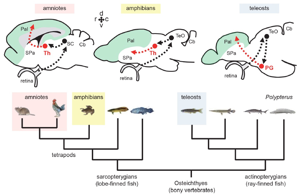
Figure1: Tectofugal pathways in amniotes, amphibians, and teleosts and their phylogenetic relationships.
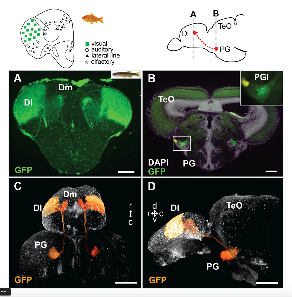
Figure2: Visualization of the preglomerular complex (PG) cells and their projection to the pallium in gene-trap transgenic fish.
1,6-hexanediol rapidly immobilizes and condenses chromatin in living human cells
Yuji Itoh, Shiori Iida, Sachiko Tamura, Ryosuke Nagashima, Kentaro Shiraki, Tatsuhiko Goto, Kayo Hibino, Satoru Ide and Kazuhiro Maeshima
Life Science Alliance 4, e202001005 (2021) DOI:10.26508/lsa.202001005
Liquid droplets formed inside the cell by liquid-liquid phase separation (LLPS) maintain membrane-less condensates/bodies (or compartments). These droplets are important for concentrating certain molecules and facilitating spatiotemporal regulation of cellular functions. 1,6-hexanediol (1,6-HD), an aliphatic alcohol, inhibits weak hydrophobic protein-protein/protein-RNA interactions required for the droplet formation (droplet melting activity) and is used here to elucidate the formation process of cytoplasmic/nuclear condensates/bodies. However, the effect of 1,6-HD on chromatin in living cells remains unclear. We found that 1,6-HD drastically suppresses chromatin motion and hyper-condenses chromatin in human cells by using live cell single-nucleosome imaging, which detects changes in the state of chromatin. These effects were enhanced in a dose-dependent manner. Chromatin was ‘frozen’ by 5%, or higher, concentrations of 1,6-HD. 1,6-HD greatly facilitated cation-dependent chromatin condensation in vitro. This 1,6-HD action is distinct from its melting activity of liquid droplets. Alcohols, such as 1,6-HD, appear to remove water molecules around chromatin and locally condense chromatin. Therefore, liquid droplet results obtained using 1,6-HD should be carefully interpreted or reconsidered when these droplets are associated with chromatin.
This work was supported by JSPS and MEXT KAKENHI grants (19K23735, 20J00572, 18K06187, 19H05273 and 20H05936), a Japan Science and Technology Agency CREST grant (JPMJCR15G2), Takeda Science Foundation, Uehara Memorial Foundation, NIG Postdoctoral Fellowship and JSPS Postdoctoral Fellowship (PD).
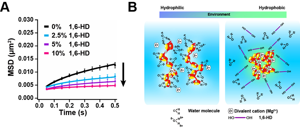
Figure: Chromatin immobilization by 1,6-HD and scheme for chromatin condensation.
(A) Mean square displacement plots (±SD among cells) of nucleosome motion in HeLa cells treated with 2.5% (light blue), 5% (purple), or 10% (pink) of 1,6-HD for 5–30 min. (B)(Left) Chromatin is associated with many water molecules with electrostatic interactions. (Right) Alcohols such as 1,6-HD can remove water molecules around chromatin, and its environment becomes more hydrophobic. This environmental change facilitates the formation of chromatin condensates. Note, this scheme is highly simplified and the molecules shown are not to scale. How 1,6-HD acts on chromatin at molecular level remains unclear.
The NIG international webinar by Prof. Kanemaki will be held on February 2nd at 4 pm, Eastern Time (on February 3rd at 6:00 am, Japan Standard Time). He will talk about a new genetic tool that enables rapid and reversible depletion of target proteins in living cells and animals. A Zoom link of the webinar will be obtained by free registration at the following URL (https://rois.zoom.us/meeting/register/tJAudOuprD4oHdV4dRlS6mAIdKX2F-jlAIEJ).
Time and Date:
Eastern Standard Time (EST): 4:00 pm, February 2nd
Pacific Standard Time (PST): 1:00 pm, February 2nd
Greenwich Mean Time (GMT): 9:00 pm, February 2nd
Central European Time (CET): 10:00 pm, February 2nd
Japan Standard Time (JST): 6:00 am, February 3rd
Registration (Zoom URL will be obtained by the free registration):
https://rois.zoom.us/meeting/register/tJAudOuprD4oHdV4dRlS6mAIdKX2F-jlAIEJ
Title:
AID2 enables rapid target protein degradation in living mammalian cells and mice
Speaker:
Prof. KANEMAKI, Masato
Molecular Cell Engineering Laboratory
National Institute of Genetics
Summary:
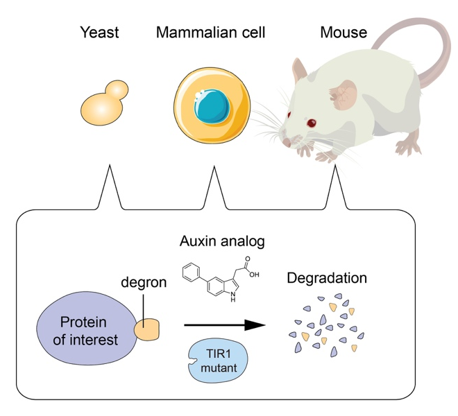
Genetic perturbation is a powerful way to analyze the function of proteins in living cells. For this purpose, we pioneered to develop the auxin-inducible degron (AID) technology by which a degron-fused protein can be rapidly degraded after the addition of the plant hormone auxin (Nishimura et al., Nat. Methods, 2009). By combining with CRISPR-based genome editing, it was possible to generate AID conditional mutants of human cells (Natsume et al., Cell Reports, 2016). The AID system became one of the popular genetic tools to study the function of proteins. However, leaky degradation and high doses of auxin for inducing degradation have been major drawbacks. Moreover, nobody has successfully applied the AID system to control protein degradation in living mice. We recently overcame these problems by taking advantage of chemical biology and successfully established the AID2 system (Yesbolatova et al. Nature Communications, 2020). By using AID2, we can now sharply control protein degradation in yeast, mammalian cells and mice.
• Link to KANEMAKI laboratory
• EurekAlert! link about the paper, Yesbolatova et al. Nature Communications, 2020
• Link to an interview of Prof. KANEMAKI
Chairperson: MAESHIMA, Kazuhiro


