


The problem that seminar information was not displayed correctly in some browser versions has been fixed.
Thank you for your understanding.
Press release
Organization of fast and slow chromatin revealed by single-nucleosome dynamics
S. S. Ashwin, Tadasu Nozaki, Kazuhiro Maeshima, and Masaki Sasai
PNAS first published September 16, 2019 DOI:10.1073/pnas.1907342116
Press release (In Japanese only)
Understanding chromatin organization and dynamics is important, since they crucially affect DNA functions. In this study, we investigate chromatin dynamics by statistically analyzing single-nucleosome movement in living human cells. Bimodal nature of the mean square displacement distribution of nucleosomes allows for a natural categorization of the nucleosomes as fast and slow. Analyses of the nucleosome–nucleosome correlation functions within these categories along with the density of vibrational modes show that the nucleosomes form dynamically correlated fluid regions (i.e., dynamic domains of fast and slow nucleosomes). Perturbed nucleosome dynamics by global histone acetylation or cohesin inactivation indicate that nucleosome–nucleosome interactions along with tethering of chromatin chains organize nucleosomes into fast and slow dynamic domains. A simple polymer model is introduced, which shows the consistency of this dynamic domain picture. Statistical analyses of single-nucleosome movement provide rich information on how chromatin is dynamically organized in a fluid manner in living cells.
This research was supported by JST CREST(JPMJCR15G2), JSPS Kakenhi (JP19H05258, JP19H05273, JP19H01860, JP16H04746) and Takeda Science Foundation, RIKEN Pioneering Project、NIG Joint (2016-A2(6))
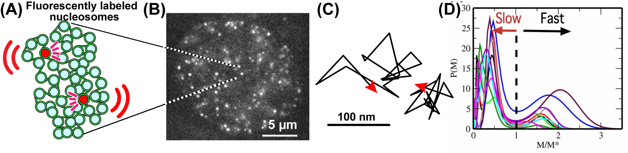
Fig: (A) A small fraction of nucleosomes, where DNA is wrapped around histone proteins, was fluorescently labeled (red). The labeled nucleosome movements can be tracked at super-resolution. (B) A single-nucleosome image of a living HeLa cell. (C) Representative two trajectories of the tracked single nucleosomes. (D) The distribution of MSD of single nucleosomes is plotted for 10-cell samples as functions of M/M*, where M* is M at the minimum between 2 peaks of the distribution.
Video: Raw video of single nucleosomes in the living HeLa cell. From Nozaki et al., (2017) Molecular Cell.
Control of homologous recombination by the HROB–MCM8–MCM9 pathway
Nicole Hustedt, Yuichiro Saito, Michal Zimmermann, Alejandro Álvarez-Quilón, Dheva Setiaputra, Salomé Adam, Andrea McEwan, Jing Yi Yuan, Michele Olivieri, Yichao Zhao, Masato T. Kanemaki, Andrea Jurisicova, and Daniel Durocher
Genes & Development online advanced publication DOI:10.1101/gad.329508.119
Most organisms including humans have two copies of genomic DNA, each of which is originated from the parents. Homologous Recombination (HR) is a reaction, which copy the sequence of donor DNA and paste it to the recipient DNA. HR plays a critical role in meiosis for gametogenesis and in damaged DNA repair during DNA replication or after irradiation, the latter of which is important to prevent cell death and cancer formation.
HR searches the donor sequence and synthesize DNA to copy and paste it to the recipient ones. Despite having a good understanding of the protein roles in the earlier steps in HR, much less is known about HR-associated DNA synthesis. We have studied MCM8–9, which is essential for HR-associated DNA synthesis. As a collaboration with the group led by Prof. Daniel Durocher at University of Toronto, we identified a new factor, HROB, which has an important role for loading MCM8–9 at the damaged DNA sites. Similar to the phenotypes of MCM8–9 loss, human cells lacking HROB showed a strong defect in DNA repair during DNA replication and HROB knockout mice were sterile, showing that, together with MCM8–9, HROB is important for genome maintenance and meiosis. We expect that elucidating HR, in which HROB and MCM8–9 are involved, would help cancer and fertility treatments in the future.
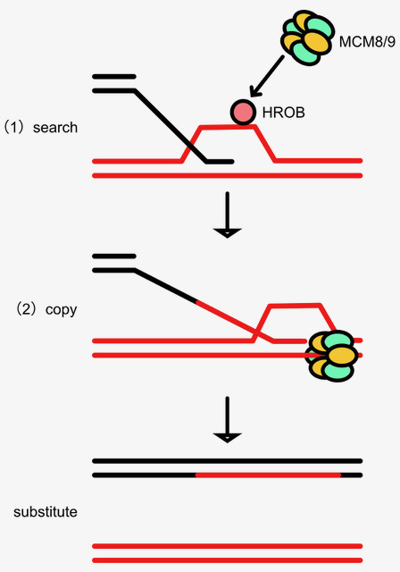
Figure: HR searches the donor sequence and uses DNA synthesis to copy and paste it to the recipient ones. In this paper, we found a novel factor, HROB, which promotes MCM8–9 loading to promote HR-associated DNA synthesis.
Neural signatures of sleep in zebrafish.
Louis C Leung, Gordon X Wang, Romain Madelaine, Gemini Skariah, Koichi Kawakami, Karl Deisseroth, Alexander E Urban, and Philippe Mourrain
Nature 571(7764) 198-204 (2019) DOI:10.1038/s41586-019-1336-7
Slow-wave sleep and rapid eye movement (or paradoxical) sleep have been found in mammals, birds and lizards, but it is unclear whether these neuronal signatures are found in non-amniotic vertebrates. Here we develop non-invasive fluorescence-based polysomnography for zebrafish, and show—using unbiased, brain-wide activity recording coupled with assessment of eye movement, muscle dynamics and heart rate—that there are at least two major sleep signatures in zebrafish. These signatures, which we term slow bursting sleep and propagating wave sleep, share commonalities with those of slow-wave sleep and paradoxical or rapid eye movement sleep, respectively. Further, we find that melanin- concentrating hormone signalling (which is involved in mammalian sleep) also regulates propagating wave sleep signatures and the overall amount of sleep in zebrafish, probably via activation of ependymal cells. These observations suggest that common neural signatures of sleep may have emerged in the vertebrate brain over 450 million years ago.
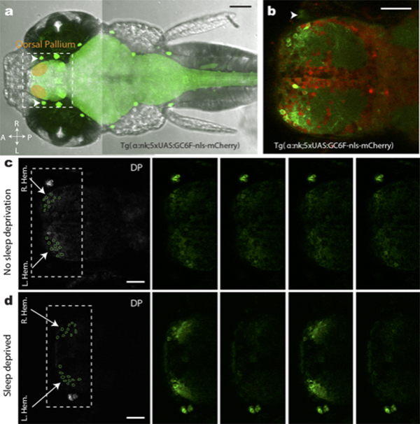
Figure: (a,b)Transgenic fish used for calcium imaging (c) Sleep non-deprived fish (d) slow bursting sleep seen in sleep deprived fish
▶This study is based on the previous study.


