


Press release
Memo1 Mediated Tiling of Radial Glial Cells Facilitates Cerebral Cortical Development
Naoki Nakagawa, Charlotte Plestant, Keiko Yabuno-Nakagawa, Jingjun Li, Janice Lee, Chu-Wei Huang, Amelia Lee, Oleh Krupa, Aditi Adhikari, Suriya Thompson, Tamille Rhynes, Victoria Arevalo, Jason L. Stein, Zoltán Molnár, Ali Badache, E. S. Anton
Neuron Published:July 02, 2019 DOI:10.1016/j.neuron.2019.05.049
Press release (In Japanese only)
In the mammalian neocortex, formation of distinct neuronal layers is important for precise information processing. During the embryonic brain development, neural progenitor cells not only generate neurons, but also play an important role in the neuronal layer formation. The process sprouted from the neural progenitor cell, termed radial process (Figure 1), is used as “guide” for migrating neurons. Each neural progenitor cell has a single radial process that extends toward the brain surface in a non-overlapping, regularly interspaced manner, therefore functioning as a “scaffolding system” for the neuronal migration and layer formation. However, the molecular and cellular mechanisms for the generation of this scaffolding system is not well understood.
A team of Dr. Naoki Nakagawa at the National Institute of Genetics and Dr. Eva S. Anton at the University of North Carolina School of Medicine identified mediator of cell motility 1 (Memo1) as a key gene for building this scaffolding system and revealed its role in the neuronal layer formation in the mouse neocortex.
By analyzing embryonic brains of Memo1-knockout mice, they found that radial processes of Memo1-deficient neural progenitor cells showed extensive branching and altered spatial distribution. These defects disrupted the scaffolding system of neural progenitor cells and resulted in neuronal misplacement and disorganized layers. These findings demonstrate that Memo1 is important for neural progenitor cells to form the regularly interspaced scaffolding system to organize cortical neurons into distinct neuronal layers (Figure 1).
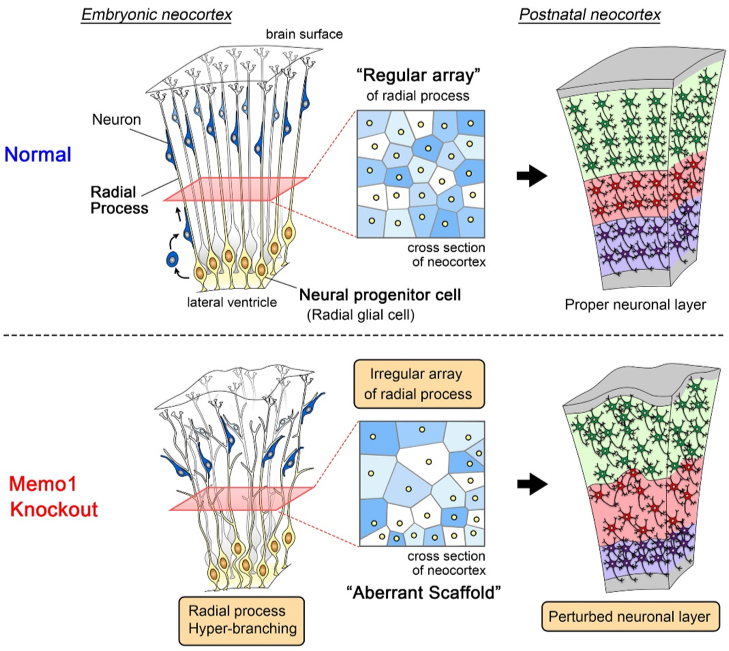
Figure 1: The scaffolding system of neural progenitor cells and neuronal layer formation
(Top) In the embryonic neocortex, neural progenitor cells are located at the ventricular surface and extend single radial processes toward the brain surface. Radial processes do not overlap with each other and are regularly interspaced. Newborn neurons migrate toward the superficial area by using this regular array of radial process as a “scaffold”, eventually forming the proper neuronal layer.
(Bottom) In the Memo1-knockout mouse neocortex, the absence of Memo1 function in neural progenitor cells causes hyper-branching and irregular distribution of radial processes. These phenotypes generate an aberrant scaffold, leading to perturbed neuronal layer formation.
NIG will be closed from August 15 and August 16, 2019 for summer holiday.
Thank you for your understanding and cooperation.
Day/Night Separation of Oxygenic Energy Metabolism and Nuclear DNA Replication in the Unicellular Red Alga Cyanidioschyzon merolae.
Shin-ya Miyagishima, Atsuko Era, Tomohisa Hasunuma, Mami Matsuda, Shunsuke Hirooka, Nobuko Sumiya, Akihiko Kondo, Takayuki Fujiwara
mBio 10(4), e00833-19, 2019 DOI:10.1128/mBio.00833-19
Eukaryotes acquired chloroplasts through an endosymbiotic event in which a cyanobacterium or a unicellular eukaryotic alga was integrated into a previously nonphotosynthetic eukaryotic cell. Photosynthesis by chloroplasts enabled algae to expand their habitats and led to further evolution of land plants. However, photosynthesis causes greater oxidative stress than mitochondrion-based respiration. In seed plants, cell division is restricted to nonphotosynthetic meristematic tissues and populations of photosynthetic cells expand without cell division. Thus, seemingly, photosynthesis is spatially sequestrated from cell proliferation. In contrast, eukaryotic algae possess photosynthetic chloroplasts throughout their life cycle. Here we show that oxygenic energy conversion (daytime) and nuclear DNA replication (night time) are temporally sequestrated in C. merolae. This sequestration enables “safe” proliferation of cells and allows coexistence of chloroplasts and the eukaryotic host cell, as shown in yeast, where mitochondrial respiration and nuclear DNA replication are temporally sequestrated to reduce the mutation rate.
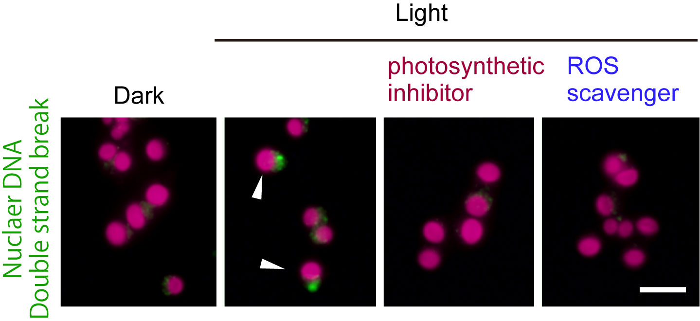
Figure1: The unicellular red alga Cyanidioschyzon merolae was cultured under a 12-h light / 12-h dark cycle. The cells early in the dark period (dark) were illuminated (light) either with or without DCMU (photosynthetic inhibitor) or TEMPOL (ROS scavenger). The green fluorescence indicates accumulation of MRE11 in the nucleus (DNA double strand break). The red is autofluorescence of the chloroplast. When the cells during the subjective night perform photosynthesis, the frequency of nuclear DSB (arrowheads) increases.
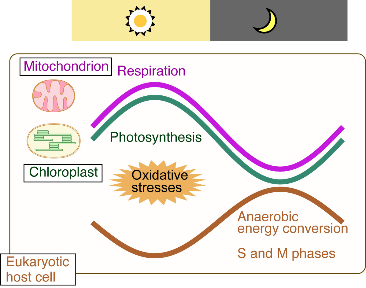
Figure2: The temporal separation of ROS-generating oxygenic energy metabolism by mitochondria and chloroplasts (endosymbiotic organelles) and nuclear DNA replication (eukaryotic host cell) ensures safe cell proliferation
NIG is proud to announce that an associate professor in the Center for Frontier Research has been awarded tenure as of July 1, 2019.
SHIMAMOTO, Yuta: Physics and Cell Biology Laboratory
Center for Frontier Research is an incubation center to simultaneously develop two elements: human resources and new research fields. Promising young scientists conduct research as principal investigator (tenure-track associate professor) to explore new frontiers in genetics and related areas, taking advantage of NIG’s research infrastructure and various support systems. Those who obtained tenure will establish new research divisions in NIG to lead the new fields that they contribute in creating.


