


A tRNA-based multiplex sgRNA expression system in zebrafish and its application to generation of transgenic albino fish.
Tomoya Shiraki and Koichi Kawakami
Scientific Reports 8, Article number: 13366 (2018) DOI:10.1038/s41598-018-31476-5
The CRISPR/Cas9 system can be introduced into zebrafish fertilized eggs by microinjection and facilitates gene knock-out, genome editing, and gene knock-in studies in zebrafish. Further, expression of gRNA and controlled expression of Cas9 in transgenic fish have enabled the study of gene functions in specific cell types in zebrafish. The transgenic Cas9 approach would be more useful if multiple sgRNAs could be expressed simultaneously since we could knock-out a gene more efficiently or disrupt multiple genes simultaneously. However, such a system had not been developed in zebrafish.
In this study, we describe a novel system to express multiple sgRNAs efficiently in zebrafish, that relies on the endogenous tRNA processing machinery. In this study, we demonstrated that active sgRNAs can be produced from tRNA-fused precursor transcripts. To show a proof of principle, we constructed a transgenic fish line expressing a single transcript containing three distinct sgRNAs, that targeted the slc45a2 (albino) gene. We found that the transgenic-albino fish showed nearly complete albino phenotypes, which were transparent enough for imaging at the cellular resolution to be performed. Thus, the tRNA-based multiplex sgRNA expression system should facilitate genetic studies of gene functions in zebrafish.
This study was supported by JSPS KAKENHI Grant Numbers (JP15H02370, JP16K20983, JP16H01651, JP18H04988), and NBRP and NBRP/Genome Information upgrading program from Japan Agency for Medical Research and Development (AMED).
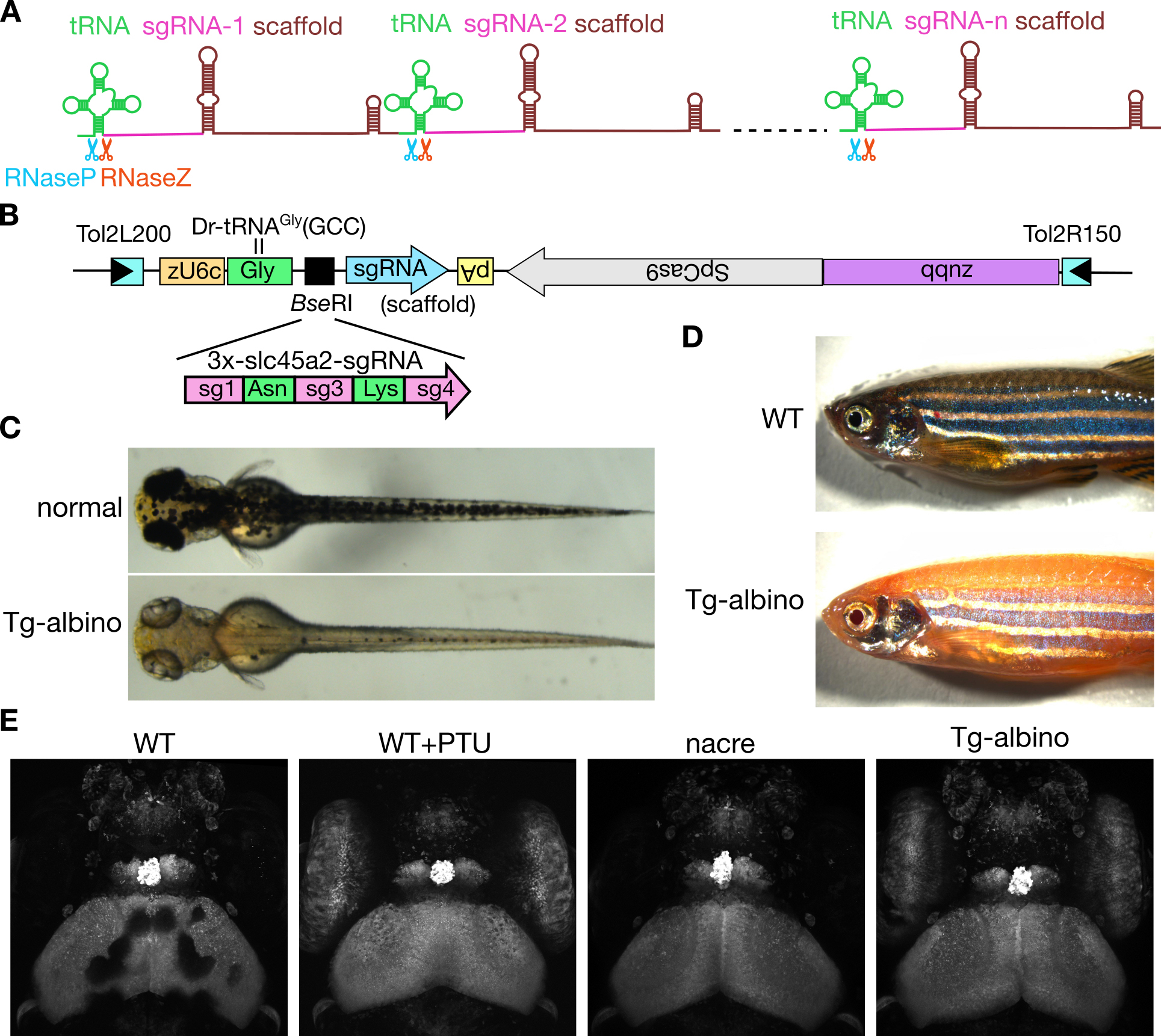
Figure: (A) Schematic diagram of the polycistronic tRNA-sgRNA system for multiplex genome editing. (B) DNA construct used for the generation of transgenic albino (Tg-albino) zebrafish. Three distinct sgRNAs targeting the albino (slc45a2) locus were expressed. (C-E) The Tg-albino fish showed nearly complete albino phenotypes, which were transparent enough for imaging at the cellular resolution. (C) Tg-albino at 3-days post-fertilization (dpf). (D)Adult Tg-albino fish. (E) Fluorescent images of a GFP-transgenic fish at 5 dpf in Tg-albino background.
![]()
Differential dynamics of cortical neuron dendritic trees revealed by long-term in vivo imaging in neonates
Shingo Nakazawa, Hidenobu Mizuno, Takuji Iwasato
Nature Communications 9, Article number: 3106 (2018) DOI:10.1038/s41467-018-05563-0
Pressrelease (In Japanese only)
Proper neuronal circuit function relies on precise dendritic projection, which is established through activity-dependent refinement during early postnatal development. Here we revealed dynamics of dendritic refinement in the mammalian brain by conducting long-term imaging of the neonatal mouse barrel cortex. By “retrospective” analyses, we identified “prospective” barrel-edge spiny stellate (SS) neurons in early neonates, which had an apical dendrite and primitive basal dendrites (BDs). These neurons retracted the apical dendrite gradually and established strong BD orientation bias through continuous “dendritic tree” turnover. A greater chance of survival was given to BD trees emerged in the barrel-center side, where thalamocortical axons (TCAs) cluster. When the spatial bias of TCA inputs to SS neurons was lost, BD tree turnover was suppressed, and most BD trees became stable and elaborated mildly. Thus, barrel-edge SS neurons could establish the characteristic BD projection pattern through differential dynamics of dendritic trees induced by spatially biased inputs.
Source: Nature Communications 9, Article number: 3106 (2018) DOI:10.1038/s41467-018-05563-0
![]()
Protocadherin-mediated cell repulsion controls the central topography and efferent projections of the abducens nucleus
Kazuhide Asakawa, Koichi Kawakami
Cell Reports Volume 24, ISSUE 6, P1562-1572, August 07, 201 DOI:10.1016/j.celrep.2018.07.024
Press release (In Japanese only)
Approximately 2-3 % of children suffer from a condition called strabismus, which is more commonly known as “lazy eye” or “crossed eyes”. The majority of cases are thought to involve both genetic and environmental factors. Knowing the genetic causes of strabismus is very important as it would help estimate risk of developing the condition and increase the chances of initiating early treatment to prevent impaired eye movement.
A major obstacle to uncovering the genetic causes of strabismus has been the physical limitation of observing the connections between the brain and eye muscles. These are formed by long cables called “axons” that are extended from brain cells deep inside the head at a very early developmental stage. Therefore it is difficult to observe the fine architecture of the brain-eye muscle connections at the cellular level and analyze how they might be affected by gene mutations.
To overcome this difficulty, researchers at the National Institute of Genetics in Japan chose as their experimental model a tropical fish called zebrafish. Zebrafish larvae are nearly transparent while they are developing, so the researchers hoped that they could make the brain-eye muscle connections visible using genetic engineering. Employing their own original techniques, the team successfully created a zebrafish in which the specific group of brain cells that send signals for outward eye movement, called abducens motor neurons, as well as the targeted eye muscles emit differently colored fluorescent light clearly visible through the developing fish’s transparent head.
These special zebrafish proved instrumental in allowing the team to make great progress in uncovering the genetic mechanism of strabismus. The team demonstrated that the abducens motor neurons developed abnormally when the gene called protocadherin 17 (pcdh17) was mutated. This gene encodes for the production of the Pcdh17 protein that is located on the cell surface of the abducens motor neurons. When an abnormal Pcdh17 protein is produced from the manipulated pcdh17 gene and located on the cell surface, the neurons did not position properly in the brain and the axons they extended failed to reach the target eye muscle. In many cases these disabled neurons clumped together forming closely-packed cellular aggregates. These results suggest that an abducens motor neuron uses repulsive forces to both position itself correctly in the brain and extend its axon correctly to the target muscle and that these forces are mediated by the Pcdh17 protein molecules on its surface interacting with itself and/or other neurons around it. This study was published in Cell Reports on 7th August.
Dr. Asakawa, who led this research project, says, “Pcdh17 creates a moderate repulsive force between abducens motor neurons presumably allowing them to behave like a fluid forming a stream and flowing into the muscle. Without them, the neurons just become frozen like ice. Since Pcdh17 protein is also present in humans, it is likely to play a similar role in our body for the development of normal eye movement. Moreover, given that many types of brain neurons connecting with the head muscles are covered with different types of proteins that are similar to Pcdh17 on their cell surface, genetic mutations in these genes might increase the risk of developing other congenital disorders of craniofacial movements”.
Asakawa continues, “Compared to the connections between the spinal cord and skeletal muscles that generate our body movements, the brain and eye muscle connections have some special characteristics making them selectively resistant to degeneration in fatal motor neuron diseases, such as amyotrophic lateral sclerosis (ALS). We expect that the visual and genetically-manipulatable brain-eye muscle connection in fish also has great potential to reveal a way to protect motor neurons from degeneration in ALS.”
This study was supported by KAKENHI (JP15H02370, JP18H04988, JP22700349, JP23115720, and research grants from The Uehara Memorial Foundation, The Kao Foundation for Arts and Sciences, The Mitsubishi Foundation, Daiichi-Sankyo Foundation of Life Science and National BioResouce Project from Japan Agency for Medical Research and Development (AMED).
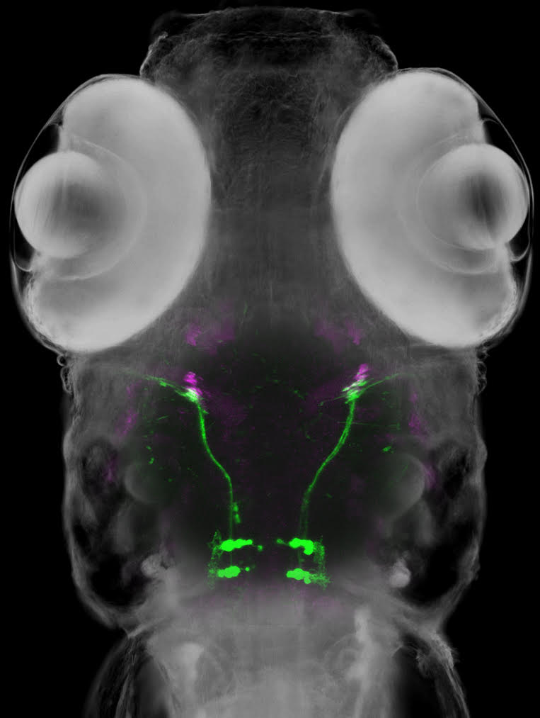
Figure1: A zebrafish larva expressing a green fluorescent protein in the abducens motor neurons and a red fluorescent protein in the eye muscle (magenta).
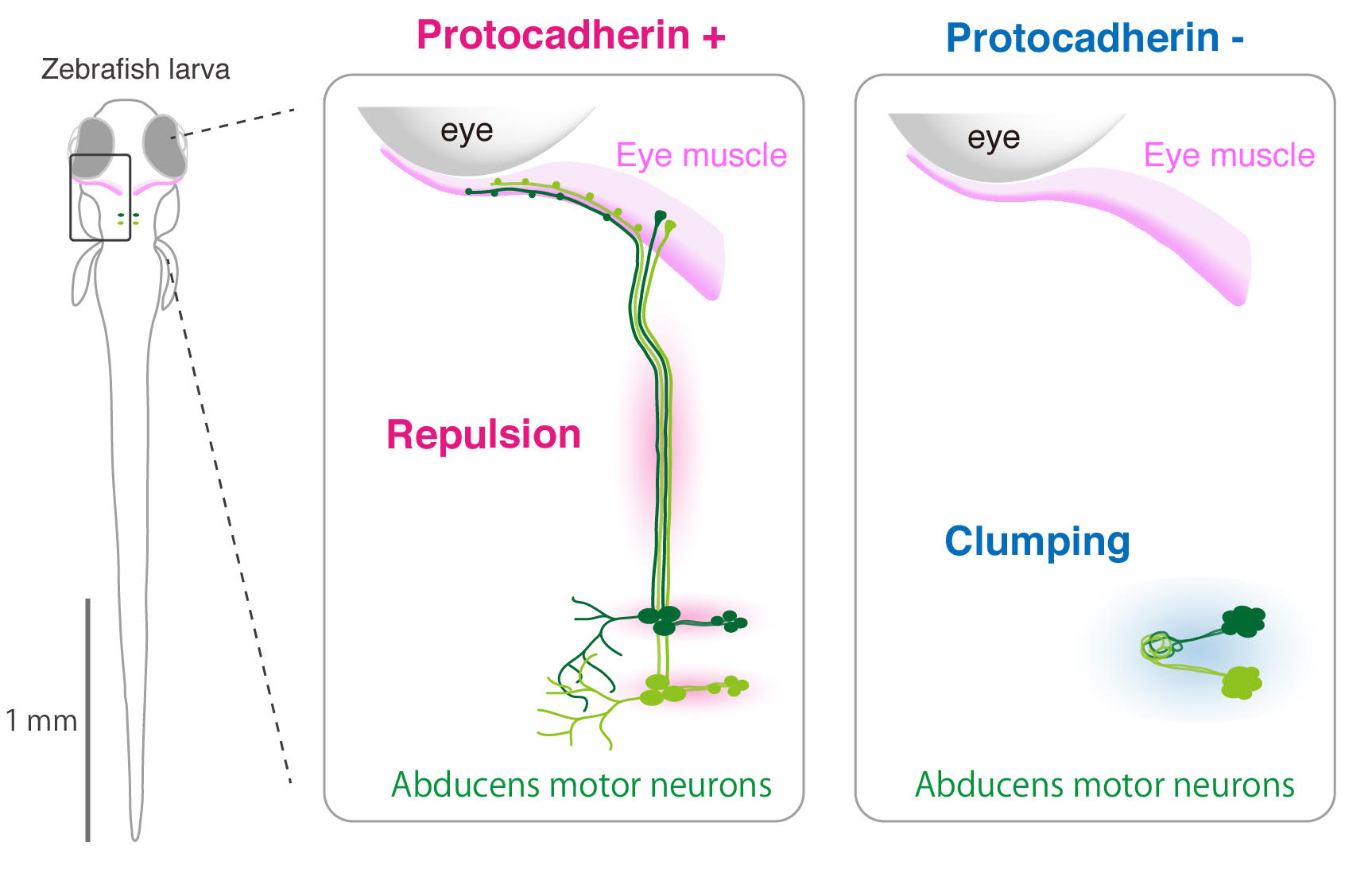
Figure2: Repulsive forces-mediated by protocadherins promote cell migration and axon ourgrowth of abducens motor neurons (left) . Loss of the rupulsive forces leads to clumping phenotype (right).
EurekAlert!, the online, global news service operated by AAAS, the science society, PUBLIC RELEASE: 7-AUG-2018
New assistant professor joins NIG as of September 1, 2018.


