


Model Fish Genomics Resource Laboratory / Sakai Group
Development and growth of organs in living whole embryo and larval grafts in zebrafish
Toshihiro Kawasaki, Akiteru Maeno, Toshihiko Shiroishi & Noriyoshi Sakai
Scientific REPORTS, 7: 16508 DOI:10.1038/s41598-017-16642-5
Age-related systemic environments influence neurogenesis and organ regeneration of heterochronic parabiotic partners; however, the difficulty of manipulating small embryos prevents the effects of aged systemic environments on primitive organs at the developmental stage from being analysed.
Here, we describe a novel transplantation system to support whole living embryos/larvae as grafts in immunodeficient zebrafish by the intrusion of host blood vessels into the grafts, allowing bodies similar to those of heterochronic parabiosis to be generated by subcutaneous grafting. Although grafted embryos/larvae formed most organs, the liver, testes and heart developed insufficiently or even occasionally disappeared. Removal of host germ cells stimulated testis development in grafted embryos. These results indicate that primitive testes are susceptible to the systemic environments that originated from the germ cells of aged hosts and imply that the primitive liver and heart are similar. This unique transplantation system will lead to new insights into the age-related systemic environments that are crucial for organogenesis in vertebrates.
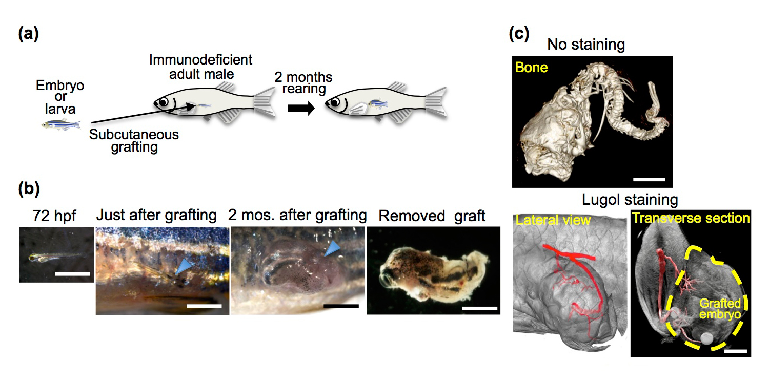
Development of grafted 72 hpf embryos in rag1 mutant hosts. (a) Schematic of whole-embryo transplantation. (b) Representative 72 hpf embryos and grafted embryos. (c) µ-CT imaging of the grafted embryo 2 months after transplantation. Cranial bones, vertebrae (No staining) and a vascular structure (Lugol staining) were observed in the grafted embryos.
Press release
An asymmetric attraction model for the diversity and robustness of cell arrangement in nematodes
Kazunori Yamamoto, Akatsuki Kimura
Development 2017 144: 4437-4449; DOI:10.1242/dev.154609
Pressrelease (In Japanese only)
During early embryogenesis in animals, cells are arranged into a species-specific pattern in a robust manner. Diverse cell arrangement patterns are observed, even among close relatives (Fig. A). In the present study, we evaluated the mechanisms by which the diversity and robustness of cell arrangements are achieved in developing embryos. We successfully reproduced various patterns of cell arrangements observed in various nematode species in Caenorhabditis elegans embryos by altering the eggshell shapes (Fig. B). The findings suggest that the observed diversity of cell arrangements can be explained by differences in the eggshell shape. Additionally, we found that the cell arrangement was robust against eggshell deformation. Computational modeling revealed that, in addition to repulsive forces, attractive forces are sufficient to achieve such robustness (Fig. C). The present model is also capable of simulating the effect of changing cell division orientation. Genetic perturbation experiments demonstrated that attractive forces derived from cell adhesion are necessary for the robustness. The proposed model accounts for both diversity and robustness of cell arrangements, and contributes to our understanding of how the diversity and robustness of cell arrangements are achieved in developing embryos.
This research article was published in the special issue of Development ‘On Growth and Form – 100 years on’, inspired by the seminal work of D’Arcy Thomson. See the authors’ comments:
Kazunori Yamamoto, the first author of the paper received Morishima Award for this research.
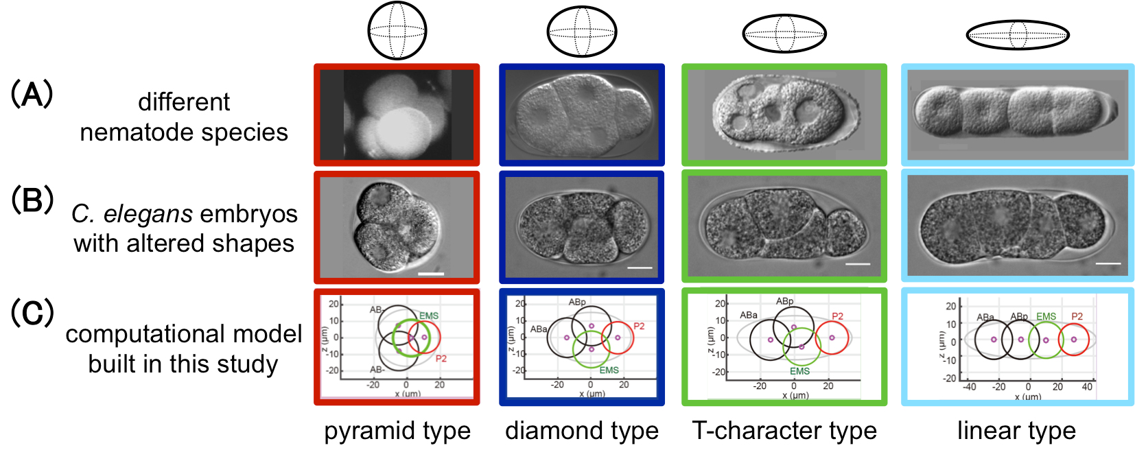
Figure: This study successfully reproduced the cell arrangement patterns of various nematode species (A) using C. elegans by altering the shape of the eggshell (B). A computational model was also built in this study that reproduces the diversity and robustness of the cell arrangement patters (C). In (A), the patterns of Enoplus brevis (red, image from Schulze and Shierenberg, Evodevo 2, 18, 2011), C. elegans (blue, this study), Cephalobus sp. (green, Goldstein, Philos. Trans. R. Soc. Lond. B Biol. Sci. 356, 1521-1531, 2001), and Belonolaimus sp. (light blue, Goldstein, 2001) are shown.
Source: Development 2017 144: 4437-4449
DOI: 10.1242/dev.154609
Division of Molecular Cell Engineering / Kanemaki Group
Conditional Degrons for Controlling Protein Expression at the Protein Level
Toyoaki Natsume and Masato T. Kanemaki
Annual Review of Genetics, DOI:https://doi.org/10.1146/annurev-genet-120116-024656
It is a common genetic analysis in many organisms to see the phenotype after depletion of a protein of interest. Technologies such as siRNA and conditional gene knockout have been employed for this purpose. However, these do not deplete a protein of interest directly. It is, therefore, the depletion effect is indirect and it takes for a relatively long time to deplete a protein of interest, depending on the half-life of the protein. This sometime causes a problem because the phenotype can be complicated by a secondary effect that was initiated by a primary defect.
Recently, conditional degron technologies, by which a degron-fused protein can be induced for rapid degradation, are seizing the attention. Protein depletion by the degron technologies work directly at the protein level, so that it is possible to analyze the direct effect after rapid protein depletion. We developed the auxin-inducible degron technology by which a degron-fused protein can be controlled by a plant hormone auxin. Others used temperature, small chemicals, light, or the expression of another protein for the similar purpose. We wrote a review paper explaining the development history of conditional degrons and their characteristics.
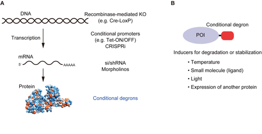
(A) Conditional technologies for controlling protein expression. From the level of gene to protein, various technologies have been developed. Conditional degrons work at the protein level. (B) Conditional degrons can be controlled by temperature, small chemicals, light, or the expression of another protein.
Microbial Genetics Laboratory / Niki Group
Release of condensin from mitotic chromosomes requires the Ran-GTP gradient in the reorganized nucleus.
Keita Aoki and Hironori Niki
Biology Open, November 15; 6(11):1614-1628, 2017 DOI:10.1242/bio.027193.
After mitosis, nuclear reorganization occurs together with decondensation of mitotic chromosomes and reformation of the nuclear envelope, thereby restoring the Ran-GTP gradient between nucleus and cytoplasm. The Ran-GTP gradient is dependent on Pim1/RCC1. Interestingly, a defect in Pim1/RCC1 in Schizosaccharomyces pombe causes post-mitotic condensation of chromatin, namely hyper-condensation, suggesting a relationship between the Ran-GTP gradient and chromosome decondensation. However, how Ran-GTP interacts with chromosome decondensation is unresolved. To examine this interaction, we used Schizosaccharomyces japonicus, which is known to undergo partial breakdown of the nuclear membrane during mitosis. We found that Pim1/RCC1 was localized on nuclear pores, but this localization failed in a temperature-sensitive mutant of Pim1/RCC1. The mutant cells exhibited hyper-condensed chromatin after mitosis due to prolonged association of condensin on the chromosomes. Conceivably, a condensin-dephosphorylation defect might cause hyper-condensed chromatin, since chromosomal localization of condensin is dependent on phosphorylation by cyclin-dependent kinase (CDK). Indeed, CDK-phospho-mimic mutation of condensin alone caused untimely condensin localization, resulting in hyper-condensed chromatin. Together, these results suggest that dephosphorylation of CDK sites of condensin might require the Ran-GTP gradient produced by nuclear pore-localized Pim1/RCC1.
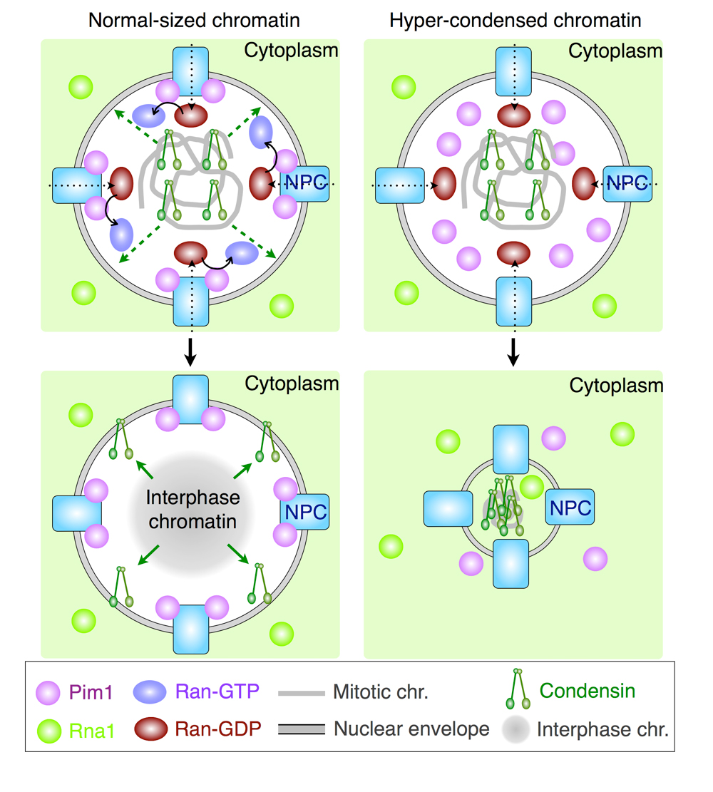
In a normal-sized nucleus (left), condensin is released from chromosomes in a Ran-GTP dependent manner. Therefore, a decondensed chromatin is formed in G1 phase. In a Pim1/RCC1-deficient nucleus, condensin is not released from chromosomes due to few Ran-GTP. Therefore, a hyper-condensed chromatin is formed in G1 phase.
Microbial Genetics Laboratory / Niki Group
Multiple cis-acting rDNAs contribute to nucleoid separation and recruit the bacterial condensin, Smc-ScpAB
Koichi Yano, and Hironori Niki
Cell Reports, Vol. 21 Iss. 5, October 31, 2017 DOI:http://dx.doi.org/10.1016/j.celrep.2017.10.014
Condensins load onto DNA to organize chromosome. Smc-ScpAB clearly loads onto the parS sites bound by Spo0J, but other loading site(s) must operate independently of parS. In this study, we asked where and how Smc-ScpAB normally selects its loading site. Our results suggest that rDNA is also a loading site. A pull-down assay revealed that Smc-ScpAB preferentially loads onto rDNA in the wild-type cell and even in a Δspo0J mutant, but not in a Δsmc mutant. Moreover, we showed that deletion mutants of rDNAs cause a defect in nucleoid separation, and at least two rDNAs near oriC are essential for separation. Full-length rDNA including promoters is required for loading and nucleoid separation. A synthetic defect by deletions of both rDNA and spo0J resulted in more aberrant nucleoid separation. We propose that a single-stranded segment DNA that is exposed at highly transcribed rRNA operons would become a target for Smc-ScpAB loading.
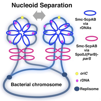
Schemes of compaction of newly replicated DNA strands. The yellow circle indicates oriC, black blue circles indicate replication forks, and magenta circles indicate rrn operons. The blue rings indicate Smc-ScpAB topologically loaded on rrn operons, and magenta rings represent Smc-ScpAB topologically loaded by ParB bound to parS as shown in Wang et al. (2017). In addition to separation of oriC, chromosome compaction would always be accompanied by proper chromosome segregation. The chromosomal region adjacent to oriC is packed by Smc in the WT so that the nucleoids are clearly separated.
Mammalian Genetics Laboratory / Shiroishi Group
SHH signaling directed by two oral epithelium-specific enhancers controls tooth and oral development
Tomoko Sagai, Takanori Amano, Akiteru Maeno, Hiroshi Kiyonari, Hyejin Seo, Sung-Won Cho and Toshihiko Shiroishi
Scientific Reports, 7, Article number: 13004 (2017) DOI:10.1038/s41598-017-12532-y
Interaction between the epithelium and mesenchyme coordinates patterning and differentiation of oral cavity structures including teeth, palatal rugae and tongue papillae. SHH is one of the key signaling molecules for this interaction. Epithelial expression of Shh in the tooth buds and tongue papillae is regulated by at least two enhancers, MRCS1 and MFCS4. However, it is unclear how the two enhancers cooperate to regulate Shh. Here, we found that simultaneous deletion of MRCS1 and MFCS4 results in the formation of a supernumerary tooth in front of the first molar. Since deletion of either single enhancer barely affects tooth development, MRCS1 and MFCS4 evidently act in a redundant fashion. Binding motifs for WNT signaling mediators are shared by MRCS1 and MFCS4, and play a central role in regulating Shh expression, indicating that the two redundant enhancers additively exert their Shh regulation by responding to WNT signal input.
This study was carried out as a collaboration of Tomoko Sagai, Takanori Amano and Toshihiko Shiroishi of National Institute of Genetics, Hiroshi Kiyonari of RIKEN Center for Life Science Technologies, and Hyejin Seo and Sung-Won Cho of Yonsei University in Korea. This study was supported by JSPS KAKENHI 24247002.
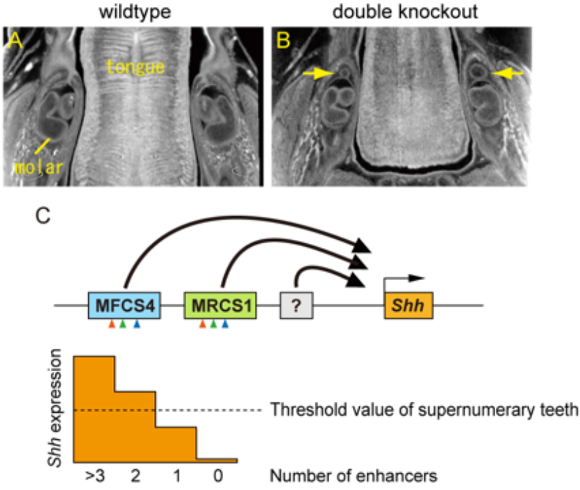
Supernumerary tooth formation in the mouse with simultaneous deletion (double knockout) of the MRCS1 and MFCS4 enhancers. (A and B) X-ray micro-CT images of transverse section of mandible in the wild type (A) and the mouse with the combinatorial deletion of MRCS1 and MFCS4 (B). Yellow arrow indicates supernumerary tooth. (C) A model of redundant action of the Shh enhancers. MRCS1 and MFCS4 share three common regulatory motifs that are located in the same order (arrowheads) and have redundant roles for the Shh expression in tooth buds. Expression levels of Shh are additively regulated by MRCS1, MFCS4, and probably unidentified enhancers. When reduction of Shh expression level falls below a threshold, supernumerary molars arise. MRCS1 and MFCS4 control different expression domains of Shh in the oropharyngeal tissues in addition to the tooth bud. Thus, the two redundant enhancers may cooperatively regulate the tooth development, while they have undergone sub-functionalization.


