


Experimental Farm / Nonomura Group
Histone H3 modifications are widely reprogrammed during male meiosis I in rice dependently on MEL1 Argonaute protein
Hua Liu, Ken-Ichi Nonomura
Journal of Cell Science, Published online, 12 August, 2016 DOI:10.1242/jcs.184937
Meiosis is a special type of cell division to halve the chromosome number, achieved by two continuous division not intervened by DNA replication. It is an indispensable mechanism to generate genetic diversity via homologous chromosome pairing and meiotic recombination, in addition to stable transmission of genetic information to the next generation.
We focused on the relation of meiosis and histone modifications (glossary), which are important for control of chromosome structure and gene expression. Generally in plants, dimethylation at the position-9 lysine of histone H3 (H3K9me2) is thought to repress gene expressions and promote the compaction of chromatin structure. In contrast, acethylation at the same position (H3K9ac) activates gene expressions. We found that H3 modifications after the meiotic entry were totally altered from the premeiotic H3 status (Fig. 1A). “Large-scale meiotic chromosome reprogramming (LMR)” named in this paper is thought to be one of the mechanisms promoting meiosis in plants.
Interestingly, LMR was completely disrupted in the mutant of MEL1 (Fig. 1B), that is an Argonaute protein (glossary) specifically expressed in rice germ cells. These results suggest possibilities that MEL1 promotes meiosis via control of LMR, and that the RNA silencing mechanism is important for plant meiosis.
This work was supported by JSPS KAKENHI (25252004), and by NIG postdoc fellowship.
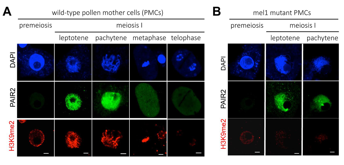
A wide reprogramming of histone H3K9me2 during meiosis I is dependent on the rice Argonaute protein MEL1.
(A) In wild-type pollen mother cells (PMCs), the level of H3K9me2 (red) is increased remarkably when cells transit from premeiosis to meiosis. Chromatin DNA is stained with DAPI (blue). PAIR2 (green) is a meiotic gene required for homologous chromosome pairing, and shows these cells undergo meiosis I. Scale bar = 5µm.
(B) mel1 mutant PMCs. PAIR2 signal (green) indicates these cells undergoing meiosis I, but no H3K9me2 reprogramming takes place.
<Glossary>
Cell Architecture Laboratory / Kimura Group
Bayesian Inference of Forces Causing Cytoplasmic Streaming in Caenorhabditis elegans Embryos and Mouse Oocytes.
Niwayama R., Nagao H., Kitajima T. S., Hufnagel L., Shinohara K., Higuchi T., Ishikawa T., Kimura A.
PLoS ONE, Vol 11, e0159917 (2016). DOI:10.1371/journal.pone.0159917
Cytoplasmic streaming is observed in wide variety of cells both in animals and plants. It is caused in many cases by cytoskeletons and molecular motors. Where these forces are exerted is difficult to identify. In this study, we developed a novel computational method to estimate the localization and amplitude of the forces generating cytoplasmic streaming. Our method infers the distribution of forces by fitting the flow field in hydrodynamics simulation to that observed in cells. We applied the method to Caenorhabditis elegans embryos and mouse oocytes. The distinct patterns of force distribution estimated in this study were consistent with the proposed distinct functions of the streaming in both of these species. We expect our method to have diverse applications, and to serve as a powerful tool for biologists who want to characterize the mechanics of biological hydrodynamic flows.
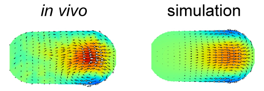
The velocity distribution in the simulation performed using the force distribution estimated in this study (right) agrees well with that measured experimentally (left). The color represents the velocity along the anterior-posterior axis, and the arrows represent the direction of the flow for cytoplasmic streaming in the C. elegans embryo.
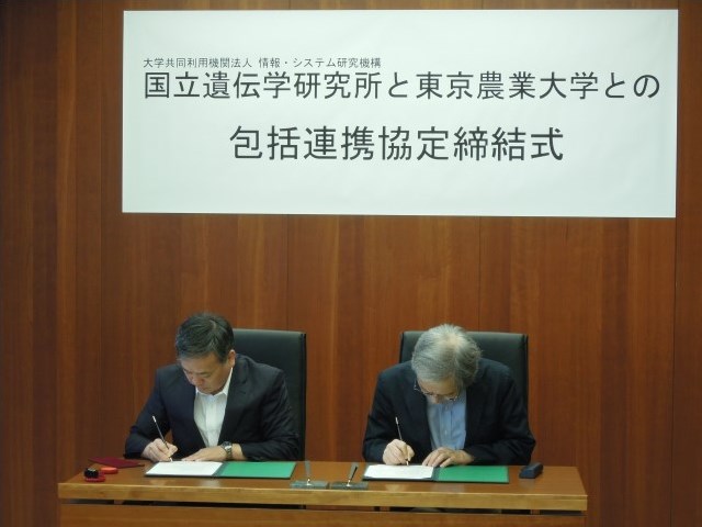
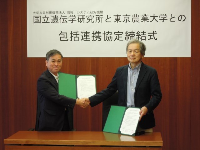
Signing ceremony
New assistant professor joins NIG as of August 1, 2016.
Kazuo HARA: Laboratory for Gene-Expression Analysis, Okubo Group


