


Mammalian Development Laboratory / Saga Group
The RNA binding protein Nanos2 organizes a post-transcriptional buffering system to retain primitive states of mouse spermatogonial stem cells
Zhi Zhou, Takayuki Shirakawa, Kazuyuki Ohbo, Aiko Sada, Wu Quan, Kazuteru Hasegawa, Rie Saba, Yumiko Saga Developmental Cell. Published Online: June 25, 2015 DOI:http://dx.doi.org/10.1016/j.devcel.2015.05.014In many adult tissues, homeostasis relies on self-renewing stem cells that are primed for differentiation. The reconciliation mechanisms of these characteristics remain a fundamental question in stem cell biology. Here, we show that Nanos2, an evolutionarily conserved RNA-binding protein, works with other cellular messenger ribonucleoprotein (mRNP) components to ensure the primitive status of SSCs through a dual mechanism that involves 1) direct recruitment and translational repression of genes that promote spermatogonial differentiation, and 2) repression of the target of rapamycin complex 1 (mTORC1), a well-known negative pathway for SSC self-renewal, by sequestration of the core factor mTOR in mRNPs. This mechanism links mRNA turnover to mTORC1 signaling through Nanos2-containing mRNPs and establishes a post-transcriptional buffering system to facilitate SSC homeostasis in the fluctuating environment within the seminiferous tubule. This research is partly supported by Grant-in-Aid for Scientific Research on Innovative Areas ”Epigenome dynamics and regulation in germ cells” and a “Data assimilation” project of the Transdisciplinary Research Integration Center in ROIS. Zhi Zhou was a NIG postdoctoral fellow 2012.
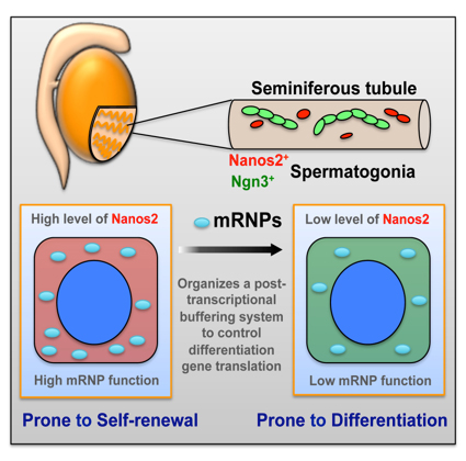
Nanos2 promotes mRNP formation and contribute to the maintenance of undifferentiated state of spermatogonial stem cells. Upon decreasing of Nanos2 level, stem cells shift to differentiation pathway.
Division of Brain Function / Hirata Group
Chemokine signalling controls integrity of radial glial scaffold in developing spinal cord and consequential proper position of boundary cap cells
Yan Zhu, Tomoko Matsumoto, Takashi Nagasawa, Fabienne Mackay, Fujio Murakami Journal of Neuroscience 17 June 2015, 35(24): 9211-9224; DOI:10.1523/JNEUROSCI.0156-15.2015Radial glial cells, the CNS neural progenitors, extend long radial processes that form the radial glial scaffold in the developing neuroepithelium. The integrity of this radial glial scaffold is well maintained throughout development. However, how this is achieved while the neuroepithelium rapidly expands and what the consequence might be when this integrity is compromised have not been well understood. In this study, we addressed these questions in the developing mouse spinal cord. We found that CXCR4, a receptor of a chemokine CXCL12 secreted from the pial meninges, is expressed in spinal cord radial glia. Conditional knockout of CXCR4 in radial glia causes disrupted radial glial scaffold with gaps at the pial endfeet layer, and consequentially lead to an invasion of boundary cap cells into the spinal cord. Since boundary cap cells are PNS cells normally positioned at the incoming and outgoing axonal roots, their invasion into the spinal cord suggests a compromised CNS/PNS boundary in the absence of CXCL12/CXCR4 signalling. By interrogating the underlying mechanisms, we then went on to show that CXCL12 signalling promotes the radial glia adhesion to basement membrane components and activates integrin β1 avidity. Our study uncovers an extrinsic molecular regulator for the maintenance of radial glial scaffold integrity during development. And our data suggest that the integrity of radial glia scaffold is important to safeguard the CNS/PNS boundary for the developing spinal cord.
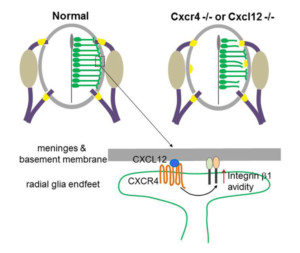
CXCL12 from the meninges regulates adhesion of radial glia to the basement membrane by enhancing avidity of integrin β1. Mice deficient in CXCL12 or its receptor CXCR4 show disrupted radial glial scaffold in their spinal cords, which consequentially causes invasion of boundary cap cells into the spinal cord.
NIG_MoG (The National Institute of Genetics Mouse Genome database)
URL: http://molossinus.lab.nig.ac.jp/msmdb/
Mammalian Genetics Laboratory / Shiroishi Group
The National Institute of Genetics Mouse Genome database (NIG_MoG; http://molossinus.lab.nig.ac.jp/msmdb/) primarily comprises the whole genome sequence data of two inbred mouse strains, MSM/Ms and JF1/Ms. These strains were established at NIG and originated from the Japanese subspecies Mus musculus molossinus. A comparative analysis of their genomes with C57BL/6J (B6: whose genome is predominantly derived from the West European subspecies M. m. domesticus), revealed a vast number of SNPs, structural variants, insertions and deletions (indels). NIG_MoG provides, especially for wet-lab biologists, visualized genome polymorphism information, browsing single-nucleotide polymorphisms and short insertions and deletions in the genomes of MSM/Ms and JF1/Ms allowing users to intuitively recognize intersubspecific genome divergence in these mouse strains using visual data. The database also supports the in silico screening of BAC clones that contain genomic DNA from MSM/Ms and the standard classical laboratory strain C57BL/6N established at RIKEN BioResorce center. NIG_MoG is thus a valuable navigator for exploring mouse genome polymorphisms and BAC clones that are useful for studies of gene function and regulation based on intersubspecific genome divergence.
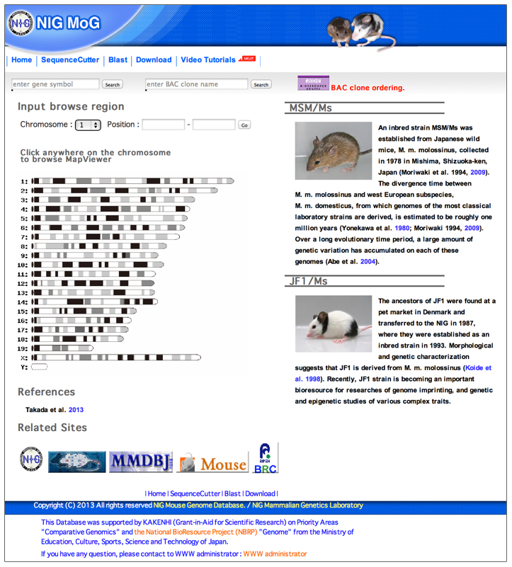
Fig. A screenshot of the top page of NIG_MoG
Biological Macromolecules Laboratory • Maeshima Group
Chromosomes Progress to Metaphase in Multiple Discrete Steps via Global Compaction/Expansion Cycles
Liang, Z., Zickler, D., Prentiss, M., Chang. F.S., Witz, G., Maeshima, K., Kleckner, N. Cell, 161, 1124-1137 (2015). DOI:10.1016/j.cell.2015.04.030Mammalian mitotic chromosome morphogenesis was analyzed by 4D live-cell and snapshot deconvolution fluorescence imaging. Prophase chromosomes, whose organization was previously unknown, are revealed to comprise co-oriented sister linear loop arrays displayed along a single, peripheral, regularly kinked topoisomeraseII /cohesin /condensinII axis (Fig a). Thereafter, rather than smooth, progressive compaction as generally envisioned, progression to metaphase is a discontinuous process involving chromosome expansion as well as compaction. At late prophase (Fig b), dependent on topoisomerase II and with concomitant cohesin release, chromosomes expand, axes split and straighten, and chromatin loops transit to a radial disposition around now-central axes. Finally, chromosomes globally compact, giving the metaphase state (Fig c). These patterns are consistent with the hypothesis that the molecular events of chromosome morphogenesis are governed by accumulation and release of chromosome stress, created by chromatin compaction and expansion. Chromosome state could evolve analogously throughout the cell cycle.
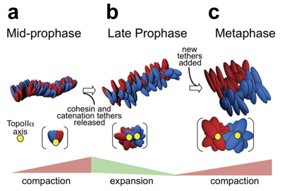
Two sister DNAs are shown in red and blue. The chromosome axis is in yellow. (a) mid-prophase; two sister chromatids are condensed with a single axis (yellow), but still mixed. (b) late-prophase; with expansion, two sisters and their axes are segregated. (c) metaphase; Further condensation and separation of the two sisters occur in metaphase.
Time & Date
9:00 ~ 16:00, Saturday, April 4th (No Reservations Required, Free Admission)
Access
Free Shuttle Buses Available to the National Institute of Genetics from the North Exit of Mishima Station. ( Service Time: 8:50 ~ 15:00)
Map
Map Free Shuttle Buses Available Parking around Mishima Station Available for Car Visitors.
No Pets Allowed except Service Dogs, No General Parking*
(*Disabled Parking Available on the Institute Premises)
Information
1111 Yata, Mishima, Shizuoka 411-8540, JAPAN TEL:+81-55-981-5873


