


Hitoshi SAWA, D. Sc., Professor,
Sawa Group, Multicellular Organization Laboratory, Structural Biology Center, National Institute of Genetics.
The human body is made up of roughly 200 types of cell or even more if cells are classified more minutely, and all of them originate from a single fertilized egg. As the fertilized egg undergoes repeated divisions, the number of cells and their types increase. Yet, one original cell splitting into two identical cells does not generate diverse types of cell. For an organism to develop, a cell must divide into two that are different from each other. This is called asymmetrical division, which is a key to cell fates. I started studying the mechanism of asymmetrical cell division 17 years ago when I went to the United States to do research, and I have been continuing to date. Asymmetrical cell division has now become a hot research theme since we have learned that it is related not only to the development of organisms and cell differentiation but also stem cell maintenance and carcinogenesis.
C. elegans is an organism that is extremely convenient for research on asymmetrical cell division. With mammals it is next to impossible to determine which cell originates from which one because there are a huge variety of types of cell, each type with a large number of cells. With C. elegans, on the other hand, its cell lineage is completely known, and it is easy to trace cell fates under a microscope since the organism’s body is transparent. Moreover, in the developmental process of C. elegans, almost all the cells undergo asymmetrical division, which is a great advantage. C. elegans is used in a variety of basic research as well as research relating to asymmetrical division.
For asymmetrical cell division, intercellular polarity, which consists of uneven cytoplasmic arrangement, is essential. Without this unevenness, each cell division only produces two identical cells. Some time ago I found C. elegans mutants some of whose cells did not undergo asymmetrical division and generated two identical cells. I analyzed the mutants, presuming that the gene responsible for this mutation could provide a key to asymmetrical division. I found that the mutation was due to abnormal Wnt receptor gene.
It is known that in mammals the Wnt signal pathway is important in asymmetrical cell division and stem cell maintenance. The Wnt signal, once inside the cell, regulates the function of APC protein. If the APC protein does not function normally, beta-catenin starts gene transcription in the nucleus, causing cell differentiation into a specific direction. Mutant APC gene is well known as the cause of colon cancer. It is also known that APC stabilizes microtubules, which form cytoskeleton.
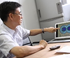
I decided to elucidate how Wnt signals are related to asymmetrical cell division in C. elegans. It was already known then that beta-catenin also functions as a transcription factor in C. elegans. I discovered that after cell division in C. elegans beta-catenin localizes to the posterior nucleus and stays there. In fact, beta-catenin also localizes to the anterior nucleus at first but is immediately ejected.
That’s correct. With the experiment, I demonstrated that Wnt signals are received into the cytoplasm via the cell posterior (tail) and localize APC protein at the cell anterior on the opposite (head). I also discovered that there are many microtubules following APC localization. In mammal cells, APC proteins exist together with microtubules to protect them. The same mechanism is found in C. elegans.
Moreover, integrating the results that I had accumulated up to that point, I constructed a model: Wnt signal -> Wnt receptor -> APC protein and microtubule localization at cell anterior -> beta-catenin localization to posterior nucleus -> asymmetrical cell division, to conduct partial verification. Firstly, a staining test enabled me to confirm the existence of APC protein at the edge of microtubules. In another experiment in which microtubules were destroyed by laser after the localization of APC protein and microtubules, the difference between the two nuclei in the quantity of beta-catenin that entering each of the nucleus after cell division became larger when microtubules were destroyed at the posterior, where their number is small from the beginning. On the other hand, the destruction of microtubules at the anterior resulted in even distribution of beta-catenin between the nuclei, that is, no quantitative asymmetry. As a result, I concluded that the numerical asymmetry of microtubules is related to the quantitative asymmetry of beta-catenin in the nuclei.
I also conducted an experiment with cells incapable of asymmetrical division because Wnt signals were not received. In those cells, numerical asymmetry of microtubules was artificially realized with laser. A clear difference was found between the two nuclei in the post-division quantity of beta-catenin. This clearly indicates that beta-catenin quantitative asymmetry is possible as long as microtubule asymmetry, which results asymmetrical cell division, is realized.
This way I succeeded in demonstrating that the model “Wnt signal -> Wnt receptor -> APC protein and microtubule localization at cell anterior -> beta-catenin localization to posterior nucleus -> asymmetrical cell division” is accurate. I am continuing with further analysis and verification because there are still unanswered questions such as why APC protein localized only at the cell anterior when Wnt signals are received. Could these research results be applied to the development of medicines? The mechanism involving Wnt signals in C. elegans is totally different from the Wnt signal pathway in mammals. However, they have one common point in that the key lies in gene transcription triggered by the function of APC and the localization in the nuclei of beta-catenin. I believe that a similar mechanism exists in mammals as well which is, I presume, hidden behind the known Wnt signal pathway. Moreover, I believe that the mechanism that I found, if destroyed, leads to oncogenesis of cells, just as the Wnt signal pathway is related to carcinogenesis. In this sense, it is quite possible that the research results leads to further clarification of the foundation of carcinogenesis.
At the moment, I am starting to examine how the mechanism involving Wnt signals is put into action in all the cells in C. eleganswith identical orientation. There are many phenomena of organisms in which all the cells are aligned or move in the same direction, but little is known as to why a particular direction is chosen. I am conducting analysis on the assumption that there is another key, in addition to Wnt signals. Through such research, I hope to elucidate eventually the whole program that governs the development of C. elegans from a fertilized egg to mature form.
(Interviewed by Naoko Nishimura on Jul. 28, 2011)
Kenji Sugioka, Kota Mizumoto and Hitoshi Sawa
Cell, 146 (6), 942-954, 16 September 2011 DOI: 10.1016/j.cell.2011.07.043


