The mechanism of cellular allometry: large spindles in large cells, small spindles in small cells

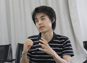
| Akatsuki KIMURA, D. Sc., Associate Professor, Cell Architecture Laboratory, Center for Frontier Research,National Institute of Genetics/ SOKENDAI. |
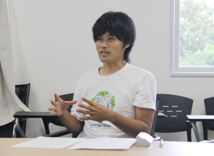
| Yuki HARA, Ph.D Student, Department of Genetics, SOKENDAI. (Graduate University for Advanced Studies) |
A human body is said to be made up of about 300 types of cells, which widely vary in size from 5 to 130 micrometers. What is invariable is the fact that organelles (small intracellular structures) such as nucleus and ribosomes are arranged similarly and that their sizes or numbers are proportional to the size of their host cell. Associate Professor Akatsuki KIMURA of the Cell Architecture Laboratory at the Center for Frontier Research has recently elucidated, together with Mr. Yuki Hara, the contribution of cell size to determine the size of an organelle called spindle, which is essential for inheritance of genetic information to progeny cells.
“Cellular allometry” means that the proportion of organelle size to cell size is maintained among cells with different sizes. Organelles such as nucleus, Golgi body, endoplasmic reticulum and mitochondria are commonly found in the cells of eukaryotic organisms, with the nucleus in the middle of the cell surrounded by the others. It has been measured that, when a cell is small, its nucleus is also small.

“I was originally interested in architecture and how a city is built, and this interest has led me to study biology,” says Dr. Kimura. He instinctively sensed some similarity between the structure of a city and that of a cell in that a city has an overall harmony of its own despite the fact that buildings are constructed there for individual occupants’ different reasons. He started his research with yeasts. “I used to study the mechanism of gene expression. Then I became strongly determined to pursue the question of spatial arrangement when I realized the importance of spatial organization of the chromosome in the process of transcription,” says Dr. Kimura, looking back on that moment in 2002.
Thinking that computational science is indispensable to work on spatial arrangement, Dr. Kimura went to acquire the basics of simulation and image analysis under Dr. Shuichi Onami, then at Keio University (presently at RIKEN). He changed his model organism to the nematode (C. elegans) and has since elucidated the mechanism of nuclear positioning to the center of the cells by analzing the time of fusion of ovum and spermium nuclei during fertilization.
He arrived at the Center for Frontier Research about three years ago in 2006. In fact, he applied for a post at the Center, which was opened to enable young researchers to carry out unique advanced research without any constraints.
“Prof. Yuji Kohara, the NIG’s Director General, said that I could freely name my laboratory when I was about to open it. So I decided to call it ‘Cell Architecture Laboratory,’” says Dr. Kimura. He has since been working with Mr. Hara, who has joined the laboratory as a doctoral student at SOKENDAI and other researchers, focusing on the dynamics of intracellular structures accompanying the cleavage (cell division) of fertilized nematode embryos. They have found that the spindle, which appears as the nucleus disappears during cell division, changes its size according to the cell size. He says, “we’ve confirmed that in the early stages of embryogenesis, in which a fertilized embryo continues to split from one cell into two cells and then four cells, with each cell becoming smaller and smaller, the length of the spindle was initially almost constant in all phase of cell division (that is, in cells of different sizes) but later it elongates proportionally to the cell size.”

Dr. Kimura and Mr. Hara have also studied the way microtubules are pulled from the cell membrane during spindle elongation. Microtubules are fibrous and radially extend from the two poles of the spindle toward the cell membrane. “We’ve found in analyses that a specific protein complex (made of heterotrimeric G protein and others) on the cell membrane regulates spindle elongation by adjusting the physical force that pulls microtubules.” They have also clarified that the spindle elongates less and more slowly in cells whose protein complex function is impaired. Moreover, they have succeeded in effectively explaining their experimentation results by computer-simulating the intracellular conditions based on the measurement data. As a result, they were the first in the world to propose that the number and location of protein complexes on the cell membrane constitute a “mechanism by which the cell learns its own size.”
It is known that abnormal segregation of genetic information during cell division trigger cancer and various other diseases. Dr. Kimura says, “our research achievements can shed new light on cell division, a fundamental phenomenon of life, and provide clues to elucidating the pathology of cancer and other diseases.” He is all the more motivated for his research in which he hopes to verify how seemingly local molecular behaviors maintain the global order in the cells by continuously building simulated models.
(Interviewed by Naoko Nishimura on Aug. 5, 2009)
Cell-size dependent spindle elongation in the Caenorhabditis elegans early embryo
Yuki Hara and Akatsuki Kimura, Current Biology, Volume 19, Issue 18, 1549-1554, 2009. DOI: 10.1016/j.cub.2009.07.050
Back


