


Sakai Group / Model Fish Genetics Laboratory
Biallelic TEDC1 variants cause a new syndrome with severe growth impairment and endocrine complications
Noriko Miyake, Kentaro Shiga, Yuya Hasegawa, Chisato Iwabuchi, Kohei Shiroshita, Hiroshi Kobayashi, Keiyo Takubo, Fabien Velilla, Akiteru Maeno, Toshihiro Kawasaki, Yukiko Imai, Noriyoshi Sakai, Tomonori Hirose, Atsushi Fujita, Hidehisa Takahashi, Nobuhiko Okamoto, Mikako Enokizono, Shiho Iwasaki, Syuichi Ito, Naomichi Matsumoto
European Journal of Human Genetics 2025 Feb 20. DOI:10.1038/s41431-025-01802-3
We encountered two affected male patients born to non-consanguineous parents, who presented with prenatal-onset severe growth impairment, primary microcephaly, developmental delay, adrenal insufficiency, congenital glaucoma, delayed bone aging, craniosynostosis, congenital tracheal stenosis, and primary hypogonadism. By exome sequencing, we identified compound heterozygous TEDC1 variants (NM_001134877.1 c.[104-5C>G];[787delG] p.[?];[(Ala263LeufsTer29)] in both affected siblings. We confirmed that the splice site variant, c.104-5C>G leads to no TEDC1 protein production via nonsense-mediated mRNA decay. The frameshift variant located in the last coding exon, c.787delG, produces a C-terminally truncated protein, which impairs the binding with TEDC2. Thus, both variants are thought to be loss-of-function. TEDC1 and TEDC2 are both required for centriole stability and cell proliferation. Our in vitro experiments using patient-derived cells revealed cell cycle abnormality. Our in vivo study using tedc1−/− zebrafish generated by CRISPR/Cas9 successfully recapitulated the growth impairment and cranial bone dysplasia as seen in our patients. The tedc1−/− mutant zebrafish were sterile and did not have developed gonads. Furthermore, we showed that biallelic TEDC1 deletion causes cilia abnormalities through defective acetylated tubulins.
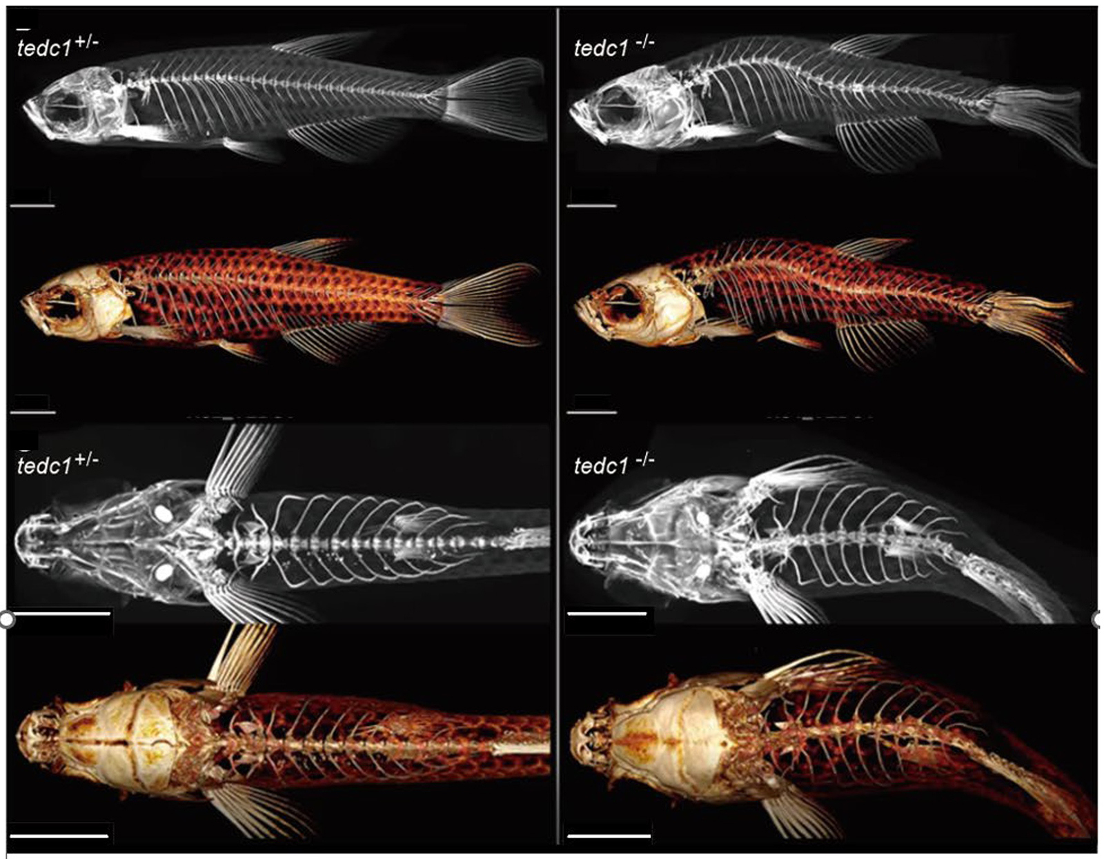
Figure: Representative lateral and upper view µCT images of tedc1+/− and tedc1−/− zebrafish


