


Press release
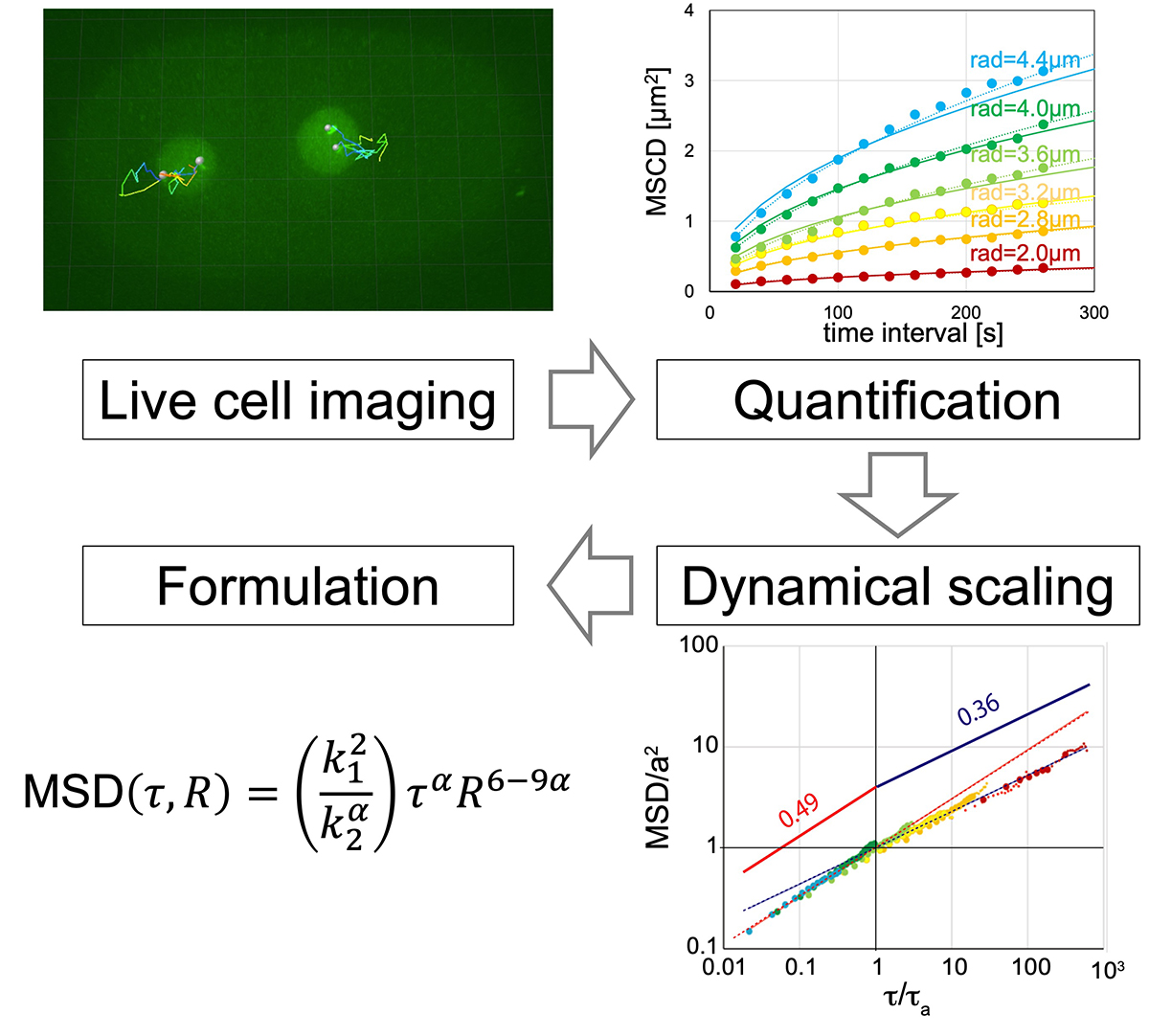
Formulation of chromatin mobility as a function of nuclear size during C. elegans embryogenesis using polymer physics theories.
Aiya K. Yesbolatova, Ritsuko Arai, *Takahiro Sakaue, and *Akatsuki Kimura. *Corresponding authors
Physical Review Letters (2022) 128, 178101 DOI:10.1103/PhysRevLett.128.178101
![]() Press release (In Japanese only)
Press release (In Japanese only)
By combining theory and experiment, Aiya K. Yesbolatova and her collaborators provided unambiguous evidence that the chromatin in early embryos obeys the universal dynamics predicted by the polymer physics. Although the chromatin dynamics is believed to play a key role in controlling the gene expression, its quantitative characterization has been elusive mainly due to the complexity in living cells. The findings and formulation help researchers quantify the chromatin motion in living cells, thus laying the foundation for future research in this biologically important problem.
Kanemaki Group / Molecular Cell Engineering Laboratory
MCMBP promotes the assembly of the MCM2–7 hetero-hexamer to ensure robust DNA replication in human cells
Yuichiro Saito, Venny Santosa, Kei-ichiro Ishiguro and Masato T. Kanemaki.
eLife (2022) 11, e77393 DOI:10.7554/eLife.77393
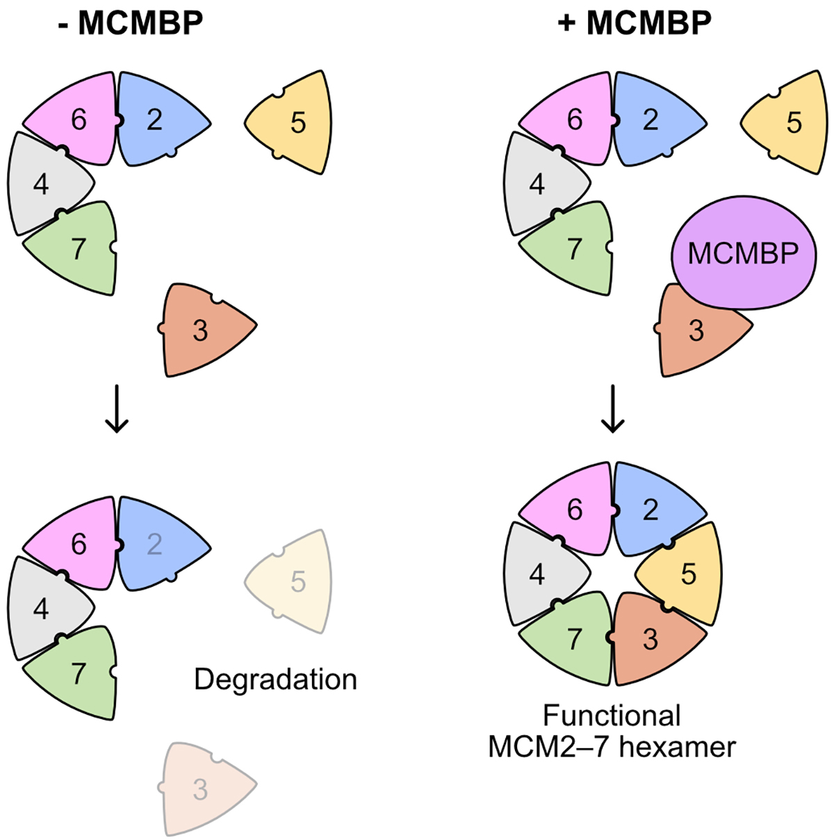
Replication of the genetic material DNA is essential for cell proliferation. The MCM2–7 hexamer, a ring-like complex composed of six subunits from MCM2 to MCM7, functions as the replicative helicase for unwinding double-stranded DNA. It is known that a large amount of the MCM2–7 hexamer is required for efficient DNA replication in the S phase. However, how the MCM2–7 hexamer is assembled has not been understood.
This paper shows that the MCM-binding protein (MCMBP) binds to the MCM subunits and plays a crucial function in incorporating MCM3 and MCM5 into the hexamer (Figure 1). Rapid degradation of MCMBP using the auxin-inducible degron 2 system (AID2) resulted in a reduced expression of the functional MCM2–7 hexamer at each cell division because newly synthesized MCM3 was not incorporated into the hexamer.
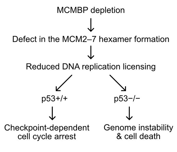
Interestingly, when the level of MCM2–7 hexamer decreased, human cells expressing the tumor suppressor gene p53 maintained genome integrity by transiently arresting the cell cycle in the G1 phase (Fig. 2). In contrast, cells lacking p53 induced cell death by entering the S phase with fewer hexamers resulting in incomplete DNA replication. These results suggest that depletion of MCMBP may specifically eliminate cancer cells with mutations in p53.
The Kanemaki Laboratory at National Institute of Genetics led this research in collaboration with Prof. Kei-ichiro Ishiguro at Kumamoto University.


