Saga Group / Mammalian Development Laboratory
Formal proof of the requirement of MESP1 and MESP2 in mesoderm specification and their transcriptional control via specific enhancers in mice
Rieko Ajima, Yuko Sakakibara, Noriko Sakurai-Yamatani, Masafumi Muraoka and Yumiko Saga
Development (2021) 148, dev194613 DOI:10.1242/dev.194613
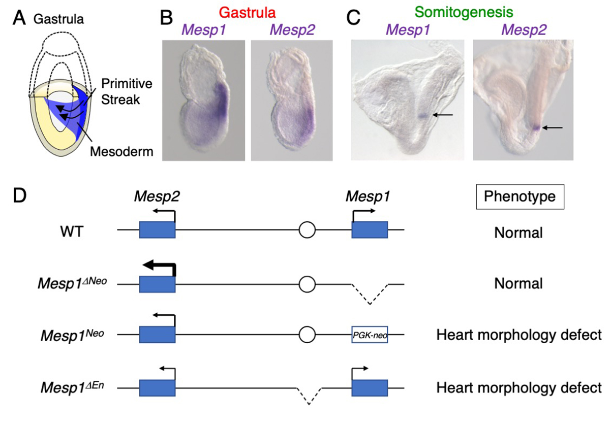
The mesodermal cells are derived from the primitive streak during gastrulation in mouse development (Figure A). These mesodermal cells give rise to the heart, somites, kidney, and limbs. MESP1 and MESP2 are transcriptional factors expressed in early mesoderm and boundary of newly forming somites (Figure B, C). The Mesp1/Mesp2 dKO embryos exhibited markedly severe mesoderm formation defects. However, MESP1 and MESP2 were thought to regulate distinct targets, because Mesp1 and Mesp2 single KO embryos display distinct phenotypes, heart morphogenesis defects and somite formation defects, respectively.
In this study, we established the Mesp1/Mesp2 mutants using genome editing techniques, and found the Mesp1/Mesp2 dKO embryos exhibited defects similar to the original Mesp1/Mesp2 dKO embryos. However, Mesp1 KO did not display any phenotypes. We noted up-regulation of Mesp2 in the Mesp1 KO embryos, suggesting that MESP2 rescues the loss of MESP1 in mesoderm specification. We also found that Mesp1 and Mesp2 expression in the early mesoderm is regulated by the common enhancer. Deletion of the enhancer caused the down-regulation of both genes, resulting in heart formation defects. This study suggests dosage-dependent roles of MESP1 and MESP2 in early mesoderm formation.