


DNA Binding by the Mis4Scc2 Loader Promotes Topological DNA Entrapment by the Cohesin Ring.
Yumiko Kurokawa and Yasuto Murayama.
Cell Reports 33, 108357(2020) DOI:10.1016/j.celrep.2020.108357
The ring-shaped cohesin complex is a member of multi-subunit SMC ATPases, which play vital roles in numerous aspects of chromosome biology including mitotic chromosome segregation, global chromosome organization, transcriptional regulation and DNA repair. Cohesin topologically encircles DNA and is thought to establish series of chromosomal interactions including sister chromatid cohesion by tethering more than one DNA molecule. Using biochemical reconstitution, we show that the ability of the loader to bind DNA plays a critical role in promoting cohesin loading. Two distinct sites within the Mis4Scc2 subunit were found to cooperatively bind DNA. Mis4Scc2 initially forms a tertiary complex with cohesin on DNA and promotes subsequent topological DNA entrapment by cohesin through its DNA binding activity. Furthermore, we show that mutations in the two DNA binding sites of Mis4 impair the chromosomal loading of cohesin. These observations demonstrate the physiological importance of DNA binding by the loader and provide mechanistic insights into the process of topological cohesin loading.
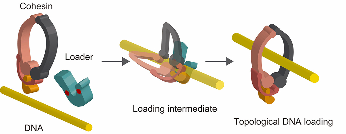
Figure: A model of topological cohesin loading onto DNA mediated by the loader complex. Cohesin forms a tertiary complex with the loader complex on DNA. This stimulates subsequent conformational change of cohesin resulting in topological DNA entrapment.
Press release
The auxin-inducible degron 2 technology provides sharp degradation control in yeast, mammalian cells, and mice
A Yesbolatova, Y Saito, N Kitamoto, H Makino-Itou, R Ajima, R Nakano, H Nakaoka, K Fukui, K Gamo, Y Tominari, H Takeuchi, Y Saga, K Hayashi, MT Kanemaki
Nature Communications 11, 5701(2020) DOI:10.1038/s41467-020-19532-z
Press release (In Japanese only)
To elucidate the role of a protein of interest in living cells, it is useful to rapidly deplete it to see what would happen to the cells. It should also be possible to control the protein function if we can timely deplete it. Because of the above reasons, techniques inducing artificial proteins degradation, known as protein knockdown, are drawing more attention.
Prof. Kanemaki at NIG and his colleagues established the AID2 system by which a protein of interest fused with a tag can be induced for rapid degradation in yeast, mammalian cells, and mice. To induce target degradation, a new chemical ligand called 5-Ph-IAA, and a mutant of the TIR1 ubiquitin ligase were used. It is now possible to induce target depletion rapidly and timely when needed. AID2 will be useful not only in basic life science, but also in medical science and drug discovery in the future.
This study was carried out by the Kanemaki and Saga groups both at NIG, Prof. Kenichiro Hayashi at Okayama University of Science, Dr Haruki Takeuchi at the University of Tokyo, Dr Hirofumi Nakaoka at Sasaki Institute, and FIMECS Inc.
This study was supported by JSPS KAKENHI grants (16K15095, 18H02170, 18H04719, 20H05396), JST A-STEP (AS2915150U), The Takeda Science Foundation, The Asahi Glass Foundation, and an AMED NBRP Fundamental Technologies Upgrading Program.
This study was published in Nature Communications on November 11th at 19:00 (JST).
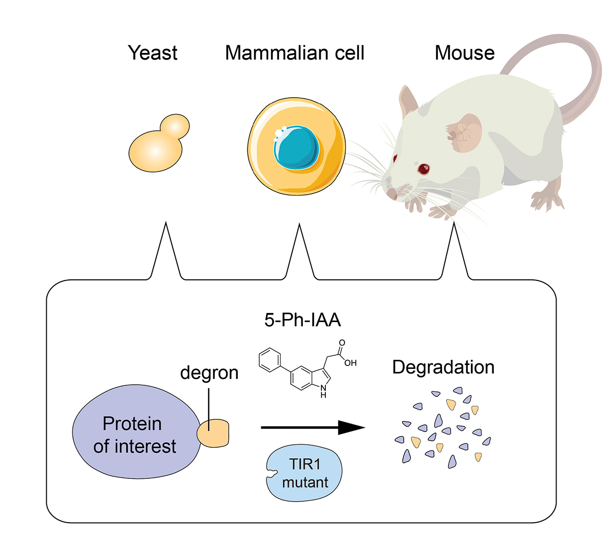
Figure: AID2 enables rapid target depletion in yeast, mammalian cells, and mice.
Press release
Genetical control of 2D pattern and depth of the primordial furrow that prefigures 3D shape of the rhinoceros beetle horn.
H Adachi, K Matsuda, T Niimi, S Kondo, H Gotoh
Scientific Reports 10, 18687 (2020) DOI:10.1038/s41598-020-75709-y
Press release (In Japanese only)
The head horn of the Asian rhinoceros beetle develops as an extensively folded primordium before unfurling into its final 3D shape at the pupal molt. The information of the final 3D structure of the beetle horn is prefigured in the folding pattern of the developing primordium. However, the developmental mechanism underlying epithelial folding of the primordium is unknown. In this study, we addressed this gap in our understanding of the developmental patterning of the 3D horn shape of beetles by focusing on the formation of furrows at the surface of the primordium that become the bifurcated 3D shape of the horn. By gene knockdown analysis via RNAi, we found that knockdown of the gene Notch disturbed overall horn primordial furrow depth without affecting the 2D furrow pattern. In contrast, knockdown of CyclinE altered 2D horn primordial furrow pattern without affecting furrow depth. Our results show how the depth and 2D pattern of primordial surface furrows are regulated at least partially independently during beetle horn development, and how both can alter the final 3D shape of the horn.
Source: H Adachi, et al., Scientific Reports 10, 18687 (2020) DOI:10.1038/s41598-020-75709-y
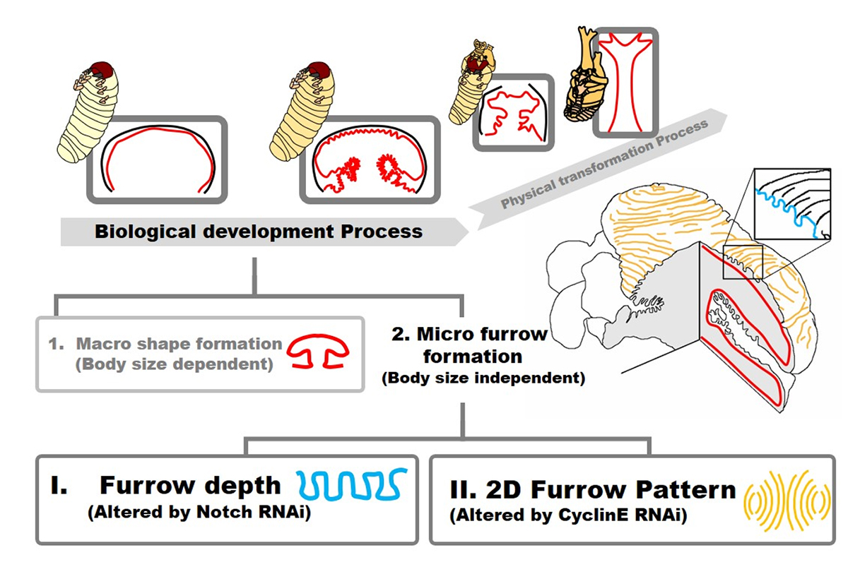
Figure: Summary of beetle horn development focusing on folding of horn primordia. The final 3D shape of horn has been already prefigured when the development of folding is done. In this study, we demonstrated that the development of primordia folding can be divided into (1) macro structure formation and (2) micro furrow formation. Micro furrow formation can be further divided into depth regulation and 2D pattern formation via Notch and CyclinE control, respectively.


