


Kawakami Group / Laboratory of Molecular and Developmental Biology
Comparative Genomics Laboratory
NOGUCHI, Hideki / Center for Genome Informatics(CGI) / Advanced Genomics Center
The Genetic Basis of Morphological Diversity in Domesticated Goldfish
Tetsuo Kon, Yoshihiro Omori, Kentaro Fukuta, Hironori Wada, Masakatsu Watanabe, Zelin Chen, Miki Iwasaki, Tappei Mishina, Shin-ichiro S. Matsuzaki, Daiki Yoshihara, Jumpei Arakawa, Koichi Kawakami, Atsushi Toyoda, Shawn M. Burgess, Hideki Noguchi, Takahisa Furukawa.
Current Biology 30, 1-15 (2020). DOI:10.1016/j.cub.2020.04.034
Although domesticated goldfish strains exhibit highly diversified phenotypes in morphology, the genetic basis underlying these phenotypes is poorly understood. Here, based on analysis of transposable elements in the allotetraploid goldfish genome, we found that its two subgenomes have evolved asymmetrically since a whole-genome duplication event in the ancestor of goldfish and common carp. We conducted whole- genome sequencing of 27 domesticated goldfish strains and wild goldfish. We identified more than 60 million genetic variations and established a population genetic structure of major goldfish strains. Genome-wide association studies and analysis of strain-specific variants revealed genetic loci associated with several goldfish phenotypes, including dorsal fin loss, long-tail, telescope-eye, albinism, and heart-shaped tail. Our results suggest that accumulated mutations in the asymmetrically evolved subgenomes led to generation of diverse phenotypes in the goldfish domestication history. This study is a key resource for understanding the genetic basis of phenotypic diversity among goldfish strains.
This work was carried out as collaboration between Osaka Univ and NIG (Lab of Molecular and Developmental Biology, Comparative Genomics Lab and Advanced Genomics Center) and supported by the NIG-JOINT program
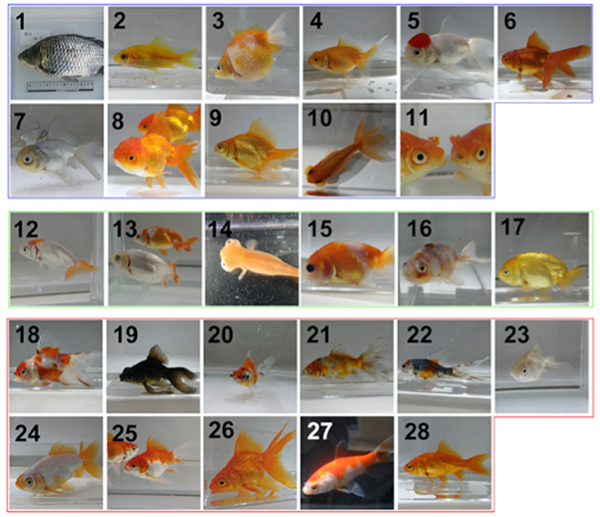
Figure: Wild type (1) and mutant (2-28) goldfish. Genome analysis revealed China (2-11), Ranchu (12-17) and Edo (18-28) groups.
RIF1 Controls Replication Initiation and Homologous Recombination Repair in a Radiation Dose-Dependent Manner
Yuichiro Saito, Junya Kobayashi, Masato T. Kanemaki, Kenshi Komatsu
Journal of Cell Science 2020 May 20. DOI:10.1242/jcs.240036
Genomic DNA is challenged by various types of stresses. Ionizing radiation (IR) cut DNA, generating a DNA damage called a DNA double-strand break (DSB). DSB is repaired via two pathways; an error-prone, ‘non-homologous end joining (NHEJ) pathway’ or a precise, ‘homologous recombination repair (HRR) pathway’. To maintain genome integrity, it is important for cells to choose an appropriate pathway for repairing DSBs.
It is believed that human cells dominantly use error-prone NHEJ to repair DSB. This idea came from the observations using high doses of IR. However, it has not been known whether the predominant use of NHEJ is applicable to DSBs induced by a low dose of IR. To answer this question, we used a low or high dose of IR and monitored the repair pathway. We found that HRR played a prominent role to repair DSBs after a low dose of IR, and it was suppressed after a high dose of IR. These results show that human cells sense radiation dose and choose an appropriate repair pathway. Furthermore, we identified RIF1 as a critical factor that controls HRR activity in an IR-dose dependent manner. We expect that these results would provide basic understandings of how radiation kills cancer cells in radiation therapy.
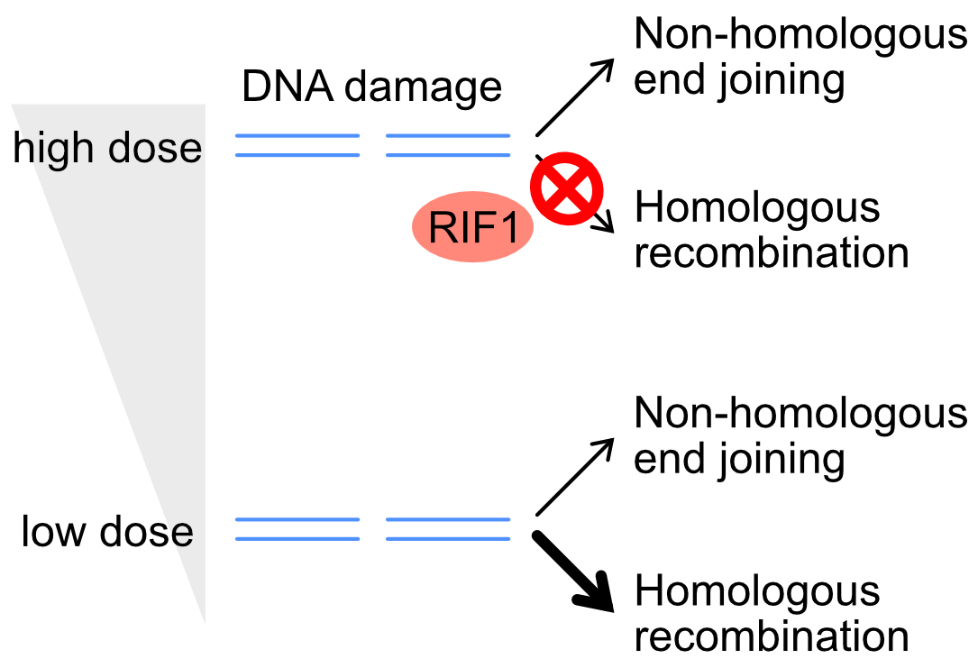
Figure: DNA double-strand break is repaired via the non-homologous end joining (NHEJ) or homologous recombination repair (HRR) pathway. RIF1 suppresses HRR only after a high dose of IR, directing cells towards using the NHEJ pathway.
Changes in the transcriptome, ploidy, and optimal light intensity of a cryptomonad upon integration into a kleptoplastic dinoflagellate
Ryo Onuma, Shunsuke Hirooka, Yu Kanesaki, Takayuki Fujiwara, Hirofumi Yoshikawa, Shin-ya Miyagishima
The ISME Journal (2020) DOI:10.1038/s41396-020-0693-4
Link to “Behind the Paper” the Nature Research Microbiology Community
Endosymbiosis of unicellular eukaryotic algae into previously nonphotosynthetic eukaryotes has established chloroplasts in several eukaryotic lineages. Additionally, certain unicellular organisms in several different lineages ingest algae and utilize them as temporal chloroplasts (kleptoplasts) for weeks to months before digesting them. Among these organisms, the dinoflagellate Nusuttodinium aeruginosum ingests the cryptomonad Chroomonas sp. and enlarges the kleptoplast with the aid of the cryptomonad nucleus. To understand how the cryptomonad nucleus is remodeled in the dinoflagellate, here we examined changes in the transcriptome and ploidy of the ingested nucleus. We show that, after ingestion, genes involved in metabolism, translation, and DNA replication are upregulated while those involved in sensory systems and cell motility are downregulated. In the dinoflagellate cell, the cryptomonad nucleus undergoes polyploidization that correlates with an increase in the mRNA levels of upregulated genes. In addition, the ingested nucleus almost loses transcriptional responses to light. Because polyploidization and loss of transcriptional regulation are also known to have occurred during the establishment of endosymbiotic organelles, these changes are probably a common trend in endosymbiotic evolution. Furthermore, we show that the kleptoplast and dinoflagellate are more susceptible to high light than the free-living cryptomonad but that the ingested nucleus reduces this damage.
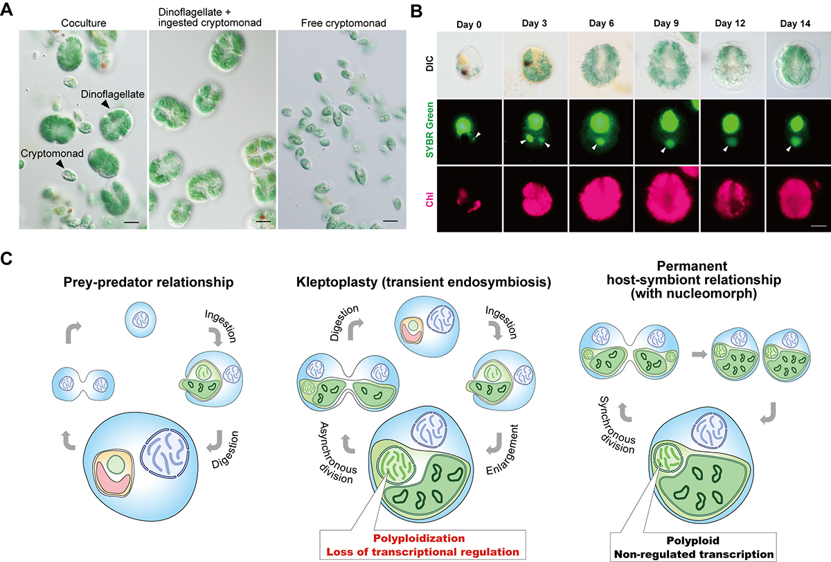
Figure: Kleptoplasty in Nusuttodinium aeruginosum.
(A) Micrographs showing the coculture of N. aeruginosum cells with Chroomonas sp.-derived kleptoplasts and free Chroomonas sp. (left). From the coculture, N. aeruginosum cells with the kleptoplasts (middle) were separated from free Chroomonas sp. (right) by filtration. Scale bar = 10 μm.
(B) Micrographs showing the change in the nucleus and kleptoplast derived from Chroomonas sp. in N. aeruginosum cells. Cells were stained with SYBR Green. Images of differential interference contrast (DIC), SYBR Green staining, and kleptoplast red fluorescence (Chl) are shown. The arrowhead indicates a nucleus derived from Chroomonas sp. The cell in the image from day 3 ingested two nuclei. Scale bar = 10 μm.
(C) Schematic comparison of prey–predator, transient endosymbiotic (kleptoplasty), and permanent endosymbiotic relationships. Predators start digesting algae immediately after phagocytic ingestion of algae (left). Kleptoplastic species retain the ingested algae for days to weeks in the cell before digesting them in some cases, including that of N. aeruginosum, with the nucleus of the algae (middle). The present study shows that the ingested nucleus is polyploidized in the host cell, which increases mRNA levels. The ingested nucleus almost loses transcriptional regulation. These changes were also common to the course of establishment of nucleomorph and chloroplast (also other types of plastids) as obligate endosymbionts in eukaryotes (right).


