


Motor Neural Circuit Laboratory • Hirata Group
RING finger protein 121 facilitates the degradation and membrane localization of voltage-gated sodium channels
Ogino, K., Low, S. E., Yamada, K., Saint-Amant, L., Zhou, W., Muto, A., Asakawa, K., Nakai, J., Kawakami, K., Kuwada, J. Y., and Hirata, H.Voltage-gated sodium channels (NaV) are known to form clusters at the membranes of excitable cells; however, what governs their transport is largely unknown. We found that the endoplasmic reticulum (ER) and cis-Golgi associated ubiquitin ligase really interesting new gene (RING) finger protein 121 (RNF121) mediates the degradation and membrane localization of NaV. This apparent quality control of NaV ensures the transport of properly folded channels to the membranes of excitable cells. To our knowledge, this is the first pathologically relevant identification of a voltage-gated ion channel as a substrate for ER-associated protein degradation, whose degradation is governed by an ER- and Golgi-associated E3-ubiquitin ligase.
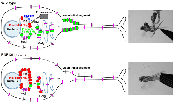
A wild-type neuron wherein RNF121 mediates ubiquitination of misfolded NaV channels marking them for proteasome-mediated degradation. Properly folded NaV channels (green) associate with NaVβ subunits (magenta) in the Golgi apparatus and are transported to the axon initial segment. Of note, some NaVβ subunits are transported to the membrane independent of NaV channels. An RNF121-deficient neuron wherein misfolded NaV channels (red) accumulates in the ER and cis-Golgi compartments, which, over time, depletes NaVβ subunits, preventing them from forming complexes with properly folded NaV channels, causing an impairment of NaV transport.
Power law relationship between cell cycle duration and cell volume in the early embryonic development of Caenorhabditis elegans
Yukinobu Arata, Hiroaki Takagi, Yasushi Sako, and Hitoshi SawaCell cycle is regulated in coordination with cell size. Yoshio Masui at Toronto University reported that the cell cycle duration is inversely proportional to the cell radius squared after the MBT in Xenopus embryo. They proposed that cell surface area is a candidate to determine cell cycle duration. However, it remains unknown whether this “second power law” is conserved in other animal embryos. Here, we found that the relationship between cell cycle duration and cell size in Caenorhabditis elegans embryos exhibited a power law distribution. The powers of the time-size relationship could be grouped into at least three classes according to C. elegans founder cell lineages. Any of the values was less than that in Xenopus embryos. Interestingly, we found that the relationship between cell and nuclear volumes exhibited a power law. Supposing interactions between nuclease and cytoplasm, we considered the volume ratio between nucleus and cell. The value of power in the volume ratio was almost identical to that of the time-size relationship in highly-size correlated lineages. We propose a new model that cell cycle duration in C. elegans embryos is determined by a geometric constraint between intercellular structures such as nucleus and cytoplasm. It remains possible that cell cycle durations in other animal embryos are determined by the same constraint.
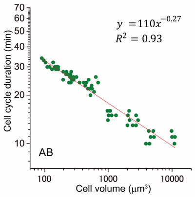
Cell cycle duration and cell volume in the AB lineage exhibited a linear relationship in a double logarithmic plot (power law).


