


Press release
Efficient open cultivation of cyanidialean red algae in acidified seawater
Shunsuke Hirooka, Reiko Tomita, Takayuki Fujiwara, Mio Ohnuma, Haruko Kuroiwa, Tsuneyoshi Kuroiwa and Shin-ya Miyagishima
Scientific Reports (2020)10: 13794 DOI:10.1038/s41598-020-70398-z
Press release (In Japanese only)
Microalgae possess high potential for producing pigments, antioxidants, and lipophilic compounds for industrial applications. However, their open pond cultures are often contaminated by other undesirable organisms, including their predators. In addition, the cost of using freshwater is relatively high, which limits the location and scale of cultivation compared with using seawater. It was previously shown that Cyanidium caldarium and Galdieria sulphuraria, but not Cyanidioschyzon merolae grew in media containing NaCl at a concentration equivalent to seawater. We found that the preculture of C. merolae in the presence of a moderate NaCl concentration enabled the cells to grow in the seawater-based medium. The cultivation of cyanidialean red algae in the seawater-based medium did not require additional pH buffering chemicals. In addition, the combination of seawater and acidic conditions reduced the risk of contamination by other organisms in the nonsterile open culture of C. merolae more efficiently than the acidic condition alone.
Source: S. Hirooka, et al., Scientific Reports (2020)10: 13794 DOI:10.1038/s41598-020-70398-z
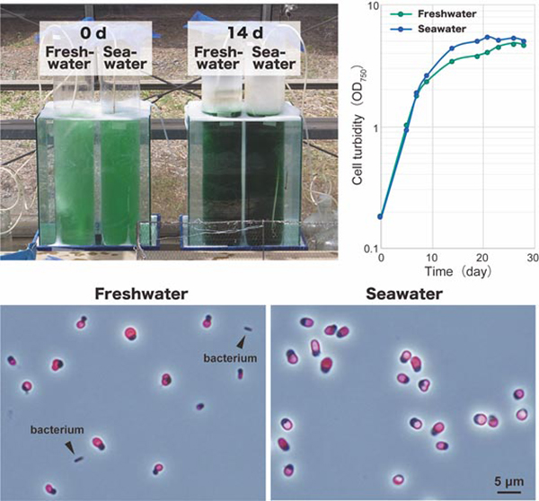
Fig: Outdoor cultivation of C. merolae in 7 L of nonsterile a freshwater-based medium and a seawater-based medium at day 0 and day 14. C. merolae cells in the seawater-based medium grew along a similar time course and to similar amounts compared with cells cultured in the freshwater-based medium. The microscopic observation of cultures 14 days after inoculation showed that the culture in the freshwater-based medium was contaminated with bacteria. In contrast, no bacteria or organisms other than C. merolae were observed in the culture in the seawater-based medium. Images were obtained by phase contrast microscopy, and the fluorescence images of chloroplasts (red) were overlaid.


