


![]()
Identification of a neuronal population in the telencephalon essential for fear conditioning in zebrafish
Pradeep Lal, Hideyuki Tanabe, Maximiliano L. Suster, Deepak Ailani, Yuri Kotani, Akira Muto, Mari Itoh, Miki Iwasaki, Hironori Wada, Emre Yaksi , and Koichi Kawakami
BMC Biology Published: 25 April 2018 DOI:10.1186/s12915-018-0502-y
EurekAlert! link about this artcle
Have you ever wondered why animals avoid dangers by sensing some “signs” possibly related to the danger? A simple form of this phenomenon is called “fear conditioning”, which is a type of learning commonly seen in every animal on the earth. By manipulating activity of specific neurons of the zebrafish brain, scientists at the National Institute of Genetics (NIG) in Japan have elucidated a neuronal population essential for fear conditioning in zebrafish. The study, published in the April 25 issue of BMC Biology, suggests that such a neural circuit essential for fear conditioning exists and is conserved during vertebrate evolution.
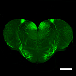
Figure: A section of the zebrafish telencephalon. The neurons essential for fear conditioning are illuminated with GFP (green fluorescence protein). Scale bars: 200 μm.
Video 1: Fear conditioning of zebrafish. The fish was placed in a plastic box with two compartments. 10 seconds after LED was on, an electric shock was given (day 1). This was repeated 10 times a day for five consecutive days. On day 5, when LED was on, the fish escaped to another compartment.
Video 2: 3D image of the neurons essential for fear conditioning. Transparent brain was created and analyzed by light-sheet microscopy.
Epidermal regulation of bone morphogenesis through the development and regeneration of osteoblasts in the zebrafish scale.
Iwasaki M., Kuroda J., Kawakami K., Wada H.
Developmental Biology 437, 105-119, 2018. DOI:10.1016/j.ydbio.2018.03.005
The scale of the teleost fish is a dermal bone embedded in the skin. We investigated the mechanisms of bone patterning in the zebrafish scale by using transgenic lines that visualize osteoblasts. We showed that the zebrafish scale contains two distinct types of osteoblasts: a monolayer sheet of central osteoblasts along the surface of scales; and marginal osteoblasts elongated along the scale edge (Fig. A). During scale growth, central osteoblasts progressively increase in size without cell proliferation. Sonic hedgehog (shh) is expressed in the epidermal cells overlying marginal osteoblasts. Inhibition of Hh signaling reduces the number of marginal osteoblasts and interferes with scale growth. Moreover, inhibition of Wnt/planar cell polarity (PCP) signaling in the epidermis caused a misorientation of scales (Fig. B), correlated to the altered expression pattern of shh. This study reveals a novel role of the epidermis in the regulation of bone patterning.
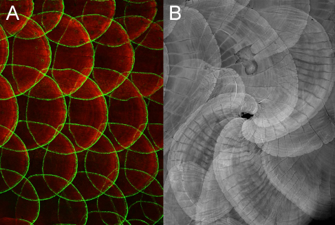
Figure: (A) Gal4 enhancer trap line (hspGFFDMC13F;UAS:GFP) expressing GFP in a specific population of osteoblasts along the scale edge. (B) Disrupted Wnt/PCP signaling in the epidermis causes a misorientation of scales, indicating a role of the epidermis in bone patterning.


