


Press release
Chromosome engineering allows the efficient isolation of vertebrate neocentromeres
Wei-Hao Shang, Tetsuya Hori, Nuno M.C. Martins, Atsushi Toyoda, Sadahiko Misu, Norikazu Monma, Ichiro Hiratani, Kazuhiro Maeshima, Kazuho Ikeo, Asao Fujiyama, Hiroshi Kimura, William C. Earnshaw, and Tatsuo FukagawaCentromeres are specified by sequence-independent epigenetic mechanisms in most organisms. Rarely, centromere repositioning results in neocentromere formation at ectopic sites. However, the mechanisms governing how and where neocentromeres form are unknown.
Here, we established a chromosome-engineering system in chicken DT40 cells that allowed us to efficiently isolate neocentromere-containing chromosomes.
Neocentromeres appear to be structurally and functionally equivalent to native centromeres. Chromatin immunoprecipitation sequencing (ChIP-seq) analysis with 18 neocentromeres revealed that the centromere-specific histone H3 variant CENP-A occupies an ∼40 kb region at each neocentromere, which has no preference for specific DNA sequence motifs. Furthermore, we found that neocentromeres were not associated with histone modifications H3K9me3, H3K4me2, and H3K36me3 or with early replication timing. Importantly, low but significant levels of CENP-A are detected around endogenous centromeres, which are capable of seeding neocentromere assembly if the centromere core is removed.
Our experimental system provides valuable insights for understanding how neocentromeres form. This was performed by Wei-Hao Shang and Tetsuya Hori (Fukagawa Lab) in collaboration with TRIC of ROIS, Fujimaya Lab. (NIG, NII), Maeshima Lab (NIG), Ikeo Lab. (NIG), Kimura Lab (Osaka U.), and Eranshaw Lab (U. Edinburgh).
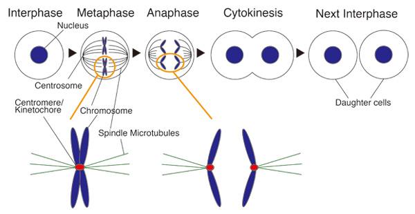
Cell division and centromere
During mitosis spindle microtubules capture a special structure of chromosome for faithful chromosome segregation. This structure is called “Kinetochore”. Centromere is a genome region in which kinetochore is formed. Dysfunction of kinetochore results in some diseases including cancer.
Division of Molecular and Developmental Biology・Kawakami Group
Various types of sense organs are distributed over the body surface. These organs are innervated by sensory axons, thereby transmit information to the central nervous system. Many sense organs emit molecules required for proper growth or guidance of the axons. However, roles of the axons for development of sense organs remain poorly understood. In this study, we reveal that proliferation of the mechanosensory organs (neuromasts) of fish is promoted by axonal innervation.
In adult zebrafish, the neuromasts give rise to new neuromasts by budding and generate a cluster of organs. The budding cells, that show high Wnt signaling activity, are associated by side branches of axons extended from the founder neuromast (Fig. 1A). To analyze the role of innervation, we ablated sensory neurons in larvae. The neuromast sent off budding cells normally, but subsequent cell proliferation to generate new sense organs did not take place (Fig. 1B,C). We propose that the axon promotes Wnt signaling activity, which is required for the proliferate phase that leads to sense organ formation.
This study was carried out in collaboration with Dr. Alain Ghysen (Montpellier University) and funded by the PRESTO of the Japan Science and Technology Agency.
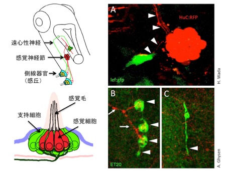
(A) The lateral line sense organ, neuromast, sends off proliferative bud cells, that show high Wnt signaling activity. Axons are indicated by arrowheads.
(B, C) The cell proliferation does not take place in the absence of the sensory axons.


