
Maeshima Group / Genome Dynamics Laboratory
Machine learning-assisted Repli-Histo labeling reveals distinct transcription-dependent constraints on chromatin motion in living cells
Katsuhiko Minami, Kako Nakazato, Kai Shimizu, Sachiko Tamura, Hiroshi Kimura, S. S. Ashwin, Kazuhiro Maeshima*
*corresponding author
Cell Reports DOI: 10.1016/j.celrep.2026.118235
Genomic DNA in the cell nucleus is wrapped around core histone proteins to form nucleosomes and is organized as chromatin together with other proteins and RNAs. Chromatin is not a static structure: nucleosomes continuously move in living cells. Previous studies have shown that transcription can influence chromatin motion (https://rupress.org/jcb/article/218/5/1511/120924/Single-nucleosome-imaging-reveals-loose-genome), but whether this effect is the same throughout the nucleus has remained unclear.
A research group led by Katsuhiko Minami (Project Researcher; formerly a SOKENDAI graduate student and currently at Harvard University), Kako Nakazato (SOKENDAI graduate student and JSPS DC2 Fellow), Kai Shimizu (SOKENDAI graduate student), Sachiko Tamura (Technical Staff), and Kazuhiro Maeshima (Professor) at the Genome Dynamics Laboratory, National Institute of Genetics, in collaboration with Hiroshi Kimura (Professor) at the Institute of Science Tokyo and S. S. Ashwin (Associate Professor) of GITAM University, investigated how different transcription systems affect the motion of distinct chromatin regions in living human cells.
The researchers combined two technologies. The first was Repli-Histo labeling, a method previously developed by the group that distinguishes four chromatin classes, ranging from euchromatin (Classes IA and IB) to heterochromatin (Classes II and III) (Minami et al., Science Advances, 2025; https://www.science.org/doi/10.1126/sciadv.adu8400). They further developed a machine-learning classifier to identify these chromatin classes objectively (Figure, upper). The second technique was single-nucleosome imaging, which allowed the team to directly track the movement of individual nucleosomes in living cells. This combination revealed that nucleosome motion becomes progressively more constrained from euchromatin toward heterochromatin. More importantly, transcription did not constrain chromatin uniformly throughout the nucleus (Figure, upper).
When RNA polymerase II (Pol II), which transcribes most protein-coding genes, was inhibited using three different inhibitors, nucleosome motion increased in euchromatin (Classes IA and IB). Increased motion was also observed in heterochromatin around nucleoli. In contrast, heterochromatin at the nuclear periphery showed little change (Figure, lower).
The researchers then examined RNA polymerase I (Pol I), which produces the major ribosomal RNAs inside nucleoli. Pol I inhibition produced a different response: it selectively increased nucleosome motion in a late-replicating Class III heterochromatin population associated with nucleoli (Figure, lower).
These results show that transcription does not simply make chromatin more or less dynamic throughout the nucleus. Instead, different transcription systems impose physical constraints on different chromatin regions, depending on both chromatin state and nuclear location. The findings also highlight the special environment surrounding nucleoli, where Pol II and Pol I transcription influence the motion of different chromatin classes.
This work was supported by JSPS and MEXT KAKENHI Grants (JP24H00061, and JP25K24664), the Takeda Science Foundation, JST SPRING (JPMJSP2104), JSPS Research Fellowships (JP23KJ0998 and JP26KJ1223), the JSPS Overseas Research Fellowship, and the Yamada Science Foundation. Sequencing and data analysis were also supported by the Platform for Advanced Genome Science (PAGS; JP22H04925).
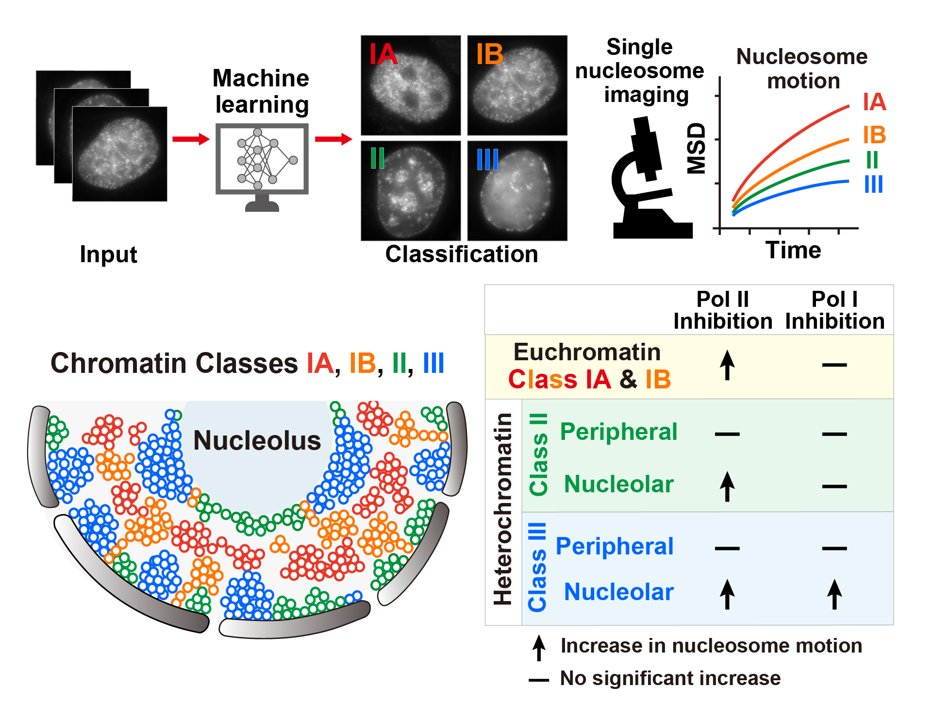
Figure: Machine learning-assisted imaging reveals distinct transcription-dependent chromatin constraints
(Upper) Machine learning-assisted Repli-Histo labeling classifies chromatin into four classes (IA, IB, II, and III), which can then be analyzed by single-nucleosome imaging to measure chromatin motion in living cells. (Lower) RNA polymerase II inhibition increases nucleosome motion in euchromatin (Classes IA and IB) and in nucleolar heterochromatin, but not in peripheral heterochromatin. In contrast, RNA polymerase I inhibition selectively increases nucleosome motion in nucleolar Class III heterochromatin.
Representative live-cell video showing Repli-Histo-labeled Class IA chromatin (left, TMR) and single-nucleosome imaging (right, JFX650) in the same cell. Individual fluorescent spots on the right correspond to single nucleosomes, allowing their motion to be tracked in living cells.